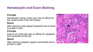
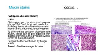
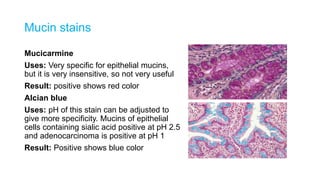
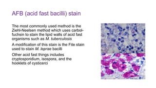
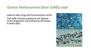
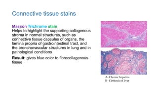
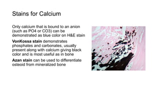
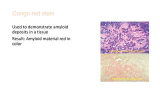
2. Hematoxylin and Eosin Staining
Principle
Hematoxylin, being a basic dye, has an affinity for
the nucleic acids in the cell nucleus
Result
After staining nuclei appear basophilic that is bluish
to purple in color
Principle
Eosin is an acidic dye with an affinity for cytoplasm
and membrane of the cell
Result
After staining cytoplasm appear eosinophilic that is
pinkish in color
3. Mucin stains contin…
PAS (peroidic acid-Schiff)
Uses:
Stains glycogen, mucins, mucoprotein,
glycoprotein and fungi and useful for
outlining tissue structures--basement
membranes, capsules, blood vessels
To differentiate between glycogen from
mucin, tissue can be pre-digested with
diastase/amylase, the glycogen will be
removed but not mucins
Fungus; further confirmed by fungal
stains
Result: Positives magenta color
4. Mucin stains
Mucicarmine
Uses: Very specific for epithelial mucins,
but it is very insensitive, so not very useful
Result: positive shows red color
Alcian blue
Uses: pH of this stain can be adjusted to
give more specificity. Mucins of epithelial
cells containing sialic acid positive at pH 2.5
and adenocarcinoma is positive at pH 1
Result: Positive shows blue color
5. Melanin stain - Fontana-Masson
Normal melanin granules brownish in color
Principle: Fontana-Masson method relies
upon the melanin granules to reduce
ammonical silver nitrate thus giving
blackish staining (but argentaffin,
chromaffin, and some lipochrome
pigments also stain black)
Schmorl's method helps in differentiating
melanin by using the reducing properties
of melanin to stain granules blue-green
Normal
6. Lipochrome (lipofucsin) pigments
Lipochrome (wear-and-tear pigments) products
of intracellular breakdown from oxidation of lipids
and lipoproteins
Found most commonly in heart, liver, CNS, and
adrenal cortex (zona reticularis)
Lipochrome can be stained by;
- Sudan black B
- Schmorl's methods
7. Iron (hemosiderin) stain
Perl's iron stain
Classic method for demonstrating iron in
tissues
Principle: Tissue section treated with
dilute HCL to release ferric ions from
binding proteins which react with
potassium ferrocyanide to produce an
insoluble blue compound (the Prussian
blue reaction) Hemosiderin, liver, iron stain.
8. Fat stains
Oil red O (ORO) is a rapid and simple
stain, can identify neutral lipids and fatty
acids in smears and tissues
It is useful in identifying fat emboli in lung
tissue or clot sections of peripheral blood
Fresh smears or cryostat sections of tissue
are necessary because fixatives
containing alcohols, or routine tissue
processing with clearing, will remove lipids
Oil red O stain of fat emboli in lung
9. Romanowsky-type" stains
Giemsa stain; can be helpful for identifying
components in a variety of tissues
Wright-Giemsa stain; utilized to stain
peripheral blood smears
Methylene blue and Toluidine blue dyes;
Have property of metachromasia, this means
that a tissue component stains a different
color than the dye itself
For example, mast cell graules, cartilage,
mucin, and amyloid will stain purple and not
blue, which is helpful in identifying these
components Cytoplasmic granules of mast cells in the
upper dermis stain metachromatically,
giving the intense purple color
10. AFB (acid fast bacilli) stain
The most commonly used method is the
Ziehl-Neelsen method which uses carbol-
fuchsin to stain the lipid walls of acid fast
organisms such as M. tuberculosis
A modification of this stain is the Fite stain
used to stain M. leprae bacilli
Other acid fast things includes
cryptosporidium, isospora, and the
hooklets of cysticerci
11. Gomori Methenamine Silver (GMS) stain
Used to stain fungi and Pneumocystis carinii
Cell walls of these organisms are stained,
so the organisms are outlined by the brown
to black stain
Cryptococcus neoformans
Candida albicans
12. Connective tissue stains
Masson Trichrome stain
Helps to highlight the supporting collagenous
stroma in normal structures, such as
connective tissue capsules of organs, the
lamina propria of gastrointestinal tract, and
the bronchovascular structures in lung and in
pathological conditions
Result: gives blue color to fibrocollagenous
tissue
A- Chronic hepatitis
B- Cirrhosis of liver
13. Stains for Calcium
Only calcium that is bound to an anion
(such as PO4 or CO3) can be
demonstrated as blue color on H&E stain
VonKossa stain demonstrates
phosphates and carbonates, usually
present along with calcium giving black
color and is most useful as in bone
Azan stain can be used to differentiate
osteoid from mineralized bone
14. Congo red stain
Used to demonstrate amyloid
deposits in a tissue
Result: Amyloid material red in
color
Positive for amyloid stain
Medullary carcinoma thyroid
15. Assignment
Q1. Name the components of the cell which appear basophilic and
eosinophilic after staining with hematoxylin and eosin, and also give reason.
Q2. Gastric biopsy shows signet ring cell carcinoma and superadded
candida infection.
a. Name the special stain to demonstrate signet ring cell carcinoma
b. Which stain can be helpful for the identification of candida albicans?
Q3. Liver biopsy of a thalassemia patient is submitted to check the iron
deposits. Which stain can be used for this purpose and what will be result of
staining?
Q4. To assess the stage of fibrosis in a liver biopsy from a patient of chronic
hepatitis ; which special stain you will apply on a liver biopsy section and
what will be the result of staining?