SIMPLE & DIFFERENTIALSTAINING
• Staining is an auxiliary technique used in
microscopy to enhance contrast in the microscopic
image.
• Stains and dyes are frequently used in biology and
medicine to highlight structures in biological tissues
for viewing, often with the aid of different
microscopes.
• Stains may be used to;
define and examine
bulk tissues (highlighting, for example,
muscle fibers or connective tissue),
cell populations (classifying different blood cells, for
instance), or organelles within individual cells
3.
SIMPLE & DIFFERENTIALSTAINING
• In biochemistry it involves adding a class-specific (
DNA, proteins, lipids, carbohydrates) dye to a
substrate to qualify or quantify the presence of a
specific compound.
• Staining and fluorescent tagging can serve similar
purposes.
• Biological staining is also used to mark cells in flow
cytometry, and to flag proteins or nucleic acids in
gel electrophoresis
4.
SIMPLE & DIFFERENTIALSTAINING
• Staining is not limited to biological
materials, it can also be used to study the
morphology of other materials for example
the lamellar structures of semi-crystalline
polymers or the domain structures of
block copolymers
5.
STAINS
• stain .a substance used to impart color to tissues
or cells, to facilitate microscopic study and
identification
• differential stain one which facilitates
differentiation of various elements in a specimen.
• supravital stain a stain introduced in living tissue
that has been removed from the body, but before
cessation of the chemical life of the cells
• vital stain a stain introduced into the living
organism, and taken up selectively by various
tissues or cellular elements
6.
Supravital staining:
• Avital stain (e.g., trypan blue) is applied
to an animal in life;
• a supravital stain (e.g., Janus green,
neutral red) is one that is applied to cells
or tissues removed from the body.
7.
Introduction
• a generaloverview of some histological
stains used to identify structures in cells
and tissues.
• This stains information should also be
considered in relation to Histology fixatives
.
8.
Staining Reactions
• Stainingreactions have both physical and chemical
characteristics.
• The mechanisms involved in staining include the
following:
• The dye may actually be dissolved in the stained
substance.
Most fat staining is accomplished in this fashion.
• A dye may be absorbed on the surface of a structure,
or
• dyes may be precipitated within the structure, simply
because environmental factors (pH, ionic strength,
temperature, etc.) favor absorption or precipitation.
9.
Staining Reactions
• Moststaining reactions involve a chemical
union between dye and stained substance
through salt linkages, hydrogen bonds, or
others.
• Staining with these dyes results in a
predictable color pattern based in part on
the acid base characteristics of the tissue.
10.
Staining Reactions
• However,color and color distribution are not
absolutely reliable for discrimination between
tissue components.
• Color will vary not only with specific stains
used, but also with the conditions that exist
during preparation of the slide
These include everything from the initial
fixing solution to the ionic strength of the
staining solution and the differentiating
solvents utilized after staining.
11.
Staining Reactions
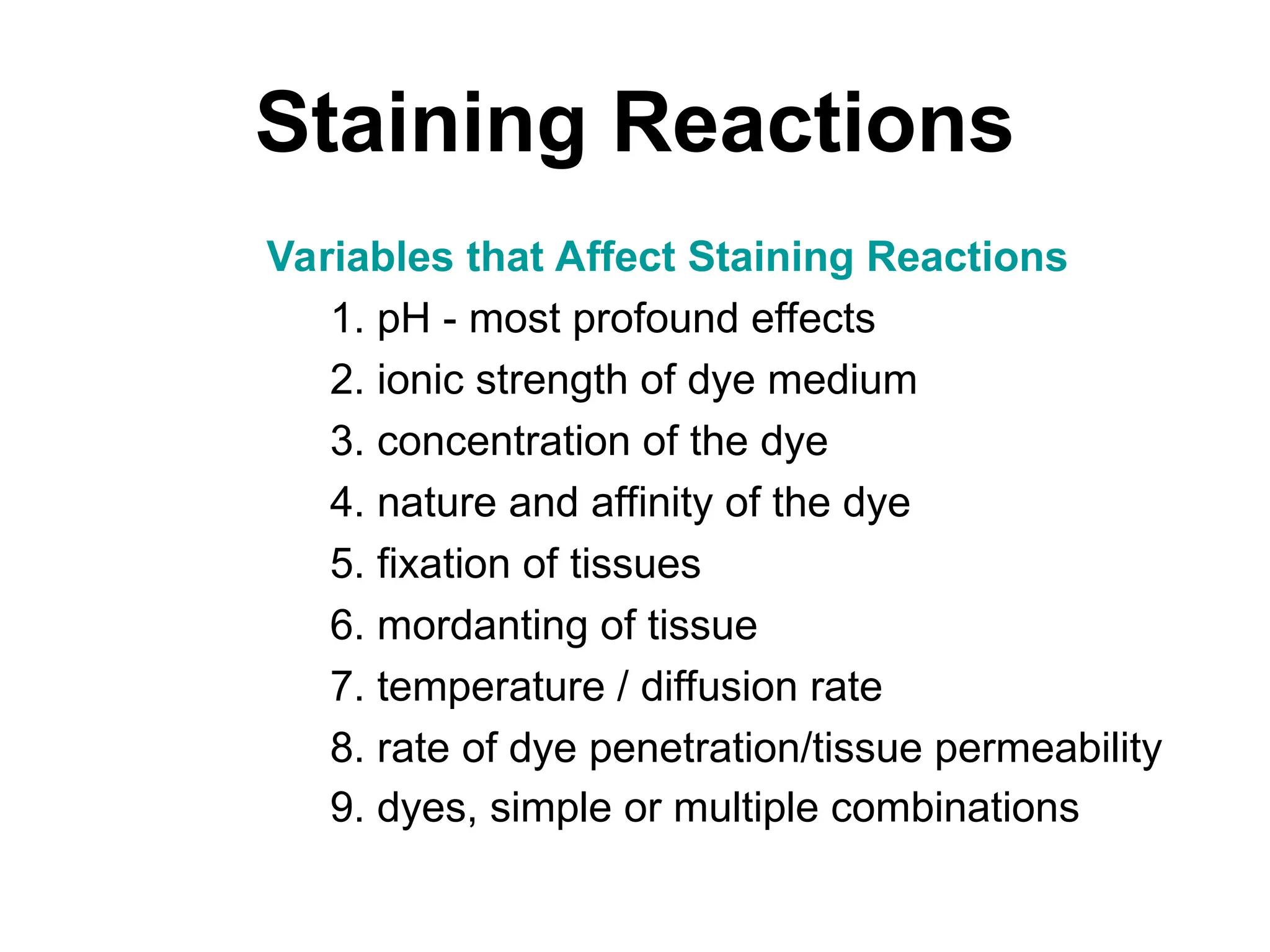
Variables thatAffect Staining Reactions
1. pH - most profound effects
2. ionic strength of dye medium
3. concentration of the dye
4. nature and affinity of the dye
5. fixation of tissues
6. mordanting of tissue
7. temperature / diffusion rate
8. rate of dye penetration/tissue permeability
9. dyes, simple or multiple combinations
12.
TYPES OF STAINING
A.Polychroming
• Process where a pure dye spontaneously forms
other dyes
• Basis for modern blood staining
• Example Methylene Blue => Polychrome
Methylene Blue
– Dye in solution oxidized to compounds of lower
methylation
– Mostly Azure A & B, more selective, more violet in color
– Accounts for differential white cell staining
13.
Polychroming cont--
• Wrightsstain = methylene Blue (basic)
& Eosin (acidic)
–Eosin added to methylene blue solution
forms insol. ppt
–Dry stain made up in alcoholic solution
–Water added during staining process on
slide
–Exact mechanism not known
14.
Metachromasia
• A puredye selectively stains a cell or tissue
component a color different from the original dye.
• MUST be a pure dye (NOT poly chrome).
• "Characteristic reversible color change that any dye
may under go by virtue of a change in its environment
NOT involving a chemical reaction of the dye
15.
Metachromasia cont--
• Commonlyused metachromatic dyes:
– toluidine Blue O
– thionine
– methylene blue
– cresyl violet
– celestine blue
– gallocyanin
– methyl violet
– safranin O
– azures
16.
Metachromasia cont---
• Consideredvaluable in the study of
specific elements of connective tissue:
mast cells, amyloid, cartilage, and mucous
material
17.
Acid and BasicDyes
• Most histologic dyes are classified either as acid
or as basic dyes.
• An acid dye exists as an anion (negatively
charged) in solution,
• while a basic dye exists as a cation (positive
charge).
• For instance, in the hematoxylin-eosin stain
(H&E), the hematoxylin-metal complex acts as a
basic dye. The eosin acts as an acid dye.
18.
Acid and BasicDyes
• Any substance that is stained by the basic
dye is considered to be basophilic;
• it carries acid groups which bind the basic
dye through salt linkages.
• When using hematoxylin, basophilic
structures in the tissue appear blue (or
purple or brown; this varies according to
the stain that is being used).
19.
Acid and BasicDyes
• A substance that is stained by an acid dye
is referred to as acidophilic;
• it carries basic groups which bind the acid
dye.
• With eosin, acidophilic structures appear
in various shades of pink.
• Since eosin is a widely used acid dye,
acidophilic substances are frequently
referred to as eosinophilic
20.
hematoxylin-eosin stain
• Awidely used, two-stage stain for cells in
which hematoxylin is followed by a
counterstain of red eosin so that the nuclei
stain a deep blue-black and the cytoplasm
stains pink
21.
hematoxylin-eosin stain
• Thisis a good general stain and is widely used.
• Most of your slides are stained with H&E. A
hematoxylin-metal complex acts a as a basic
dye, staining nucleic acids in the nucleus and
the cytoplasm blue, brown, or black.
• Eosin is an acid aniline dye which stains the
more basic proteins within cells (cytoplasm) and
in extracellular spaces (collagen) pink to
22.
Eosin
• Stains cytoplasmpink to red; red blood
cells are also bright red.
• Common counterstain to hematoxylin.
• Stain intensity varies with the formula as
well as the fixative.
23.
Hematoxylin
• Stains nucleiblue to dark-blue.
• Stains the matrix of hyaline cartilage,
myxomatous, and mucoid material pale
blue.
• Stains myelin weakly but is not noticeable
if combined with eosin stain.
Periodic acid-Schiff (PAS)
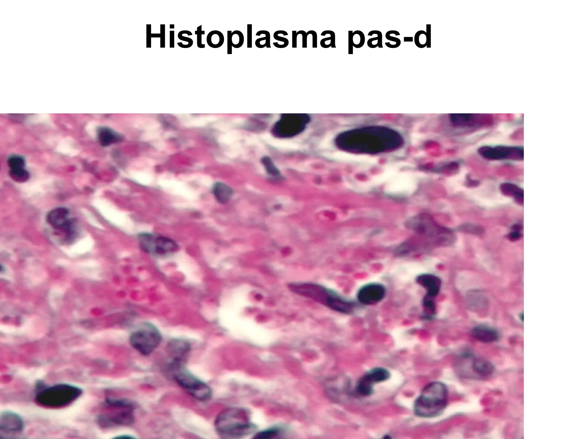
•Periodic acid-Schiff (PAS)
• Stains glycogen, mucin, fungus, basement
membrane and other substances.
• Stain used to detect fungal organisms and
cytoplasmic accumulation of glycogen.
• Stains lysosomes granules red-purple, can
be used in recognition of macrophages.
26.
PAS staining
• Periodicacid-Schiff staining is used to
mark carbohydrates (glycogen,
glycoprotein, proteoglycans).
• It is used to distinguish different types of
glycogen storage diseases
27.
Periodic acid-Schiff (PAS)
•Adjacent hydroxyl groups
(1, 2 glycols) or amino
and hydroxyl groups are
oxidized to aldehyde
groups with periodic acid.
Schiff's Reagent then
produces a red or
magenta addition product
with the aldehyde groups
and this technique
identifies a number of
polysaccharides and
carbohydrate-containing
compounds
Alcian Blue
• Stainsmucopolysaccharides or
glycosaminoglycans
• cationic dye (positively charged molecule)
for the demonstration of
glycosaminoglycans.
• binds anionic (negative) sites on the
polysaccharide.
.
30.
Alcian Blue Stain
•Bronchus. Alcian blue
staining of the bronchus
highlights the presence of
goblet cells in the mucosa
(slightly increased in this
case), as well as the
bronchial
submucosalglands.
• Beneath the epithelial
basement membrane,
there is a vascularized
layer of connective tissue
with wisps of smooth
muscle above the
submucosal glands
31.
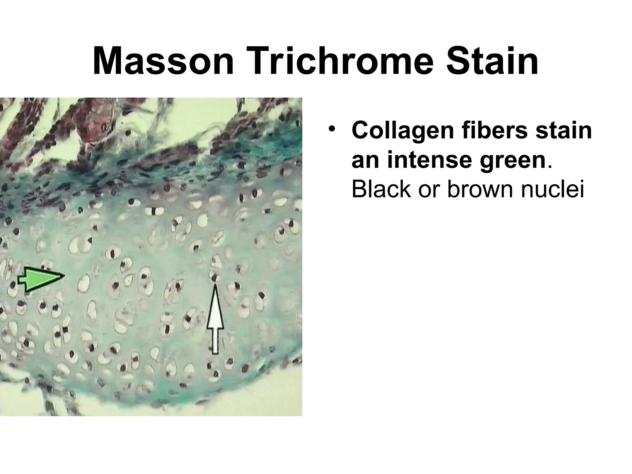
Masson’s Trichrome Stain
•Masson’s Trichrome Stain
• Stains nuclei deep blue, skeletal and smooth
muscles red, collagen and mucin blue.
• Stains brain and spinal cord parenchymal tissue
dusky pink to red.
• Used to evaluate fibrosis
• Striations in skeletal muscles also shows up much
better in Masson’s trichrome than in hematoxylin
and eosin stain.
– Although called a trichrome, four dyes (hematoxylin,
Biebrich scarlet, acid fuchsin, and analine blue) are
utilized
32.
Masson's trichrome
• Masson'strichrome is (as the name implies) a
three-colour staining protocol. The recipe has
evolved from Masson's original technique for
different specific applications, but all are well-
suited to distinguish cells from surrounding
connective tissue. Most recipes produce red
keratin and muscle fibers, blue or green staining
of collagen and bone, light red or pink staining of
cytoplasm, and black cell nuclei
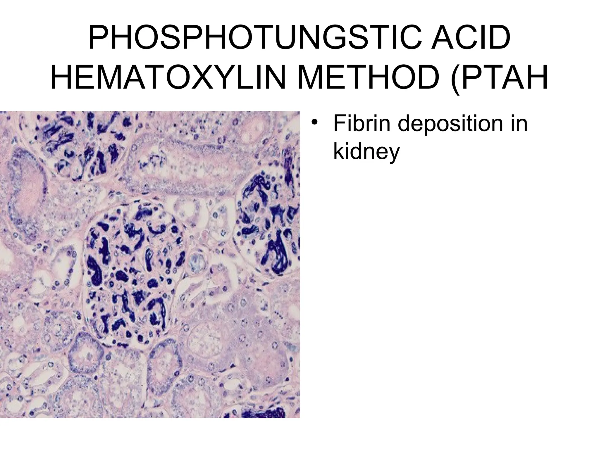
PhosphoTungstic Acid Hematoxylin
(PTAH)
•stains nucleus and cytoplasm detail and
connective tissue fibers.
• Stains collagen pink, fibrin blue, and
striated muscle blue.
• Historic stain used to show CNS reactive
astrocytes now used immunochemistry for
glial fibrillary acidic protein (GFAP).
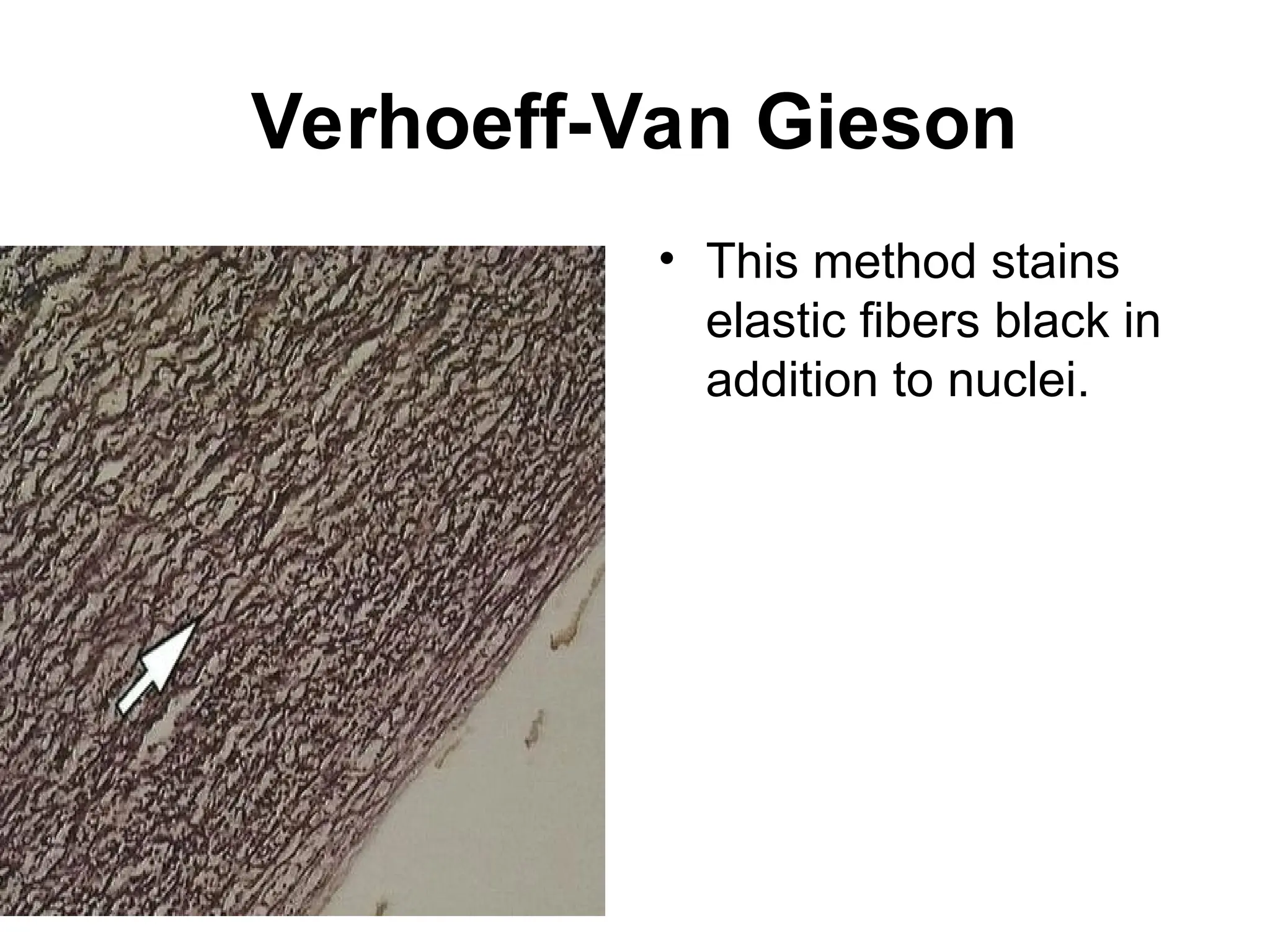
Verhoeff-Van Gieson
• Verhoeff-VanGieson or elastic-Van Gieson
(EVG) stain.
• This is a combination of Verhoeff’s elastic
stain which is a hematoxylin stain containing
ferric chloride and Wright’s iodine solution
and Van Gieson stain which contains acid
fuchsin, picric acid, and hematoxylin.
• Stains elastic fibers blue-black to black,
collagen pale red, other tissue elements
yellow, and nuclei blue to black
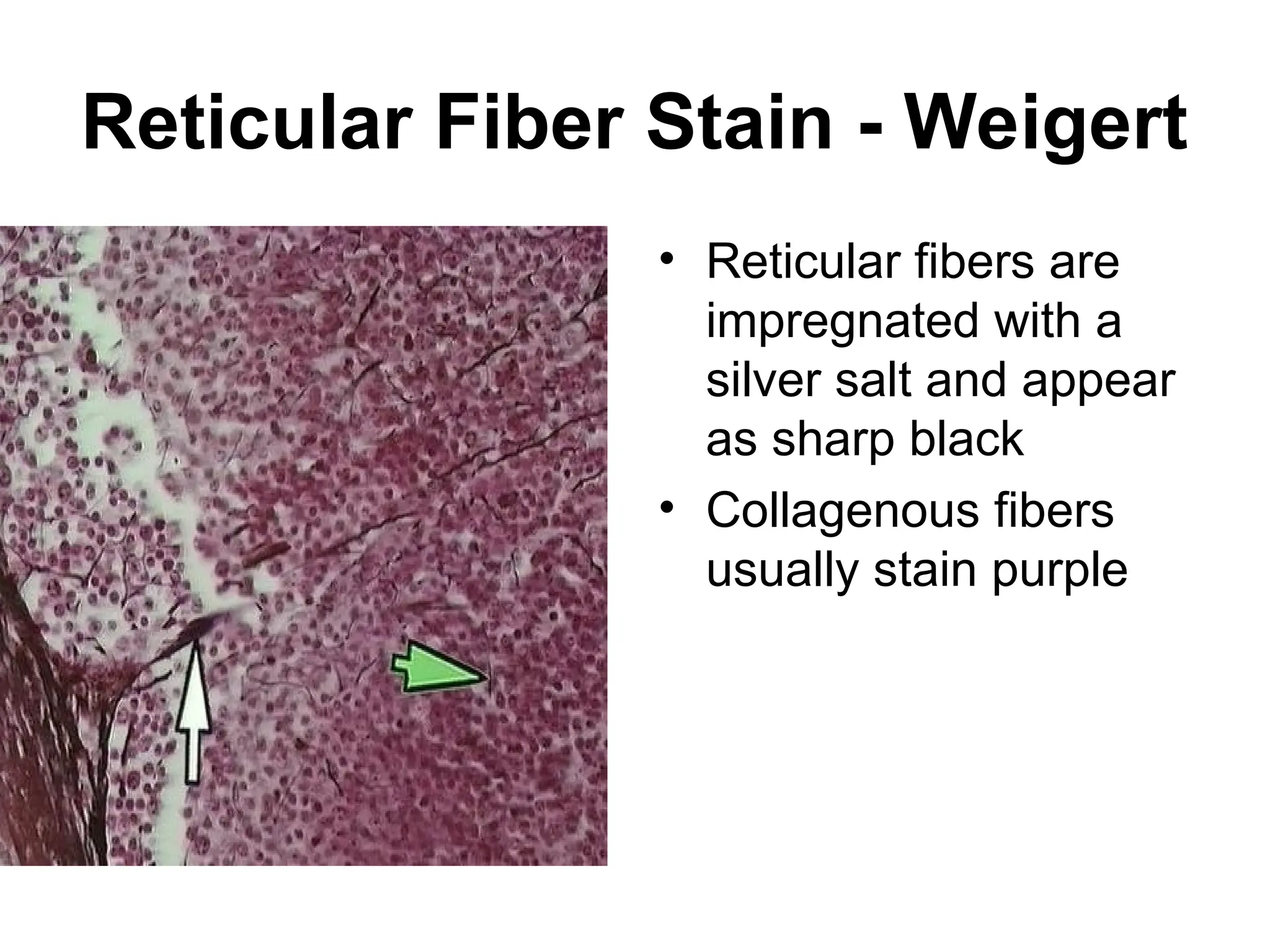
Reticular Fiber Stain- Weigert
• Reticular fibers are
impregnated with a
silver salt and appear
as sharp black
• Collagenous fibers
usually stain purple
40.
Silver staining
• Silverstaining is the use of silver to stain histologic
sections.
• This kind of staining is important especially to show
proteins (for example type III collagen) and DNA.
• It is used to show both substances inside and
outside cells.
• Silver staining is also used in
temperature gradient gel electrophoresis.
• Some cells are argentaffin.
These reduce silver solution to metallic silver after
formalin fixation.
.
41.
Silver staining----cont---
• Othercells are argyrophilic.
These reduce silver solution to
metallic silver after being exposed
to the stain that contains a
reductant, for example
hydroquinone or formalin
42.
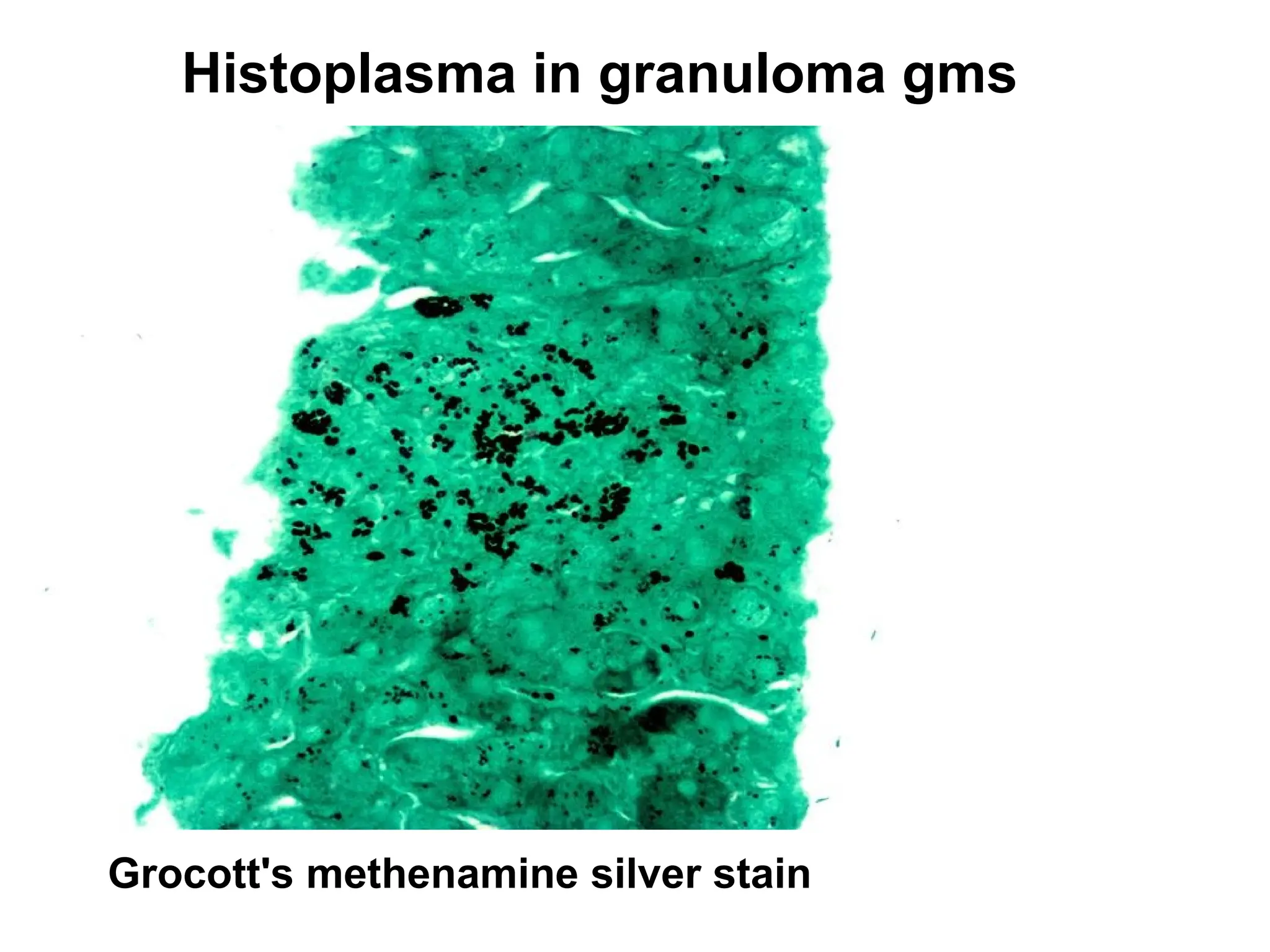
Silver staining----cont---
• Inpathology,
the Grocott-Gomori's (or Gömöri),
methenamine silver stain, abbreviated GMS, is a
popular staining method in histology.
• It is used widely as a screen for fungal organisms.
Particularly useful in staining carbohydrates.
• It can be used to identify the yeast-like fungus
Pneumocystis jiroveci which causes a form of
pneumonia called Pneumocystis Pneumonia (PCP)
or Pneumocystosis.
• The cell walls of these organisms are outlined by
the brown to black stain
Sudan staining
• Sudanstaining is the use of Sudan dyes to stain
sudanophilic substances, usually lipids.
Sudan III,
Sudan IV,
Oil Red O,
Osmium tetroxide, and
Sudan Black B are often used.
• Sudan staining is often used to determine the
level of fecal fat to diagnose steatorrhea
45.
Papanicolaou staining
• Papanicolaoustaining, or Pap staining, is a
frequently used method for examining cell
samples from various bodily secretions.
• It is frequently used to stain Pap smear
specimens.
• It uses a combination of
• haematoxylin,
• Orange G, eosin Y,
• Light Green SF yellowish, and sometimes
Bismarck Brown Y
46.
BIOLOGICAL STAINS
• StainingTechniques
• Because microbial cytoplasm is usually
transparent, it is necessary to stain microorganisms
before they can be viewed with the light
microscope.
• In some cases, staining is unnecessary, for
example when microorganisms are very large or
when motility is to be studied, and a drop of the
microorganisms can be placed directly on the slide
and observed.
A preparation such as this is called a wet mount.
47.
wet mount.
• Awet mount can also be prepared by
placing a drop of culture on a cover slip (a
glass cover for a slide) and then inverting
it over a hollowed out slide.
This procedure is called the hanging
drop.
48.
Simple stain techniques
Simple stain techniques.
• Staining can be performed with basic dyes such as
crystal violet or
methylene blue, positively charged dyes that are attracted to
the negatively charged materials of the microbial cytoplasm.
Such a procedure is the simple stain procedure.
• An alternative is to use a dye such as nigrosin or Congo red,
acidic, negatively charged dyes.
They are repelled by the negatively charged cytoplasm and
gather around the cells, leaving the cells clear and
unstained.
This technique is called the negative stain technique.
49.
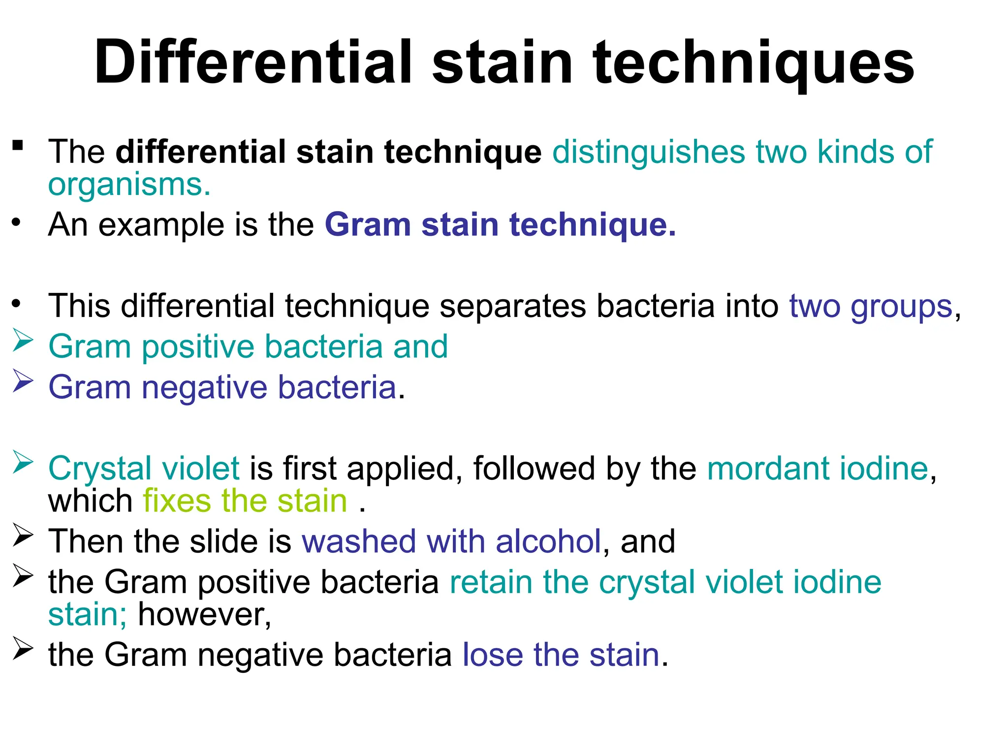
Differential stain techniques
The differential stain technique distinguishes two kinds of
organisms.
• An example is the Gram stain technique.
• This differential technique separates bacteria into two groups,
Gram positive bacteria and
Gram negative bacteria.
Crystal violet is first applied, followed by the mordant iodine,
which fixes the stain .
Then the slide is washed with alcohol, and
the Gram positive bacteria retain the crystal violet iodine
stain; however,
the Gram negative bacteria lose the stain.
50.
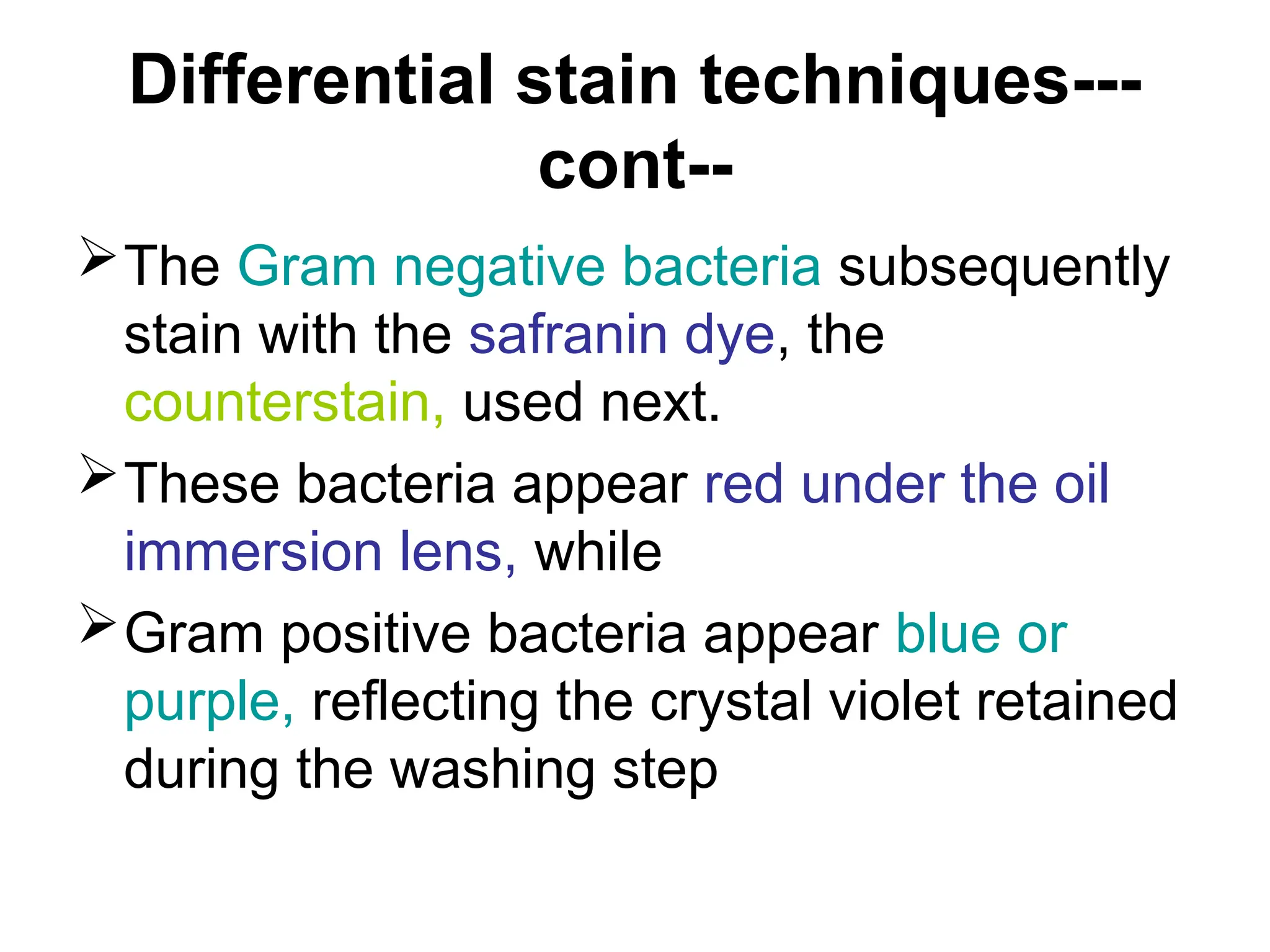
Differential stain techniques---
cont--
TheGram negative bacteria subsequently
stain with the safranin dye, the
counterstain, used next.
These bacteria appear red under the oil
immersion lens, while
Gram positive bacteria appear blue or
purple, reflecting the crystal violet retained
during the washing step
51.
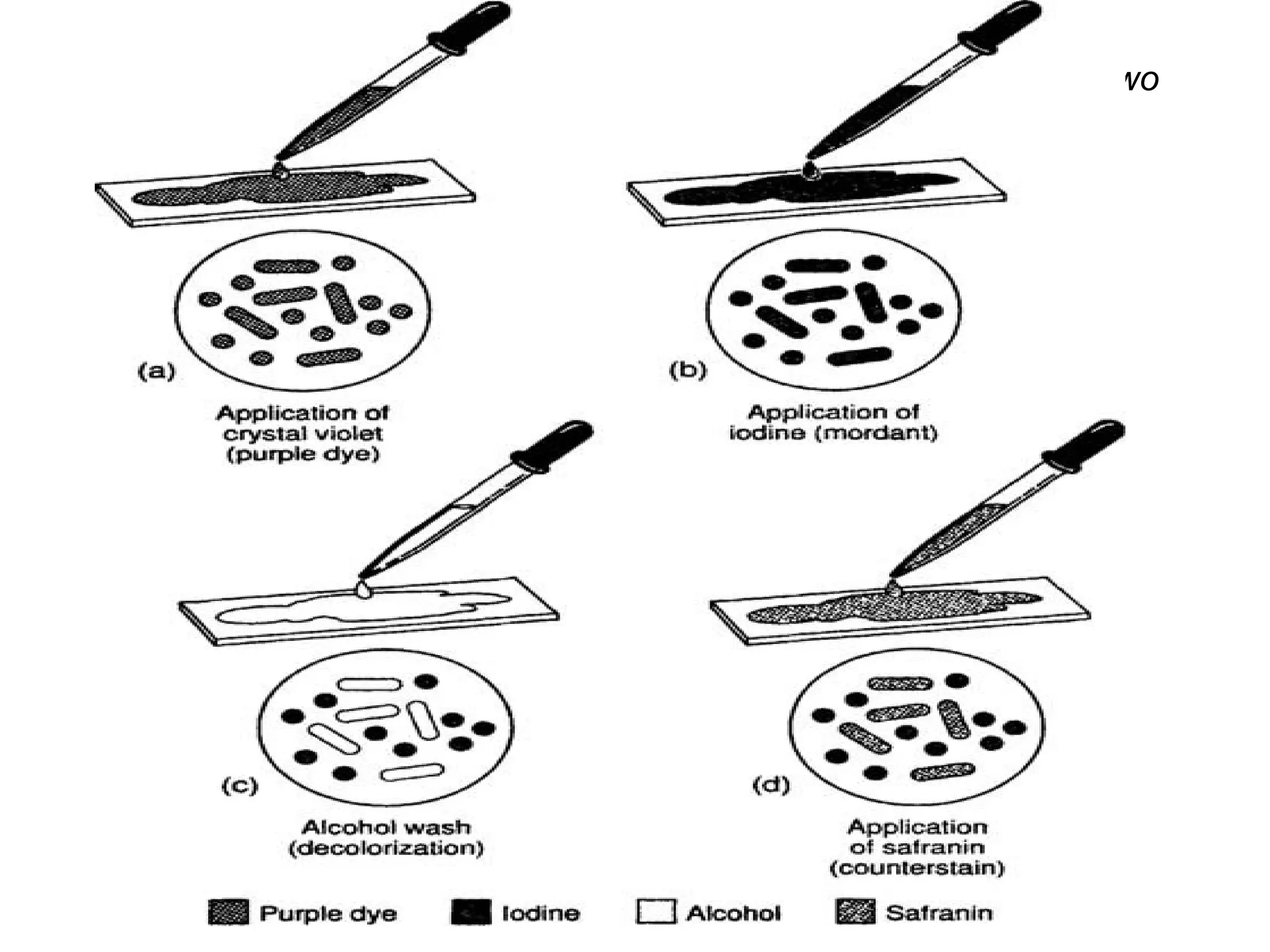
The Gram stainprocedure used for differentiating bacteria into two
groups
52.
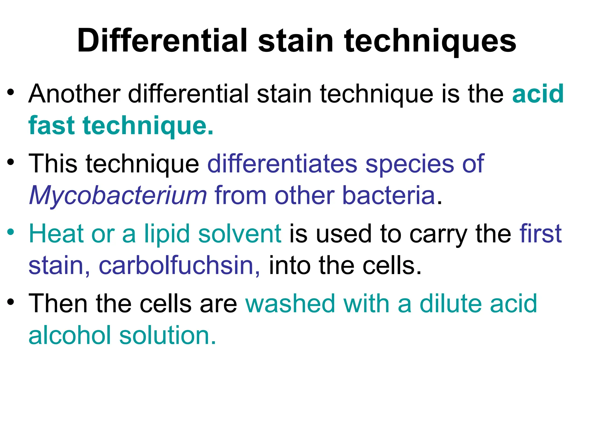
Differential stain techniques
•Another differential stain technique is the acid
fast technique.
• This technique differentiates species of
Mycobacterium from other bacteria.
• Heat or a lipid solvent is used to carry the first
stain, carbolfuchsin, into the cells.
• Then the cells are washed with a dilute acid
alcohol solution.
53.
Differential stain techniques
•Mycobacterium species resist the effect of
the acid alcohol and retain the carbolfuchsin
stain (bright red).
• Other bacteria lose the stain and take on the
subsequent methylene blue stain (blue).
• Thus, the acid fast bacteria appear bright
red, while the nonacid fast bacteria appear
blue when observed under oil immersion
microscopy.
54.
Differential stain techniques
Other stain techniques seek to identify various
bacterial structures of importance.
• For instance, a special stain technique highlights the
flagella of bacteria by coating the flagella with dyes or
metals to increase their width.
Flagella so stained can then be observed.
• A special stain technique is used to examine bacterial
spores.
Malachite green is used with heat to force the stain
into the cells and give them color.
A counterstain, safranin, is then used to give color to
the non sporeforming bacteria.
At the end of the procedure, spores stain green and
other cells stain red.
55.
BIOLOGICAL STAINS
• Invivo staining ( Intra Vital Staining ):
is the process of dyeing living tissues—in
vivo means "in life" (compare with in vitro
staining).
By causing certain cells or structures to
take on contrasting colour(s), their form
(morphology) or position within a cell or
tissue can be readily seen and studied..
56.
BIOLOGICAL STAINS- Invivo
staining ( Intra Vital Staining
• The usual purpose is to reveal cytological
details that might otherwise not be
apparent; however, staining can also
reveal where certain chemicals or specific
chemical reactions are taking place within
cells or tissues
57.
BIOLOGICAL STAINS
• Invitro staining involves ;
colouring cells or structures that have been
removed from their biological context.
Certain stains are often combined to reveal
more details and features than a single stain
alone.
Combined with specific protocols for fixation
and sample preparation, scientists and
physicians can use these standard techniques
as consistent, repeatable diagnostic tools.
58.
BIOLOGICAL STAINS-- Invitro
staining
• A counterstain is stain that makes cells or
structures more visible, when not
completely visible with the principal stain
• For example, crystal violet stains only
Gram-positive bacteria in Gram staining.
• A safranin counterstain is applied that
stains all cells, allowing identification of
Gram-negative bacteria
59.
BIOLOGICAL STAINS
• Whileex vivo, many cells continue to live
and metabolize until they are "fixed".
Some staining methods are based on this
property.
• Those stains excluded by the living cells
but taken up by the already dead cells are
called vital stains (e.g. trypan blue or
propidium iodide for eukaryotic cells).
60.
BIOLOGICAL STAINS
• Thosethat enter and stain living cells are called
supravital stains (e.g. New Methylene Blue and
Brilliant Cresyl Blue for reticulocyte staining).
However, these stains are eventually toxic to the
organism, some more so than others.
• Partly due to their toxic interaction inside a living
cell, when supravital stains enter a living cell, they
might produce a characteristic pattern of staining
different from the staining of an already fixed cell
(e.g. "reticulocyte" look versus diffuse
"polychromasia")..
61.
Endospore staining
• Endosporestaining is used to identify the
presence or absence of endospores,
which make bacteria very difficult to kill.
This is particularly useful for identifying
endospore-forming bacterial pathogens
like Clostridium difficile.
62.
Common biological stains
•There are several staining methods that are
used routinely with bacteria.
• These methods may be classified as
• 1) simple (nonspecific) and
• 2) differential (specific).
1) Simple stains will react with all microbes in
an identical fashion.
They are useful solely for increasing
contrast so that morphology, size, and
arrangement of organisms can be
determined.
63.
Common biological stains
2)Differential stains give varying results
depending on the organism being treated.
These results are often helpful in
identifying the microbe
64.
Simple Stains: Directand
Indirect Staining
• Stains (dyes) are chemicals containing
chromophores, groups that impart color.
• Their specificity is determined by their chemical
structure.
• Stains are generally salts in which one of the ions is
colored. (A salt is a compound composed of a
positively charged ion and a negatively charged
ion.)
For example, the dye methylene blue is actually the
salt methylene blue chloride which will dissociate in
water into a positively charged methylene blue ion
which is blue in color and a negatively charged
chloride ion which is colorless
65.
Simple Stains: Directand
Indirect Staining
• Commonly used microbiological stains
generally fall into one of two categories –
• 1.)Basic stains or
2.) acidic stains
( although there are a few stains such as
India Ink) which are neutral).
• A basic dye is a stain that is cationic
(positively charged) and will therefore react
with material that is negatively charged
66.
Simple Stains: Directand
Indirect Staining
• The cytoplasm of all bacterial cells have a
slight negative charge when growing in a
medium of near neutral pH and will
therefore attract and bind with basic dyes
• Some examples of basic dyes are
crystal violet,
safranin,
basic fuchsin and
methylene blue
67.
Simple Stains: Directand
Indirect Staining
• Acid dyes have negatively charged
chromophores and are repelled by the
bacterial surface forming a deposit aroung
the organism.
• They stain the background and leave the
microbe transparent.
Nigrosine and
congo red are examples of acid dyes.
68.
• Note: Thedyes used for bacteriological
staining are generally aniline dyes, derived
from coal tar, which means they are
POTENTIALLY CARCINOGENIC and
should be handled carefully.
• Avoid contact with them by keeping them
off skin, clothing and benches.
69.
Common biological stains
•Acridine orange (AO)
is a nucleic acid selective fluorescent
cationic dye useful for cell cycle
determination.
It is cell-permeable, and interacts with
DNA and RNA by intercalation or
electrostatic attractions.
• When bound to DNA, it is very similar
spectrally to fluorescein.
70.
Common biological stains
Cresyl violet
• Cresyl violet stains the acidic components
of the neuronal cytoplasm a violet colour,
specifically nissl bodies.
• Often used in brain research
• Crystal violet, when combined with a
suitable mordant, stains cell walls purple.
Crystal violet is an important component in
Gram staining
71.
Common biological stains
•Eosin
• Eosin is most often used as a counterstain
to haematoxylin, imparting a pink or red
colour to
cytoplasmic material,
cell membranes, and
some extracellular structures.
• It also imparts a strong red colour to red
blood cells..
72.
Eosin
• Most oftenused is eosin Y (also known as
eosin Y ws or eosin yellowish); it has a
very slightly yellowish cast.
• The other eosin compound is eosin B
(eosin bluish or imperial red); it has a very
faint bluish cast.
• The two dyes are interchangeable, and the
use of one or the other is more a matter of
preference and tradition
73.
Common biological stains
•Haematoxylin
is a nuclear stain.
Used with a mordant, haematoxylin stains
nuclei blue-violet or brown.
It is most often used with eosin in H&E
(haematoxylin and eosin) staining—one of
the most common procedures in histology
74.
Common biological stains
Acid fuchsine
• Acid fuchsine may be used to stain
collagen,
smooth muscle, or
mitochondria.
• Acid fuchsine is used as the nuclear and cytoplasmic
stain in Mallory's trichrome method.
• Acid fuchsine stains cytoplasm in some variants of
Masson's trichrome.
• In Van Gieson's picro-fuchsine, acid fuchsine imparts its
red colour to collagen fibres.
• Acid fuchsine is also a traditional stain for mitochondria
(Altmann's method)
75.
Romanowsky stains
• TheRomanowsky stains are all based on a combination of
eosinate (chemically reduced eosin) and methylene blue
(sometimes with its oxidation products azure A and azure B
).
• Common variants include
Wright's stain,
Jenner's stain,
May-Grunwald stain,
Leishman stain and
Giemsa stain.
All are used to examine blood or bone marrow samples.
They are preferred over H&E for inspection of blood cells
because different types of leukocytes (white blood cells) can
be readily distinguished.
All are also suited to examination of blood to detect blood-
borne parasites like malaria
76.
Stainability of tissues
•Tissues which take up stains are called
chromatic. Chromosomes were so named
because of their ability to absorb a violet
stain.
• Positive affinity for a specific stain may be
designated by the suffix -philic. For example,
tissues that stain with an azure stain may be
referred to as azurophilic.
77.
Stainability of tissues—cont--
•This may also be used for more
generalized staining properties, such as
acidophilic for tissues that stain by acidic
stains (most notably eosin),
basophilic when staining in basic dyes,
and
amphophilic when staining with either acid
or basic dyes.
• In contrast, Chromophobic tissues do not
take up coloured dye readily