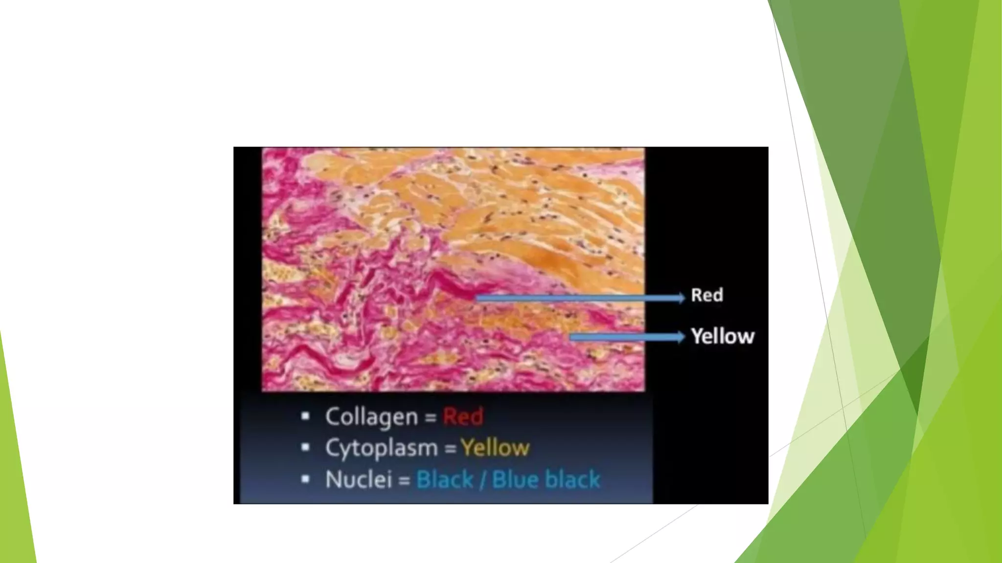
Special stains are used in dermatopathology to enhance contrast in microscopic images and selectively stain cells, structures, and molecules. Common special stains include PAS for glycogen and basement membranes, Alcian blue for mucins, trichrome stains for collagen, Verhoeff-Van Gieson for collagen and muscle, Congo red for amyloid, Sudan stains for lipids, Giemsa and toluidine blue for mast cells, GMS and Fontana-Masson for fungi and melanin, Ziehl-Neelsen for acid-fast bacteria, and Gram staining for classifying bacteria. Each stain colors specific tissues or molecules to aid in diagnosis and examination of cellular components and structures.
Pathology Special Stains for FFPE Tissue StainingBioGenex
Special stains refer to alternative staining techniques that are used when H&E stains do not provide all the cellular information required. These techniques use a variety of dyes and methods so that pathologists can visualize tissue morphology and detect the presence of particular cell types, structures or pathogens (e.g. bacteria).BioGenex offers broadest special stain menu anywhere (over 30 special stains), including:
Grocott’s Methenamine Silver (GMS) Stain
Reticulin Stains
Trichrome Stains
Giemsa Stain
Periodic Acid-Schiff (PAS) Stains
Human colors. color of normal and pathologic tissueManan Shah
Colors are important to all living organisms
They are crucial for protection, metabolism, sexual behavior, and communication.
Human organs obviously have color, that is, the liver is brown, the heart is red, bones are white, and so on.
Hematoxylin and Eosin (H&E) staining is a routine staining technique that reveals exceptional detail of tissue structure and makeup of the cells. Stained cell structures (e.g. nucleus, cytoplasm, organelles, extra-cellular components) provide important information for tissue-based cancer diagnosis. Special stains refer to alternative staining techniques that are used when H&E stains do not provide all the cellular information required. These techniques use a variety of dyes and methods so that pathologists can visualize tissue morphology and detect the presence of particular cell types, structures or pathogens (e.g. bacteria). We have the broadest special stain† menu anywhere (over 30 special stains), including:
Grocott’s Methenamine Silver (GMS) Stain
Reticulin Stains
Trichrome Stains
Giemsa Stain
Periodic Acid-Schiff (PAS) Stains
For More information Contact Customer support at customer.service@biogenex.com or follow the link http://biogenex.com/us/applications/special-stains/special-stains-controls.html
These lecture slides, by Dr Sidra Arshad, offer a quick overview of physiological basis of a normal electrocardiogram.
Learning objectives:
1. Define an electrocardiogram (ECG) and electrocardiography
2. Describe how dipoles generated by the heart produce the waveforms of the ECG
3. Describe the components of a normal electrocardiogram of a typical bipolar leads (limb II)
4. Differentiate between intervals and segments
5. Enlist some common indications for obtaining an ECG
Study Resources:
1. Chapter 11, Guyton and Hall Textbook of Medical Physiology, 14th edition
2. Chapter 9, Human Physiology - From Cells to Systems, Lauralee Sherwood, 9th edition
3. Chapter 29, Ganong’s Review of Medical Physiology, 26th edition
4. Electrocardiogram, StatPearls - https://www.ncbi.nlm.nih.gov/books/NBK549803/
5. ECG in Medical Practice by ABM Abdullah, 4th edition
6. ECG Basics, http://www.nataliescasebook.com/tag/e-c-g-basics
NVBDCP.pptx Nation vector borne disease control programSapna Thakur
NVBDCP was launched in 2003-2004 . Vector-Borne Disease: Disease that results from an infection transmitted to humans and other animals by blood-feeding arthropods, such as mosquitoes, ticks, and fleas. Examples of vector-borne diseases include Dengue fever, West Nile Virus, Lyme disease, and malaria.
Explore natural remedies for syphilis treatment in Singapore. Discover alternative therapies, herbal remedies, and lifestyle changes that may complement conventional treatments. Learn about holistic approaches to managing syphilis symptoms and supporting overall health.
Tom Selleck Health: A Comprehensive Look at the Iconic Actor’s Wellness Journeygreendigital
Tom Selleck, an enduring figure in Hollywood. has captivated audiences for decades with his rugged charm, iconic moustache. and memorable roles in television and film. From his breakout role as Thomas Magnum in Magnum P.I. to his current portrayal of Frank Reagan in Blue Bloods. Selleck's career has spanned over 50 years. But beyond his professional achievements. fans have often been curious about Tom Selleck Health. especially as he has aged in the public eye.
Follow us on: Pinterest
Introduction
Many have been interested in Tom Selleck health. not only because of his enduring presence on screen but also because of the challenges. and lifestyle choices he has faced and made over the years. This article delves into the various aspects of Tom Selleck health. exploring his fitness regimen, diet, mental health. and the challenges he has encountered as he ages. We'll look at how he maintains his well-being. the health issues he has faced, and his approach to ageing .
Early Life and Career
Childhood and Athletic Beginnings
Tom Selleck was born on January 29, 1945, in Detroit, Michigan, and grew up in Sherman Oaks, California. From an early age, he was involved in sports, particularly basketball. which played a significant role in his physical development. His athletic pursuits continued into college. where he attended the University of Southern California (USC) on a basketball scholarship. This early involvement in sports laid a strong foundation for his physical health and disciplined lifestyle.
Transition to Acting
Selleck's transition from an athlete to an actor came with its physical demands. His first significant role in "Magnum P.I." required him to perform various stunts and maintain a fit appearance. This role, which he played from 1980 to 1988. necessitated a rigorous fitness routine to meet the show's demands. setting the stage for his long-term commitment to health and wellness.
Fitness Regimen
Workout Routine
Tom Selleck health and fitness regimen has evolved. adapting to his changing roles and age. During his "Magnum, P.I." days. Selleck's workouts were intense and focused on building and maintaining muscle mass. His routine included weightlifting, cardiovascular exercises. and specific training for the stunts he performed on the show.
Selleck adjusted his fitness routine as he aged to suit his body's needs. Today, his workouts focus on maintaining flexibility, strength, and cardiovascular health. He incorporates low-impact exercises such as swimming, walking, and light weightlifting. This balanced approach helps him stay fit without putting undue strain on his joints and muscles.
Importance of Flexibility and Mobility
In recent years, Selleck has emphasized the importance of flexibility and mobility in his fitness regimen. Understanding the natural decline in muscle mass and joint flexibility with age. he includes stretching and yoga in his routine. These practices help prevent injuries, improve posture, and maintain mobilit
263778731218 Abortion Clinic /Pills In Harare ,sisternakatoto
263778731218 Abortion Clinic /Pills In Harare ,ABORTION WOMEN’S CLINIC +27730423979 IN women clinic we believe that every woman should be able to make choices in her pregnancy. Our job is to provide compassionate care, safety,affordable and confidential services. That’s why we have won the trust from all generations of women all over the world. we use non surgical method(Abortion pills) to terminate…Dr.LISA +27730423979women Clinic is committed to providing the highest quality of obstetrical and gynecological care to women of all ages. Our dedicated staff aim to treat each patient and her health concerns with compassion and respect.Our dedicated group ABORTION WOMEN’S CLINIC +27730423979 IN women clinic we believe that every woman should be able to make choices in her pregnancy. Our job is to provide compassionate care, safety,affordable and confidential services. That’s why we have won the trust from all generations of women all over the world. we use non surgical method(Abortion pills) to terminate…Dr.LISA +27730423979women Clinic is committed to providing the highest quality of obstetrical and gynecological care to women of all ages. Our dedicated staff aim to treat each patient and her health concerns with compassion and respect.Our dedicated group of receptionists, nurses, and physicians have worked together as a teamof receptionists, nurses, and physicians have worked together as a team wwww.lisywomensclinic.co.za/
Title: Sense of Smell
Presenter: Dr. Faiza, Assistant Professor of Physiology
Qualifications:
MBBS (Best Graduate, AIMC Lahore)
FCPS Physiology
ICMT, CHPE, DHPE (STMU)
MPH (GC University, Faisalabad)
MBA (Virtual University of Pakistan)
Learning Objectives:
Describe the primary categories of smells and the concept of odor blindness.
Explain the structure and location of the olfactory membrane and mucosa, including the types and roles of cells involved in olfaction.
Describe the pathway and mechanisms of olfactory signal transmission from the olfactory receptors to the brain.
Illustrate the biochemical cascade triggered by odorant binding to olfactory receptors, including the role of G-proteins and second messengers in generating an action potential.
Identify different types of olfactory disorders such as anosmia, hyposmia, hyperosmia, and dysosmia, including their potential causes.
Key Topics:
Olfactory Genes:
3% of the human genome accounts for olfactory genes.
400 genes for odorant receptors.
Olfactory Membrane:
Located in the superior part of the nasal cavity.
Medially: Folds downward along the superior septum.
Laterally: Folds over the superior turbinate and upper surface of the middle turbinate.
Total surface area: 5-10 square centimeters.
Olfactory Mucosa:
Olfactory Cells: Bipolar nerve cells derived from the CNS (100 million), with 4-25 olfactory cilia per cell.
Sustentacular Cells: Produce mucus and maintain ionic and molecular environment.
Basal Cells: Replace worn-out olfactory cells with an average lifespan of 1-2 months.
Bowman’s Gland: Secretes mucus.
Stimulation of Olfactory Cells:
Odorant dissolves in mucus and attaches to receptors on olfactory cilia.
Involves a cascade effect through G-proteins and second messengers, leading to depolarization and action potential generation in the olfactory nerve.
Quality of a Good Odorant:
Small (3-20 Carbon atoms), volatile, water-soluble, and lipid-soluble.
Facilitated by odorant-binding proteins in mucus.
Membrane Potential and Action Potential:
Resting membrane potential: -55mV.
Action potential frequency in the olfactory nerve increases with odorant strength.
Adaptation Towards the Sense of Smell:
Rapid adaptation within the first second, with further slow adaptation.
Psychological adaptation greater than receptor adaptation, involving feedback inhibition from the central nervous system.
Primary Sensations of Smell:
Camphoraceous, Musky, Floral, Pepperminty, Ethereal, Pungent, Putrid.
Odor Detection Threshold:
Examples: Hydrogen sulfide (0.0005 ppm), Methyl-mercaptan (0.002 ppm).
Some toxic substances are odorless at lethal concentrations.
Characteristics of Smell:
Odor blindness for single substances due to lack of appropriate receptor protein.
Behavioral and emotional influences of smell.
Transmission of Olfactory Signals:
From olfactory cells to glomeruli in the olfactory bulb, involving lateral inhibition.
Primitive, less old, and new olfactory systems with different path
Ethanol (CH3CH2OH), or beverage alcohol, is a two-carbon alcohol
that is rapidly distributed in the body and brain. Ethanol alters many
neurochemical systems and has rewarding and addictive properties. It
is the oldest recreational drug and likely contributes to more morbidity,
mortality, and public health costs than all illicit drugs combined. The
5th edition of the Diagnostic and Statistical Manual of Mental Disorders
(DSM-5) integrates alcohol abuse and alcohol dependence into a single
disorder called alcohol use disorder (AUD), with mild, moderate,
and severe subclassifications (American Psychiatric Association, 2013).
In the DSM-5, all types of substance abuse and dependence have been
combined into a single substance use disorder (SUD) on a continuum
from mild to severe. A diagnosis of AUD requires that at least two of
the 11 DSM-5 behaviors be present within a 12-month period (mild
AUD: 2–3 criteria; moderate AUD: 4–5 criteria; severe AUD: 6–11 criteria).
The four main behavioral effects of AUD are impaired control over
drinking, negative social consequences, risky use, and altered physiological
effects (tolerance, withdrawal). This chapter presents an overview
of the prevalence and harmful consequences of AUD in the U.S.,
the systemic nature of the disease, neurocircuitry and stages of AUD,
comorbidities, fetal alcohol spectrum disorders, genetic risk factors, and
pharmacotherapies for AUD.
Recomendações da OMS sobre cuidados maternos e neonatais para uma experiência pós-natal positiva.
Em consonância com os ODS – Objetivos do Desenvolvimento Sustentável e a Estratégia Global para a Saúde das Mulheres, Crianças e Adolescentes, e aplicando uma abordagem baseada nos direitos humanos, os esforços de cuidados pós-natais devem expandir-se para além da cobertura e da simples sobrevivência, de modo a incluir cuidados de qualidade.
Estas diretrizes visam melhorar a qualidade dos cuidados pós-natais essenciais e de rotina prestados às mulheres e aos recém-nascidos, com o objetivo final de melhorar a saúde e o bem-estar materno e neonatal.
Uma “experiência pós-natal positiva” é um resultado importante para todas as mulheres que dão à luz e para os seus recém-nascidos, estabelecendo as bases para a melhoria da saúde e do bem-estar a curto e longo prazo. Uma experiência pós-natal positiva é definida como aquela em que as mulheres, pessoas que gestam, os recém-nascidos, os casais, os pais, os cuidadores e as famílias recebem informação consistente, garantia e apoio de profissionais de saúde motivados; e onde um sistema de saúde flexível e com recursos reconheça as necessidades das mulheres e dos bebês e respeite o seu contexto cultural.
Estas diretrizes consolidadas apresentam algumas recomendações novas e já bem fundamentadas sobre cuidados pós-natais de rotina para mulheres e neonatos que recebem cuidados no pós-parto em unidades de saúde ou na comunidade, independentemente dos recursos disponíveis.
É fornecido um conjunto abrangente de recomendações para cuidados durante o período puerperal, com ênfase nos cuidados essenciais que todas as mulheres e recém-nascidos devem receber, e com a devida atenção à qualidade dos cuidados; isto é, a entrega e a experiência do cuidado recebido. Estas diretrizes atualizam e ampliam as recomendações da OMS de 2014 sobre cuidados pós-natais da mãe e do recém-nascido e complementam as atuais diretrizes da OMS sobre a gestão de complicações pós-natais.
O estabelecimento da amamentação e o manejo das principais intercorrências é contemplada.
Recomendamos muito.
Vamos discutir essas recomendações no nosso curso de pós-graduação em Aleitamento no Instituto Ciclos.
Esta publicação só está disponível em inglês até o momento.
Prof. Marcus Renato de Carvalho
www.agostodourado.com
Couples presenting to the infertility clinic- Do they really have infertility...Sujoy Dasgupta
Dr Sujoy Dasgupta presented the study on "Couples presenting to the infertility clinic- Do they really have infertility? – The unexplored stories of non-consummation" in the 13th Congress of the Asia Pacific Initiative on Reproduction (ASPIRE 2024) at Manila on 24 May, 2024.
These simplified slides by Dr. Sidra Arshad present an overview of the non-respiratory functions of the respiratory tract.
Learning objectives:
1. Enlist the non-respiratory functions of the respiratory tract
2. Briefly explain how these functions are carried out
3. Discuss the significance of dead space
4. Differentiate between minute ventilation and alveolar ventilation
5. Describe the cough and sneeze reflexes
Study Resources:
1. Chapter 39, Guyton and Hall Textbook of Medical Physiology, 14th edition
2. Chapter 34, Ganong’s Review of Medical Physiology, 26th edition
3. Chapter 17, Human Physiology by Lauralee Sherwood, 9th edition
4. Non-respiratory functions of the lungs https://academic.oup.com/bjaed/article/13/3/98/278874
- Video recording of this lecture in English language: https://youtu.be/lK81BzxMqdo
- Video recording of this lecture in Arabic language: https://youtu.be/Ve4P0COk9OI
- Link to download the book free: https://nephrotube.blogspot.com/p/nephrotube-nephrology-books.html
- Link to NephroTube website: www.NephroTube.com
- Link to NephroTube social media accounts: https://nephrotube.blogspot.com/p/join-nephrotube-on-social-media.html
2. WHAT IS STAIN?
Staining is an auxiliary technique used in microscopy to enhance contrast in the microscopic
images.
Stains may be used to define and examine the bulk tissue
1. Cell population
2. Muscle fibers
3. Connective tissues
4. Organelles
3. SPECIAL STAINS
Used in addition to H & E staining to selectively
Stain cells & cellular components
Gives information on:
Presence of certain class of molecules
Their localization
Number of molecules
5. PAS
Most commonly used special stain
Stains neutral mucopolysaccharides (Glycogen)
Basement membranes (e.g SLE)
Fungi, Parasides
Fibrin, Inclusion bodies
6.
7.
8.
9. ALCIAN BLUE
Best stain for mucin
Alcian blue (pH 2.5)
- Acid mucopolysaccharides (glycosaminoglycain’s)
- Light blue
Alcian blue (pH 0.5)
- Sulfated mucopolysaccharides (heparin sulfate)
- Blue
Does NOT stain neutral mucins
12. MUCICARMINE
To demonstrate acid & neutral mucopolysaccharides
- Epithelial mucin
- Capsule of cryptococcus neoformans
Results
Acid epithelial mucins : Deep rose to red
Nuclei : Black
Others tissue elements : Light yellow
13.
14. COLLOIDAL IRON
A mucin stain for dermal/connective tissue mucin
Stains acid mucopolysaccharides
RESULTS
Proteoglycans , Acid mucins : BRIGHT BLUE
Collagen : RED
Muscle & cytoplasm : YELLOW
15.
16. STAINS FOR PIGMENTS &
MINERALS
1. Fontana-Masson
2. Von-kossa
3. Alzarin red S
4. Prussian blue stain
17. FONTANA-MASSON
Stains melanin & argentaffin granules – BLACK (nuclei will be red)
Useful for quantifying melanocytes (e.g. in vitiligo) & in cases of minocycline pigmentary
alteration
Also stains cryptococcus
RESULTS
Melanin , Argentaffin, Chromaffin : BLACK
Nuclei : RED
18. USES
To identify melanin & argentaffin granules
In diagnosis of malignant melanoma
Argentaffin granules are found in carcinoid tumors
24. PRUSSIAN BLUE STAIN (PERLS IRON)
Hemosiderin and ferric ions
Useful for identifying iron as the source of pigment
Stains iron – BLUE
RESULT
Ferric iron : Blue
Nuclei : Red
25.
26. ALIZARIN RED
Stain for calcium salts
RESULTS
Calcium : ORANGE RED
Background : GREEN
29. TRICHROME-MASSON
Smooth muscle , cytoplasm, keratin – Red
Useful for distinguishing leiomyoma’s from dermatofibromas & neural tumors
Collagen – Blue/green
Useful in evaluating the characteristics of dermal collagen
Nucleus - Black
30. USES
It is used to differentiate b/w collagen & smooth muscle in tumor
To identified the increased collagen deposition : Keloid
RESULT
Nuclei : Blue/Black
Cytoplasm, Muscle, & Erythrocytes : Red
Collagen : Blue
31.
32. VERHOEFF-VAN GIESON OR WEIGERT
Used to differentiate collagen and smooth muscle
Can be used to demonstrate the presence of collagen in pathological conditions( DLE, EN, RA,
SLE, MORPHOEA)
Stains nuclei : Blue
Cytoplasm, muscle, fibrin : Yellow
Collagen : Bright
Elastic fibers : Black
42. SUDAN BLACK B
Most sensitive of all fat dyes
Stains neutral fats – Blue-Black
Stains phospholipid – Grey
43.
44. OIL RED O
Stains fat red
Frozen/Fresh tissue ( once tissue is fixed &processed into paraffin blocks, this method does not
work)
This may be very helpful in seeing the fat globules in sebaceous carcinoma
51. GRAM STAINING
Differentiates bacteria by the chemical & physical properties of their cell well by detecting
peptidoglycan
Gram +ve bacteria retain the crystal violet dye & thus are stained violet : BLUE
Gram –ve bacteria : PINK
52.
53. GOMORI METHENAMINE SILVER (GMS)
Fungal cell walls
Stains fungi & parasites brown or black with a green background
Pneumocystis carnii, histoplasma , leshmania
54.
55.
56. GIEMSA STAIN
Used to stain bacteria & protozoa
H. pylori, rickettsia & chlamydia
Bacteria stains : BLUE
Cytoplasm stains from : Pink to rose
Nuclei : BLUE
Eosinophils are also easily detected
60. ZIEHL-NEELSEN
Acid fast refers to cell walls containing high lipid content
Used to stain Mycobacteria, oocysts of Cryptosporidium parvum, Cyclospora, Isospora
Acid fast cells stain : RED
Non acid fast cells stain : BLUE
61.
62. ACID FAST STAINING
TECHNIQUES
Ziehl-Neelsen stain ( classic type)
Kinyoun
Auramine-rhodamine (fluorescent dyes)
Fite : for leprosy bacillus which is less acid fast