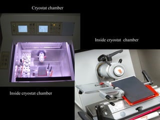
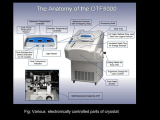
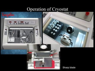
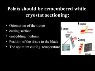
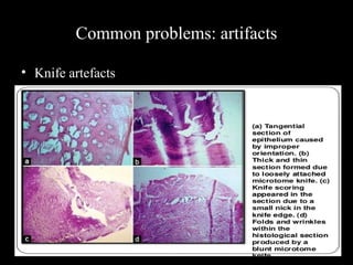
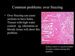
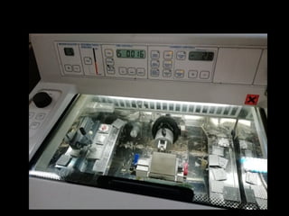
Frozen
Section
Presented By:
Dr. Nafisa Abedin
MD, Pathology,Resident Phase-A
;Dept. of Pathology, BSMMU.
Supervised by
Dr. Ferdousy Begum
Associate Professor
Department of Pathology,
BSMMU.
What is frozen section?
The frozen section procedure is a pathological
laboratory procedure to perform rapid microscopic
analysis of a specimen. The technical name for this
procedure is Cryo section
History:
Sir Louis B. Wilson
Frozen Section Resulted From
Need for Rapid Intra operative
Diagnosis.
Mayo Clinic Chief of Pathology
Louis B. Wilson pioneered the
frozen section at the Rochester,
Minnesota clinic in 1905 .
Frozen section–indications
1.Urgent diagnosis during surgery ( benign and
malignant)
2. Involvement of resection margins by malignancy, eg
basal cell carcinomas
3.Ganglion cells in Hirschprung disease
4. Enzyme histochemistry (ATPase, NADPH in muscle
biopsy samples)
5.Non enzyme histochemistry (lipid,carbohydrate)
6. For DIF, silver and gold impregnation methods
7. Identification of the type of tissue, eg. parathyroid
gland
What pathologist should know before
frozen section:
 
1)Cryostat machine
2)Request form
3)What surgeon wants to know?
4)what surgeons have sent?
5)How to contact them?
Request form for frozen
section,Pathology
department ,BSMMU
What pathologist should know during
frozen section
1)Whether tissue selection is appropriate or not
2) Type of tissue should cut at proper temperature.
3)Time consideration(About 20-30 min)
4) Communication with surgeon.
Flow chart of standard
operating procedure at BSMMU
Grossing station of BSMMU
Personal protection
Section of the tissue:
1)Tissue sample should represent the specimen
2)should not contain any necrotic area
3)3-4mm is ideal section
4)Sample once collected should be frozen immediately
Fixation:
Frozen section procedure requires unfixed tissue
Post fixation: for enzyme histochemistry(in controlled
condition)
Principle
Tissue
is
Frozen
Water in
Tissue
Turns to
Ice
Tissue
Becomes firm
And water
Act as
embedding
medium
Freezing techniques:
• Liquid nitrogen (-1900
c)
• isopentane cooled by liquid nitrogen (-1500
c)
• dry ice(-700
c)
• carbon dioxide gas(-700
c)
• aerosol sprays(-500
c)
Ideal temperature chart
Methods for suitable freezing
1)cryostat:
-microtome is inside the chamber
-microtome in under constant temperature control
2)Freezing microtome:
-Tissue fixed separately
-Microtome is not under temperature control
Cryostat Machine
Cryostat:
Types:
1)Closed-cycle cryostats
2)continuous-flow cryostat
3)Bath cryostat
4)Multistage cryostats
Cryostat chamber
Inside cryostat chamber
Fig: Various electronically controlled parts of cryostat
Operation of Cryostat
Sharp blade
Points should be remembered while
cryostat sectioning:
• Orientation of the tissue
• cutting surface
• embedding medium.
• Position of the tissue to the blade
• The optimum cutting temperature
Cryostat sectioning technique
Using the brush
Using the anti
role plate
Frozen section embedding medium
Taking of sections from stage
The cut section will rest on the surface of
the blade holder after cutting ,a room
temperature slide is held above the section,
where electrostatic attraction causes the
tissue to adhere to the slide.
Staining:
1)Rapid H-E method
2)Toludine method
3)Methylene Blue
4)Methyl violet for amyloid
5)Oil red O for fat
6)PAS
Fig: Different stains
Technique of staining
• Immediately fix frozen section in 95% ethyl alchohol
• Rinse well in distilled water
• Stain with Hematoxylin Stain
• Wash well in distilled water
• Counterstain in Eosin Y Working Solution
• Dehydrate in ethyl alcohol
• Subsequent treatment
Pros and cons of frozen section
Pros:
• Fastest of all methods
• Excellent for IHC, IF, ISH.
• Often easiest to section-depending upon the tissue.
Cons:
• Poorest morphology
• Prone to freezing artifact-must be snap frozen
Frozen section Vs routine section:
Poor morphology in frozen section in comparison to routine section
Common problems: artifacts
• Ice crystal artifacts:
- Cause: slow freezing of tissue
- Solution:Freeze fast(flash/snap)
- It is a chemical property of
water, that water will expand on
freezing.
Fig: Ice crystal artifacts (Clefts
and vacuoles in the specimen)
Common problems: artifacts
• Knife artefacts
Common problems: over freezing
• Over freezing can cause
sections to have holes.
Tissues with high water
content eg. edematous or
bloody tissue will show this
problem.
Kidney tumor is a good example
of water content causing this
problem
Quality assurance
• Never two cases in one grossing area
• System of identification of block and slide should be
established
• Never two cases in one cryostat
• Unlabeled slides should never be used
Quality assurance
• Correlation has to be done in each case.
• The cryostat must be clean periodically.
• Less frequently used machine can be decontaminated
on a longer cycle
• Records keeping.
• Sometimes the procedure is misused by surgeons to
satisfy their curiosity
• To compensate their deficiency in recognize normal
anatomical structure.
• To communicate the results immediately to the
patient’s relative.
Misuse of frozen section
Take home message
• Definitive diagnosis is not possible in all cases.
• Important for surgeons to influence the mode of
surgery
• Misuse must be avoided.
• It is necessary but never substitutes routine tissue
processing.
Frozen Section Basics

Frozen Section Basics

  • 2.
    Frozen Section Presented By: Dr. NafisaAbedin MD, Pathology,Resident Phase-A ;Dept. of Pathology, BSMMU. Supervised by Dr. Ferdousy Begum Associate Professor Department of Pathology, BSMMU.
  • 3.
    What is frozensection? The frozen section procedure is a pathological laboratory procedure to perform rapid microscopic analysis of a specimen. The technical name for this procedure is Cryo section
  • 4.
    History: Sir Louis B.Wilson Frozen Section Resulted From Need for Rapid Intra operative Diagnosis. Mayo Clinic Chief of Pathology Louis B. Wilson pioneered the frozen section at the Rochester, Minnesota clinic in 1905 .
  • 5.
    Frozen section–indications 1.Urgent diagnosisduring surgery ( benign and malignant) 2. Involvement of resection margins by malignancy, eg basal cell carcinomas 3.Ganglion cells in Hirschprung disease 4. Enzyme histochemistry (ATPase, NADPH in muscle biopsy samples) 5.Non enzyme histochemistry (lipid,carbohydrate) 6. For DIF, silver and gold impregnation methods 7. Identification of the type of tissue, eg. parathyroid gland
  • 6.
    What pathologist shouldknow before frozen section:   1)Cryostat machine 2)Request form 3)What surgeon wants to know? 4)what surgeons have sent? 5)How to contact them?
  • 7.
    Request form forfrozen section,Pathology department ,BSMMU
  • 8.
    What pathologist shouldknow during frozen section 1)Whether tissue selection is appropriate or not 2) Type of tissue should cut at proper temperature. 3)Time consideration(About 20-30 min) 4) Communication with surgeon.
  • 9.
    Flow chart ofstandard operating procedure at BSMMU
  • 10.
  • 11.
  • 12.
    Section of thetissue: 1)Tissue sample should represent the specimen 2)should not contain any necrotic area 3)3-4mm is ideal section 4)Sample once collected should be frozen immediately
  • 13.
    Fixation: Frozen section procedurerequires unfixed tissue Post fixation: for enzyme histochemistry(in controlled condition)
  • 14.
  • 15.
    Freezing techniques: • Liquidnitrogen (-1900 c) • isopentane cooled by liquid nitrogen (-1500 c) • dry ice(-700 c) • carbon dioxide gas(-700 c) • aerosol sprays(-500 c)
  • 16.
  • 17.
    Methods for suitablefreezing 1)cryostat: -microtome is inside the chamber -microtome in under constant temperature control 2)Freezing microtome: -Tissue fixed separately -Microtome is not under temperature control
  • 18.
  • 19.
  • 20.
    Cryostat chamber Inside cryostatchamber Inside cryostat chamber
  • 21.
    Fig: Various electronicallycontrolled parts of cryostat
  • 22.
  • 23.
    Points should beremembered while cryostat sectioning: • Orientation of the tissue • cutting surface • embedding medium. • Position of the tissue to the blade • The optimum cutting temperature
  • 24.
    Cryostat sectioning technique Usingthe brush Using the anti role plate
  • 25.
  • 26.
    Taking of sectionsfrom stage The cut section will rest on the surface of the blade holder after cutting ,a room temperature slide is held above the section, where electrostatic attraction causes the tissue to adhere to the slide.
  • 27.
    Staining: 1)Rapid H-E method 2)Toludinemethod 3)Methylene Blue 4)Methyl violet for amyloid 5)Oil red O for fat 6)PAS Fig: Different stains
  • 28.
    Technique of staining •Immediately fix frozen section in 95% ethyl alchohol • Rinse well in distilled water • Stain with Hematoxylin Stain • Wash well in distilled water • Counterstain in Eosin Y Working Solution • Dehydrate in ethyl alcohol • Subsequent treatment
  • 29.
    Pros and consof frozen section Pros: • Fastest of all methods • Excellent for IHC, IF, ISH. • Often easiest to section-depending upon the tissue. Cons: • Poorest morphology • Prone to freezing artifact-must be snap frozen
  • 30.
    Frozen section Vsroutine section: Poor morphology in frozen section in comparison to routine section
  • 31.
    Common problems: artifacts •Ice crystal artifacts: - Cause: slow freezing of tissue - Solution:Freeze fast(flash/snap) - It is a chemical property of water, that water will expand on freezing. Fig: Ice crystal artifacts (Clefts and vacuoles in the specimen)
  • 32.
  • 33.
    Common problems: overfreezing • Over freezing can cause sections to have holes. Tissues with high water content eg. edematous or bloody tissue will show this problem. Kidney tumor is a good example of water content causing this problem
  • 34.
    Quality assurance • Nevertwo cases in one grossing area • System of identification of block and slide should be established • Never two cases in one cryostat • Unlabeled slides should never be used
  • 35.
    Quality assurance • Correlationhas to be done in each case. • The cryostat must be clean periodically. • Less frequently used machine can be decontaminated on a longer cycle • Records keeping.
  • 37.
    • Sometimes theprocedure is misused by surgeons to satisfy their curiosity • To compensate their deficiency in recognize normal anatomical structure. • To communicate the results immediately to the patient’s relative. Misuse of frozen section
  • 38.
    Take home message •Definitive diagnosis is not possible in all cases. • Important for surgeons to influence the mode of surgery • Misuse must be avoided. • It is necessary but never substitutes routine tissue processing.