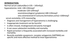
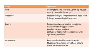
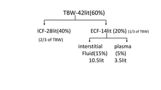
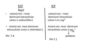
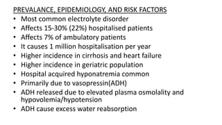
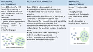
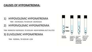
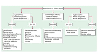
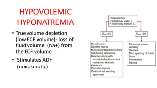
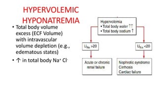
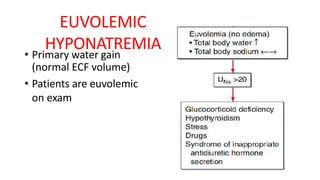
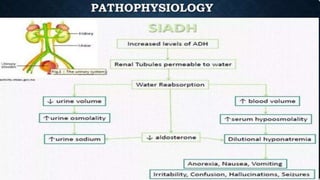
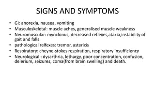
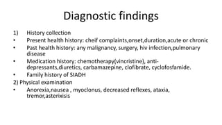
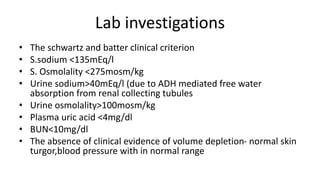
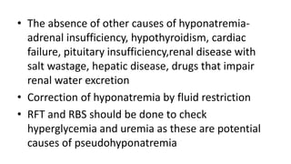
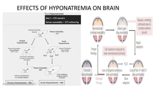
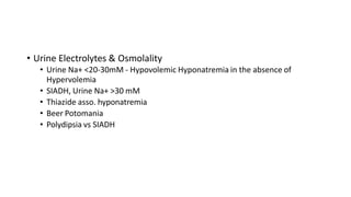
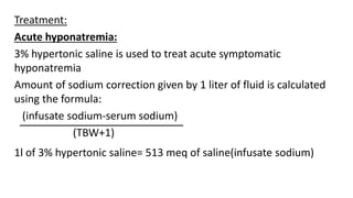
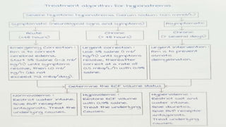
HYPONATREMIA
BY
Dr. K. Dasaradh Jr-1
INTRODUCTION:
Normal serum sodium(Na+)=135 – 145mEq/L
hyponatremia: mild- 130-134meq/l
moderate-120-129meq/l
severe(neurological symptoms)-100-119meq/l
very severe(raised ICT,seizures,herniation,coma)-<100meq/l
serum osmolality <275 mosmol/kg
• Diagnosis and management of hyponatremia is challenging
• Inappropriate treatment can be harmful
• Is often missed, misdiagnosed and poorly managed
• It can cause substantial morbidity and mortality
• Rapid correction is frequently associated with increased morbidity and
mortatlity
• Recently available vasopressin- receptor antagonists (VAPTANS) are
specific and more effective method to treat hyponatremia
Stage Symptoms
Mild GI symptoms like anorexia, vomiting, nausea,
apathy, headache, lethargy
Moderate Predominantly GI symptoms+ mild head ache,
lethargy as neurological symptoms
Severe Predominantly neurological symptoms
classically affecting gait+ataxia
Another pattern involves
confusion/disorientation(associated with
agitation), psychosis
Very severe Features of raised intracranial tension
Stupor,coma,tentorial herniation, Cheyne-
stokes respiration,death
TBW-42lit(60%)
ICF-28lit(40%) ECF-14lit (20%) (1/3 of TBW)
(2/3 of TBW)
interstitial plasma
Fluid(15%) (5%)
10.5lit 3.5lit
ECF ICF
Nacl
• cation(+ve) – most cation(+ve) – most
dominant extracellular dominant intracellular
cation is sodium(Na+) cation is k+,mg2+
• Anion(-ve)- most dominant Anion(-ve)- most dominant
extracullular anion is chloride(cl-) intracellular anion is po4
2-
f/by
PH- 7.4 proteins
PH-7.1
Physiology of sodium:
• Water is a major contributor of body weight
• Eg. Adult male – 70kg
60% exclusively made up of water
total body water(TBW) =42 lit
18% of our body weight proteins
15% of our body weight lipids
7% of our body weight minerals
• In female 50% of body weight is made up of water, as female have
more adipose tissue(more fat content)
• Water inversely proportional to fat
• Children 70% body weight is made up of water
PREVALANCE, EPIDEMIOLOGY, AND RISK FACTORS
• Most common electrolyte disorder
• Affects 15-30% (22%) hospitalised patients
• Affects 7% of ambulatory patients
• It causes 1 million hospitalisation per year
• Higher incidence in cirrhosis and heart failure
• Higher incidence in geriatric population
• Hospital acquired hyponatremia common
• Primarily due to vasopressin(ADH)
• ADH released due to elevated plasma osmolality and
hypovolemia/hypotension
• ADH cause excess water reabsorption
HYPERTONIC
HYPONATREMIA
ISOTONIC HYPONATREMIA HYPOTONIC
HYPONATREMIA
Posm > 290 mOsmol/kg H2O
•Results due to presence of
another effective osmole that
causes free water to move from
the ICF to the ECF
• Examples:
 Mannitol (osmoticdiuretic),
glycine, marked hyperglycemia
•Treatment correct the
underlying condition or
removal
of the offending agent
Posm 275-290 mOsmol/kg H2O
• Pseudohyponatremia= laboratory artifact
• Due to marked increase in serum lipids or
proteins
•Results in reduction of fraction of serum that is
water and an artificially low serum Na+
•Plasma water Na+ concentration and osmolality
are unchanged but the measured Na+
concentration in the total plasma volume will be
reduced given the specimen contains less plasma
water
• Only occurs when flame photometry or
indirect potentiometry are used
• Direct potentiometry and ion-specific
electrodes avoids this misdiagnosis
Posm < 275 mOsmol/kg
H2O
•True physiologic
hyponatremia that results
from excess water either
due
to ADH stimulation or
impaired water excretion
CAUSES OF HYPONATREMIA:
1) HYPOVOLEMIC HYPONATREMIA
TBW – DECREASED, TB SODIUM –DECREASED
2) HYPERVOLEMIC HYPONATREMIA
TBW- MARKEDLY INCREASED, TB SODIUM –NEAR NORMAL BUT DILUTED
3) EUVOLEMIC HYPONATREMIA
TBW- NORMAL, TB SODIUM -LOW
HYPOVOLEMIC
HYPONATREMIA
• True volume depletion
(low ECF volume)- loss of
fluid volume (Na+) from
the ECF volume
• Stimulates ADH
(nonosmotic)
HYPERVOLEMIC
HYPONATREMIA
• Total body volume
excess (ECF Volume)
with intravascular
volume depletion (e.g.,
edematous states)
• ↑ in total body Na+ Cl-
EUVOLEMIC
HYPONATREMIA
• Primary water gain
(normal ECF volume)
• Patients are euvolemic
on exam
SIADH
• MCC of Euvolemic hyponatremia
• It is a condition in which the body makes too much ADH. This hormone helps the
kidneys control the amount of water your body looses through the urine, causes the
body to retain too much water
• The resulting impairment of water secretion and consequent water retention produces
the hyponatremia(i.e, s.sodium <135mmol/l) with concomitant
hypoosmolality(s.osmolality <275mosm/kg) and high urine osmolality that are the
hallmark of SIADH
• Generation of hyponatremia requires intake of free water
• Four distinct patterns of AVP(arginine vasopressin) secretion
I. Unregulated, erratic AVP secretion
II. Failure to suppress AVP at lower serum osmolalities
III. Reset Osmostat
IV. No detectable circulating AVP
• Subclinically Volume expanded
• s Uric Acid <4 mg/dl
• Causes of SIADH
• MALIGNANT CAUSES: carcinoma of lungs(small cell),
mestothelioma,endocrine thymoma,
lymphoma,sarcoma(ewing)
• PULMONARY CAUSES: bacterial and viral
pneumonis,pulmonary abscess, tuberculosis,
asthma,cystic fibrosis
• DISORDERS OF CNS:encephalitis,meningitis,brain
abscess,brain tumours,hydrocephalus
• Drugs that stimulate AVP:
SSRI’s,TCA’s,clofibrate,carbamazepine,cylcofosfamide
SIGNS AND SYMPTOMS
• GI: anorexia, nausea, vomiting
• Musculoskeletal: muscle aches, generalised muscle weakness
• Neuromuscular: myoclonus, decreased reflexes,ataxia,instability of
gait and falls
• pathological relfexes: tremor, asterixis
• Respiratory: cheyne-stokes respiration, respiratory insufficiency
• Neurological : dysarthria, lethargy, poor concentration, confusion,
delerium, seizures, coma(from brain swelling) and death.
Diagnostic findings
1) History collection
• Present health history: cheif complaints,onset,duration,acute or chronic
• Past health history: any malignancy, surgery, hiv infection,pulmonary
disease
• Medication history: chemotherapy(vincristine), anti-
depressants,diuretics, carbamazepine, clofibrate, cyclofosfamide.
• Family history of SIADH
2) Physical examination
• Anorexia,nausea , myoclonus, decreased reflexes, ataxia,
tremor,asterixisis
Lab investigations
• The schwartz and batter clinical criterion
• S.sodium <135mEq/l
• S. Osmolality <275mosm/kg
• Urine sodium>40mEq/l (due to ADH mediated free water
absorption from renal collecting tubules
• Urine osmolality>100mosm/kg
• Plasma uric acid <4mg/dl
• BUN<10mg/dl
• The absence of clinical evidence of volume depletion- normal skin
turgor,blood pressure with in normal range
• The absence of other causes of hyponatremia-
adrenal insufficiency, hypothyroidism, cardiac
failure, pituitary insufficiency,renal disease with
salt wastage, hepatic disease, drugs that impair
renal water excretion
• Correction of hyponatremia by fluid restriction
• RFT and RBS should be done to check
hyperglycemia and uremia as these are potential
causes of pseudohyponatremia
Cerebral salt wasting syndrome
• Caused by :
1) Loss of adrenergic tone to the nephrons
Inhibition of RAS axis(B1 receptors needed for renin production)
Decreased in aldosterone production
Decreased sodium and water reabsorption
2)Increased natriuretic peptides can also cause CSW.
Markers SIADH CSW
Intravascular volume Euvolemic Hypovolemic
Central venous pressure/
PCWP
Normal Decreased
Serum potassium Normal Increased
Serum uric acid Extremely low < 2mg/dl(ADH
is a uricosuric substancer)
Marginal decrease
Urine sodium Increased increased
EFFECTS OF HYPONATREMIA ON BRAIN
• Persistent Chronic Hyponatremia:
• Efflux of organic osmolytes(Creatine, Betaine, Glutamate, Myoinositol,
Taurine)
• Rapid correction(>8-10 mM in 24 hr or 18 mM in 48 hr) is dangerous and can
lead to ODS
• Osmotic Demyelination Syndrome:
• MC site –PONS
• Pathophysiology: cerebral apoptosis and loss of myelin due to osmotic stress
accordingly the brain area rich in oligodendrocytes and myelin tend to be the most
frequently affected
• C/F- confusion, delirium, hallucinations, tremor, problem in swallowing,slurred speech,
reduced alertness, weakness in face, arms and legs
• Other sites: Cerebellum, Lateral Geniculate Body, Thalamus, Putamen, Cerebral cortex
Treatment: relowering of sodium using 5% dextrose and desmopressin
Acute cerebral edema Osmotic demyelination
syndrome(central pontine
myelinolysis)
Postoperative menstruating
women
Elderly women taking thiazides
Children
Patients with polydipsia,
secondary to psychiatric disorders
Hypoxemic patients
Marathon runners
Liver transplant recipients
Alcoholic patients
Malnourished patients
Hypokalemic patients
Burn victims
Elderly women taking thiazides
Hypoxemic patients
Severe hyponatremia(Na+
<105meq/l)
DIAGNOSTIC EVALUATION
• Detailed Drug History
• Clinical assesment of volume status
• Radiological Imaging -to r/o pulmonary or CNS cause of hyponatremia
• Blood Investigations
• Serum osmolality
• BUN & s Creat
• s K+
• s. Glucose
• s. Uric Acid
• Thyroid, Adrenal, Pituitary Function
• Urine Electrolytes & Osmolality
• Urine Na+ <20-30mM - Hypovolemic Hyponatremia in the absence of
Hypervolemia
• SIADH, Urine Na+ >30 mM
• Thiazide asso. hyponatremia
• Beer Potomania
• Polydipsia vs SIADH
Treatment:
Acute hyponatremia:
3% hypertonic saline is used to treat acute symptomatic
hyponatremia
Amount of sodium correction given by 1 liter of fluid is calculated
using the formula:
(infusate sodium-serum sodium)
(TBW+1)
1l of 3% hypertonic saline= 513 meq of saline(infusate sodium)
• Chronic hyponatremia:
• If the patient presents after 48hrs /if the cause is not
certain/time of onset of symptoms is unknowm, then it is
considered as chronic hyponatremia.
• RATE OF CORRECTION
8-10mmol/24hr
18mmol/48hr
Drugs used in hyponatremia:
Lixivaptan,tolvaptan,mozavaptan,satvaptan are used
Tolvaptan most commonly used
All these are hepatotoxic, regular LFT should be done
V1+V2 blocker: conivaptan
Selective v1a blocker: relcovaptan is used
Selection v1b or v3 blocker: nelivaptan
Other uses of vaptans are
Relcovaptan is used in Raynaud’s syndrome
adrenal hyperplasia
preterm labour
Tolvaptan is used in autosomal dominant polycystic
kidney disease
Vaptans have also been tried in congestive heart failure

hyponatremia.pptx

  • 1.
  • 2.
    INTRODUCTION: Normal serum sodium(Na+)=135– 145mEq/L hyponatremia: mild- 130-134meq/l moderate-120-129meq/l severe(neurological symptoms)-100-119meq/l very severe(raised ICT,seizures,herniation,coma)-<100meq/l serum osmolality <275 mosmol/kg • Diagnosis and management of hyponatremia is challenging • Inappropriate treatment can be harmful • Is often missed, misdiagnosed and poorly managed • It can cause substantial morbidity and mortality • Rapid correction is frequently associated with increased morbidity and mortatlity • Recently available vasopressin- receptor antagonists (VAPTANS) are specific and more effective method to treat hyponatremia
  • 3.
    Stage Symptoms Mild GIsymptoms like anorexia, vomiting, nausea, apathy, headache, lethargy Moderate Predominantly GI symptoms+ mild head ache, lethargy as neurological symptoms Severe Predominantly neurological symptoms classically affecting gait+ataxia Another pattern involves confusion/disorientation(associated with agitation), psychosis Very severe Features of raised intracranial tension Stupor,coma,tentorial herniation, Cheyne- stokes respiration,death
  • 4.
    TBW-42lit(60%) ICF-28lit(40%) ECF-14lit (20%)(1/3 of TBW) (2/3 of TBW) interstitial plasma Fluid(15%) (5%) 10.5lit 3.5lit
  • 5.
    ECF ICF Nacl • cation(+ve)– most cation(+ve) – most dominant extracellular dominant intracellular cation is sodium(Na+) cation is k+,mg2+ • Anion(-ve)- most dominant Anion(-ve)- most dominant extracullular anion is chloride(cl-) intracellular anion is po4 2- f/by PH- 7.4 proteins PH-7.1
  • 6.
    Physiology of sodium: •Water is a major contributor of body weight • Eg. Adult male – 70kg 60% exclusively made up of water total body water(TBW) =42 lit 18% of our body weight proteins 15% of our body weight lipids 7% of our body weight minerals • In female 50% of body weight is made up of water, as female have more adipose tissue(more fat content) • Water inversely proportional to fat • Children 70% body weight is made up of water
  • 7.
    PREVALANCE, EPIDEMIOLOGY, ANDRISK FACTORS • Most common electrolyte disorder • Affects 15-30% (22%) hospitalised patients • Affects 7% of ambulatory patients • It causes 1 million hospitalisation per year • Higher incidence in cirrhosis and heart failure • Higher incidence in geriatric population • Hospital acquired hyponatremia common • Primarily due to vasopressin(ADH) • ADH released due to elevated plasma osmolality and hypovolemia/hypotension • ADH cause excess water reabsorption
  • 8.
    HYPERTONIC HYPONATREMIA ISOTONIC HYPONATREMIA HYPOTONIC HYPONATREMIA Posm> 290 mOsmol/kg H2O •Results due to presence of another effective osmole that causes free water to move from the ICF to the ECF • Examples:  Mannitol (osmoticdiuretic), glycine, marked hyperglycemia •Treatment correct the underlying condition or removal of the offending agent Posm 275-290 mOsmol/kg H2O • Pseudohyponatremia= laboratory artifact • Due to marked increase in serum lipids or proteins •Results in reduction of fraction of serum that is water and an artificially low serum Na+ •Plasma water Na+ concentration and osmolality are unchanged but the measured Na+ concentration in the total plasma volume will be reduced given the specimen contains less plasma water • Only occurs when flame photometry or indirect potentiometry are used • Direct potentiometry and ion-specific electrodes avoids this misdiagnosis Posm < 275 mOsmol/kg H2O •True physiologic hyponatremia that results from excess water either due to ADH stimulation or impaired water excretion
  • 9.
    CAUSES OF HYPONATREMIA: 1)HYPOVOLEMIC HYPONATREMIA TBW – DECREASED, TB SODIUM –DECREASED 2) HYPERVOLEMIC HYPONATREMIA TBW- MARKEDLY INCREASED, TB SODIUM –NEAR NORMAL BUT DILUTED 3) EUVOLEMIC HYPONATREMIA TBW- NORMAL, TB SODIUM -LOW
  • 11.
    HYPOVOLEMIC HYPONATREMIA • True volumedepletion (low ECF volume)- loss of fluid volume (Na+) from the ECF volume • Stimulates ADH (nonosmotic)
  • 12.
    HYPERVOLEMIC HYPONATREMIA • Total bodyvolume excess (ECF Volume) with intravascular volume depletion (e.g., edematous states) • ↑ in total body Na+ Cl-
  • 13.
    EUVOLEMIC HYPONATREMIA • Primary watergain (normal ECF volume) • Patients are euvolemic on exam
  • 14.
    SIADH • MCC ofEuvolemic hyponatremia • It is a condition in which the body makes too much ADH. This hormone helps the kidneys control the amount of water your body looses through the urine, causes the body to retain too much water • The resulting impairment of water secretion and consequent water retention produces the hyponatremia(i.e, s.sodium <135mmol/l) with concomitant hypoosmolality(s.osmolality <275mosm/kg) and high urine osmolality that are the hallmark of SIADH • Generation of hyponatremia requires intake of free water • Four distinct patterns of AVP(arginine vasopressin) secretion I. Unregulated, erratic AVP secretion II. Failure to suppress AVP at lower serum osmolalities III. Reset Osmostat IV. No detectable circulating AVP • Subclinically Volume expanded • s Uric Acid <4 mg/dl
  • 15.
    • Causes ofSIADH • MALIGNANT CAUSES: carcinoma of lungs(small cell), mestothelioma,endocrine thymoma, lymphoma,sarcoma(ewing) • PULMONARY CAUSES: bacterial and viral pneumonis,pulmonary abscess, tuberculosis, asthma,cystic fibrosis • DISORDERS OF CNS:encephalitis,meningitis,brain abscess,brain tumours,hydrocephalus • Drugs that stimulate AVP: SSRI’s,TCA’s,clofibrate,carbamazepine,cylcofosfamide
  • 18.
    SIGNS AND SYMPTOMS •GI: anorexia, nausea, vomiting • Musculoskeletal: muscle aches, generalised muscle weakness • Neuromuscular: myoclonus, decreased reflexes,ataxia,instability of gait and falls • pathological relfexes: tremor, asterixis • Respiratory: cheyne-stokes respiration, respiratory insufficiency • Neurological : dysarthria, lethargy, poor concentration, confusion, delerium, seizures, coma(from brain swelling) and death.
  • 19.
    Diagnostic findings 1) Historycollection • Present health history: cheif complaints,onset,duration,acute or chronic • Past health history: any malignancy, surgery, hiv infection,pulmonary disease • Medication history: chemotherapy(vincristine), anti- depressants,diuretics, carbamazepine, clofibrate, cyclofosfamide. • Family history of SIADH 2) Physical examination • Anorexia,nausea , myoclonus, decreased reflexes, ataxia, tremor,asterixisis
  • 20.
    Lab investigations • Theschwartz and batter clinical criterion • S.sodium <135mEq/l • S. Osmolality <275mosm/kg • Urine sodium>40mEq/l (due to ADH mediated free water absorption from renal collecting tubules • Urine osmolality>100mosm/kg • Plasma uric acid <4mg/dl • BUN<10mg/dl • The absence of clinical evidence of volume depletion- normal skin turgor,blood pressure with in normal range
  • 21.
    • The absenceof other causes of hyponatremia- adrenal insufficiency, hypothyroidism, cardiac failure, pituitary insufficiency,renal disease with salt wastage, hepatic disease, drugs that impair renal water excretion • Correction of hyponatremia by fluid restriction • RFT and RBS should be done to check hyperglycemia and uremia as these are potential causes of pseudohyponatremia
  • 22.
    Cerebral salt wastingsyndrome • Caused by : 1) Loss of adrenergic tone to the nephrons Inhibition of RAS axis(B1 receptors needed for renin production) Decreased in aldosterone production Decreased sodium and water reabsorption 2)Increased natriuretic peptides can also cause CSW.
  • 23.
    Markers SIADH CSW Intravascularvolume Euvolemic Hypovolemic Central venous pressure/ PCWP Normal Decreased Serum potassium Normal Increased Serum uric acid Extremely low < 2mg/dl(ADH is a uricosuric substancer) Marginal decrease Urine sodium Increased increased
  • 25.
  • 26.
    • Persistent ChronicHyponatremia: • Efflux of organic osmolytes(Creatine, Betaine, Glutamate, Myoinositol, Taurine) • Rapid correction(>8-10 mM in 24 hr or 18 mM in 48 hr) is dangerous and can lead to ODS • Osmotic Demyelination Syndrome: • MC site –PONS • Pathophysiology: cerebral apoptosis and loss of myelin due to osmotic stress accordingly the brain area rich in oligodendrocytes and myelin tend to be the most frequently affected • C/F- confusion, delirium, hallucinations, tremor, problem in swallowing,slurred speech, reduced alertness, weakness in face, arms and legs • Other sites: Cerebellum, Lateral Geniculate Body, Thalamus, Putamen, Cerebral cortex Treatment: relowering of sodium using 5% dextrose and desmopressin
  • 27.
    Acute cerebral edemaOsmotic demyelination syndrome(central pontine myelinolysis) Postoperative menstruating women Elderly women taking thiazides Children Patients with polydipsia, secondary to psychiatric disorders Hypoxemic patients Marathon runners Liver transplant recipients Alcoholic patients Malnourished patients Hypokalemic patients Burn victims Elderly women taking thiazides Hypoxemic patients Severe hyponatremia(Na+ <105meq/l)
  • 28.
    DIAGNOSTIC EVALUATION • DetailedDrug History • Clinical assesment of volume status • Radiological Imaging -to r/o pulmonary or CNS cause of hyponatremia • Blood Investigations • Serum osmolality • BUN & s Creat • s K+ • s. Glucose • s. Uric Acid • Thyroid, Adrenal, Pituitary Function
  • 29.
    • Urine Electrolytes& Osmolality • Urine Na+ <20-30mM - Hypovolemic Hyponatremia in the absence of Hypervolemia • SIADH, Urine Na+ >30 mM • Thiazide asso. hyponatremia • Beer Potomania • Polydipsia vs SIADH
  • 30.
    Treatment: Acute hyponatremia: 3% hypertonicsaline is used to treat acute symptomatic hyponatremia Amount of sodium correction given by 1 liter of fluid is calculated using the formula: (infusate sodium-serum sodium) (TBW+1) 1l of 3% hypertonic saline= 513 meq of saline(infusate sodium)
  • 31.
    • Chronic hyponatremia: •If the patient presents after 48hrs /if the cause is not certain/time of onset of symptoms is unknowm, then it is considered as chronic hyponatremia.
  • 33.
    • RATE OFCORRECTION 8-10mmol/24hr 18mmol/48hr Drugs used in hyponatremia: Lixivaptan,tolvaptan,mozavaptan,satvaptan are used Tolvaptan most commonly used All these are hepatotoxic, regular LFT should be done V1+V2 blocker: conivaptan Selective v1a blocker: relcovaptan is used Selection v1b or v3 blocker: nelivaptan
  • 34.
    Other uses ofvaptans are Relcovaptan is used in Raynaud’s syndrome adrenal hyperplasia preterm labour Tolvaptan is used in autosomal dominant polycystic kidney disease Vaptans have also been tried in congestive heart failure