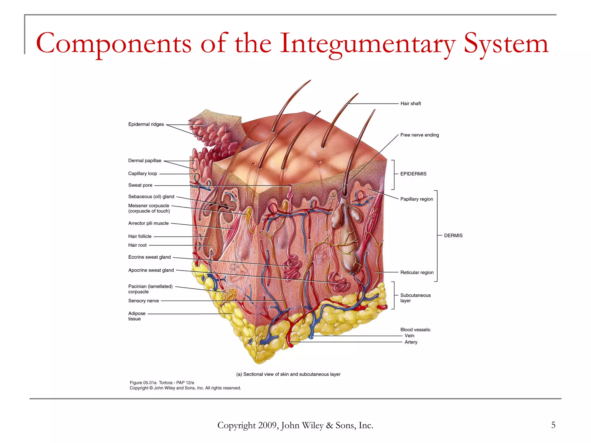
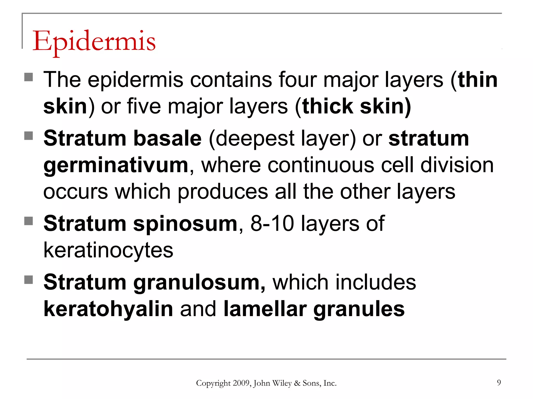
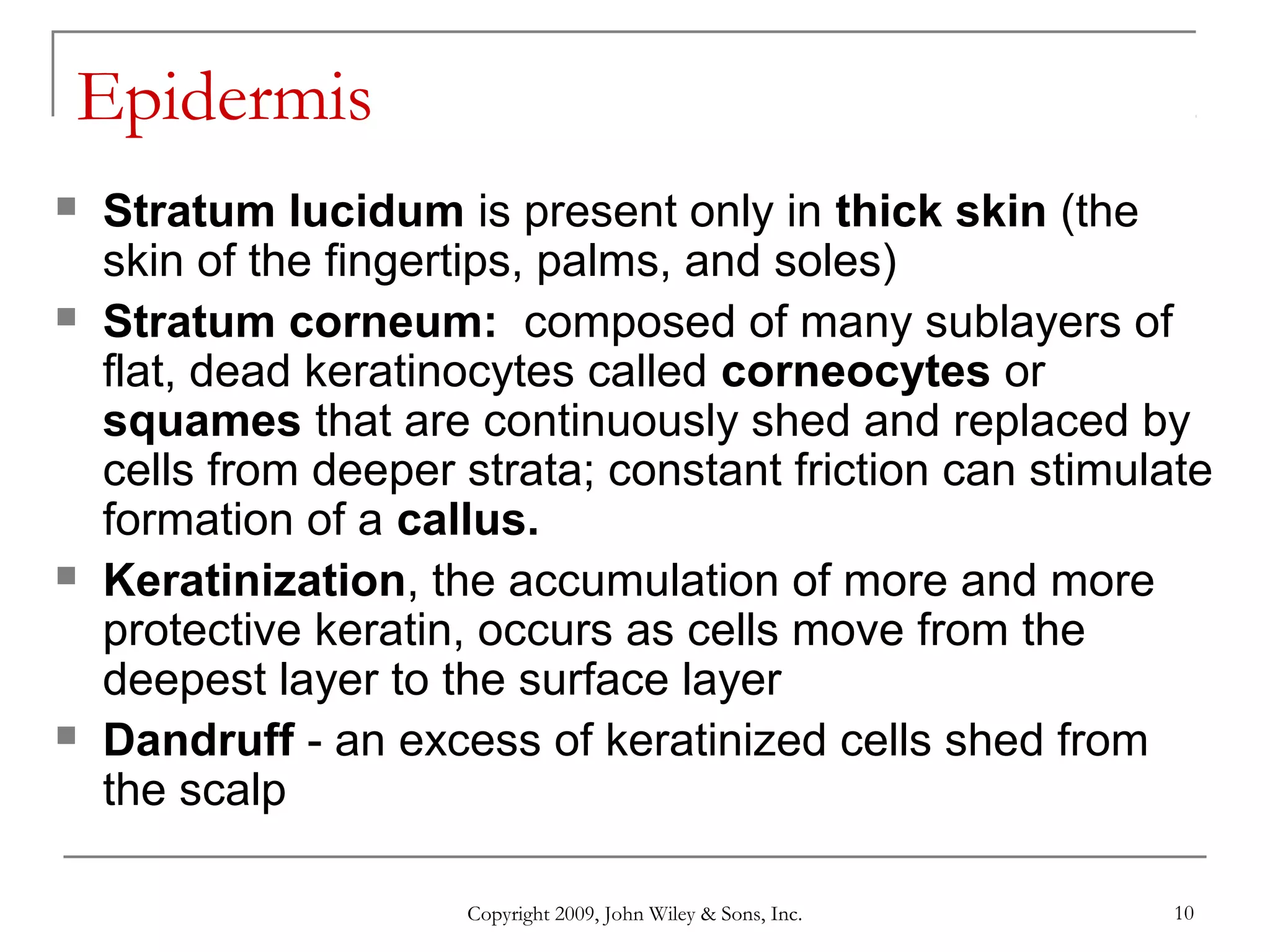
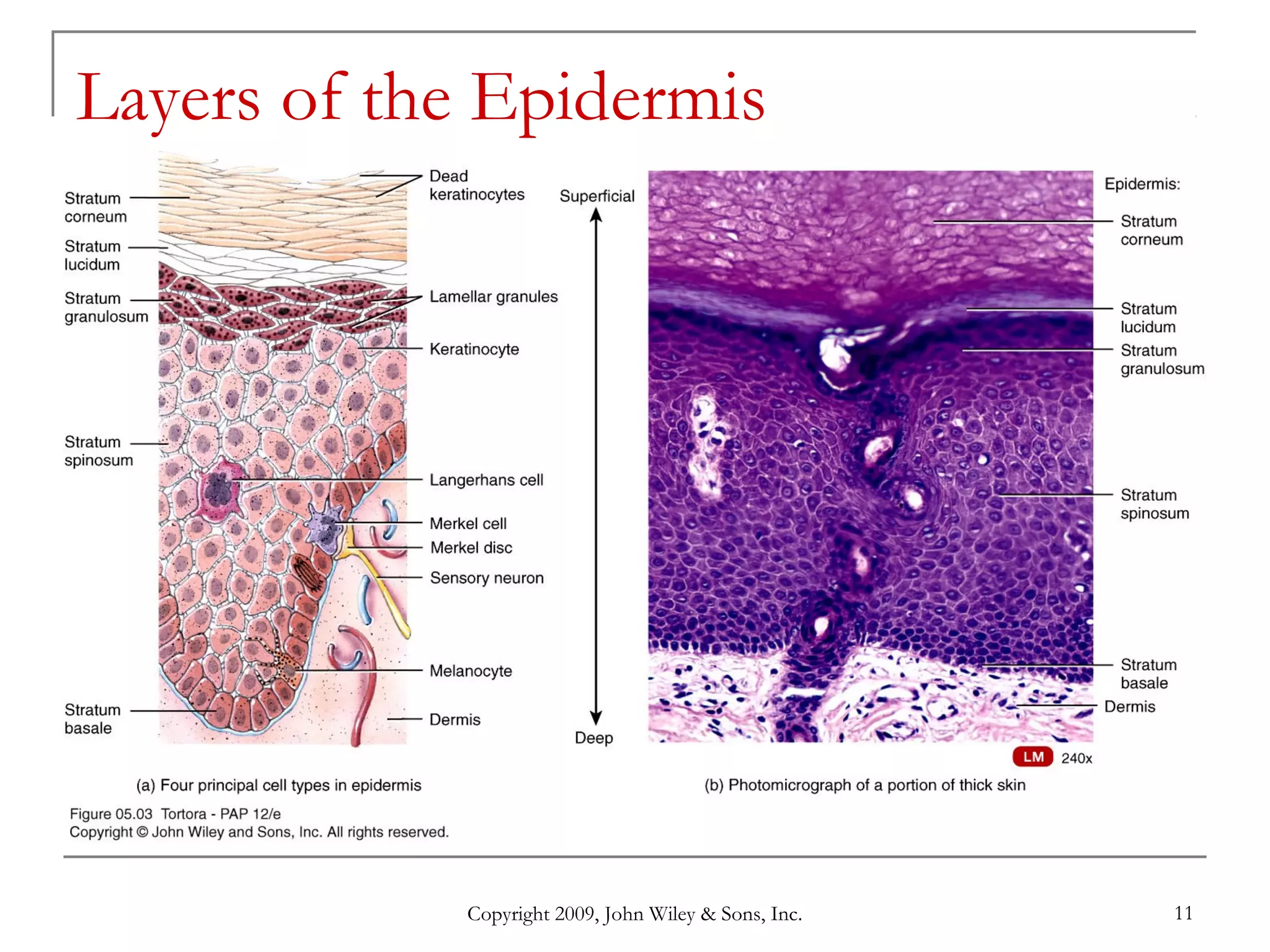
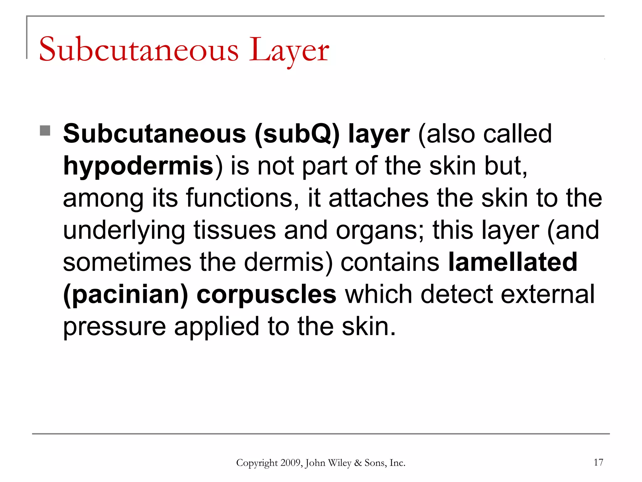
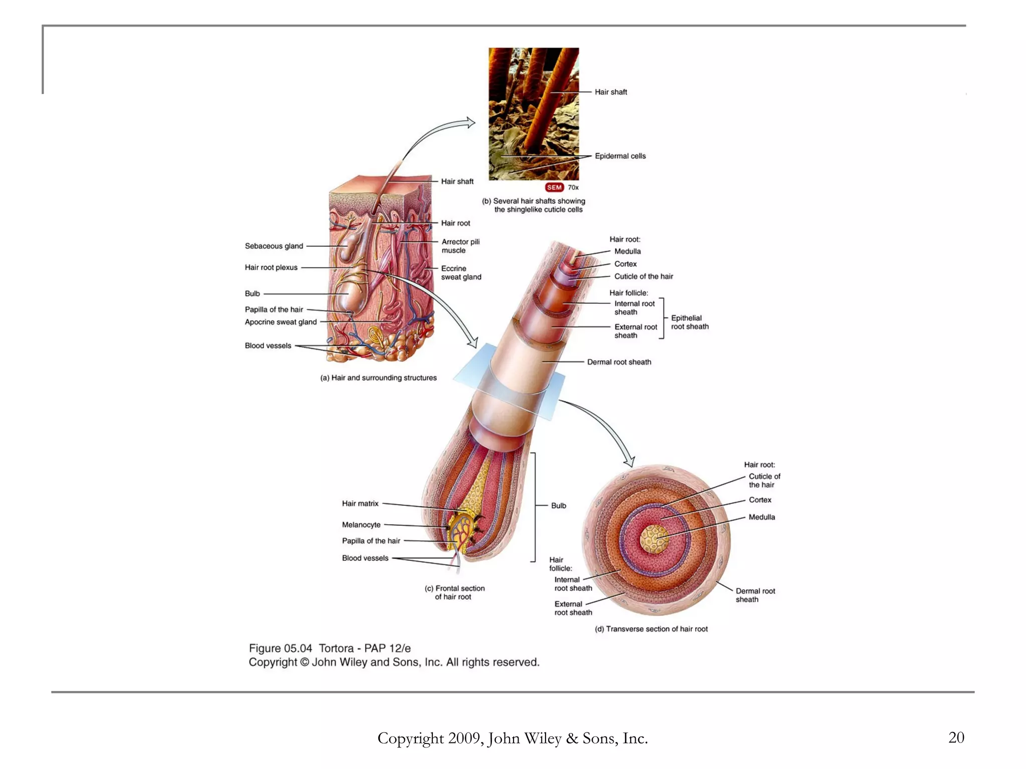
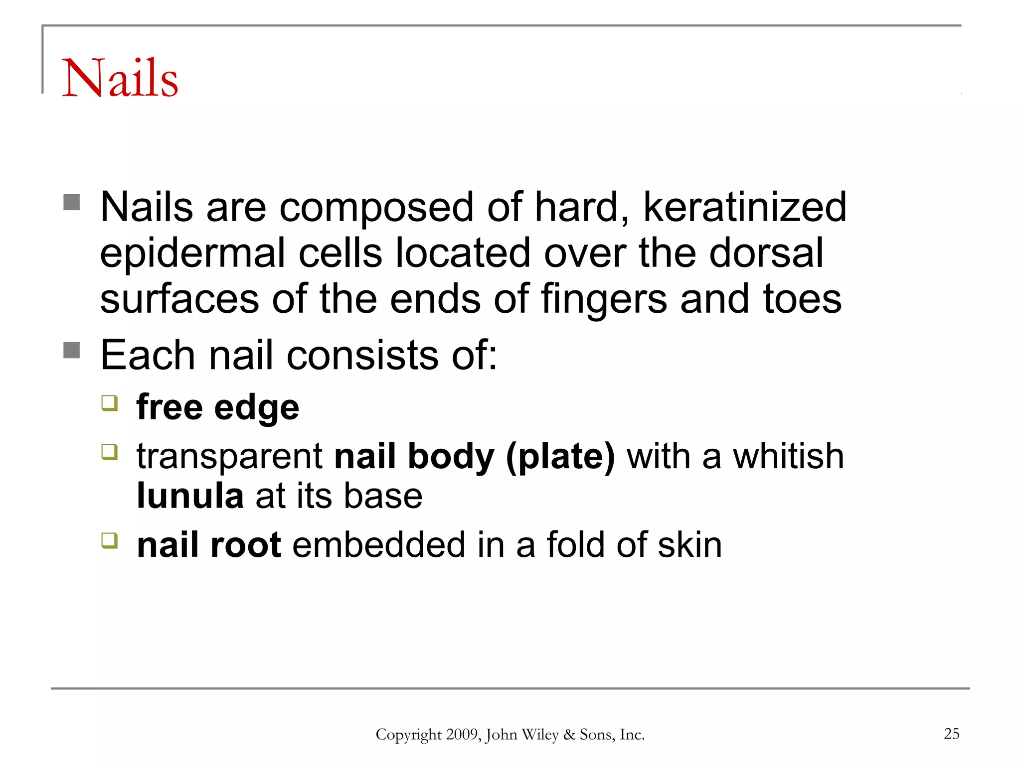
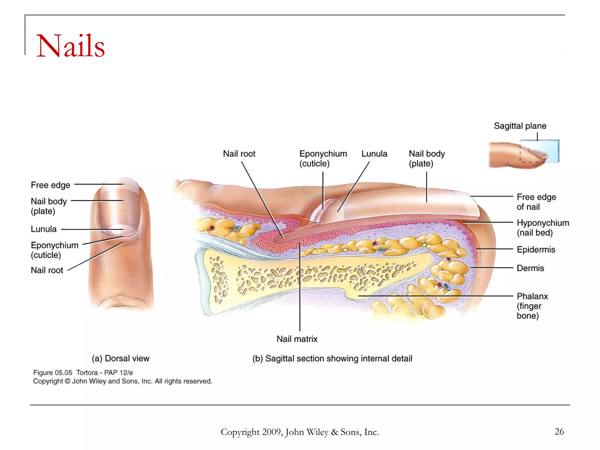
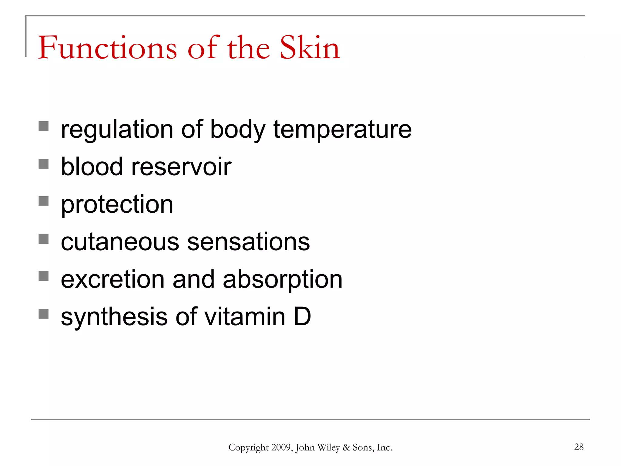
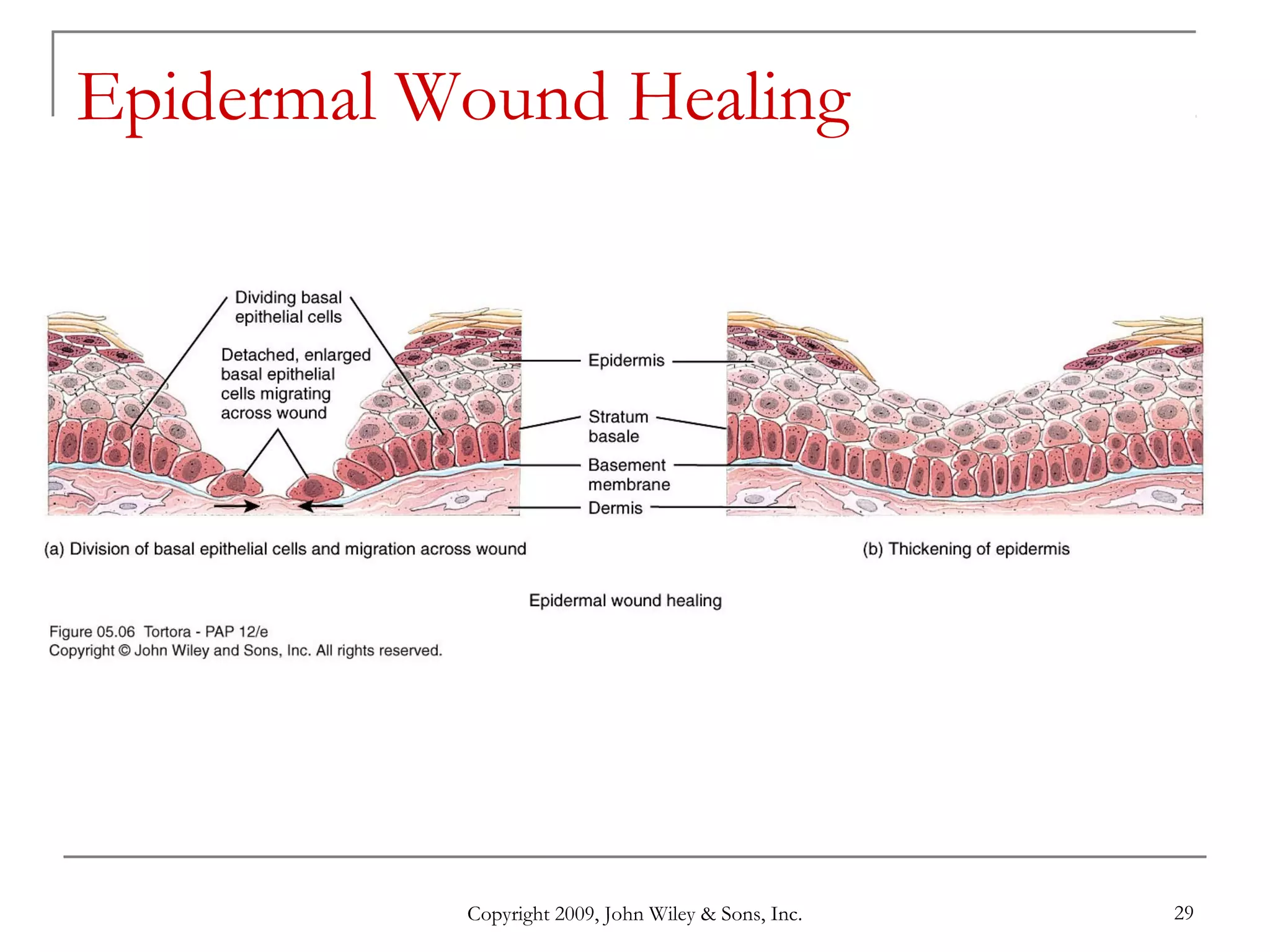
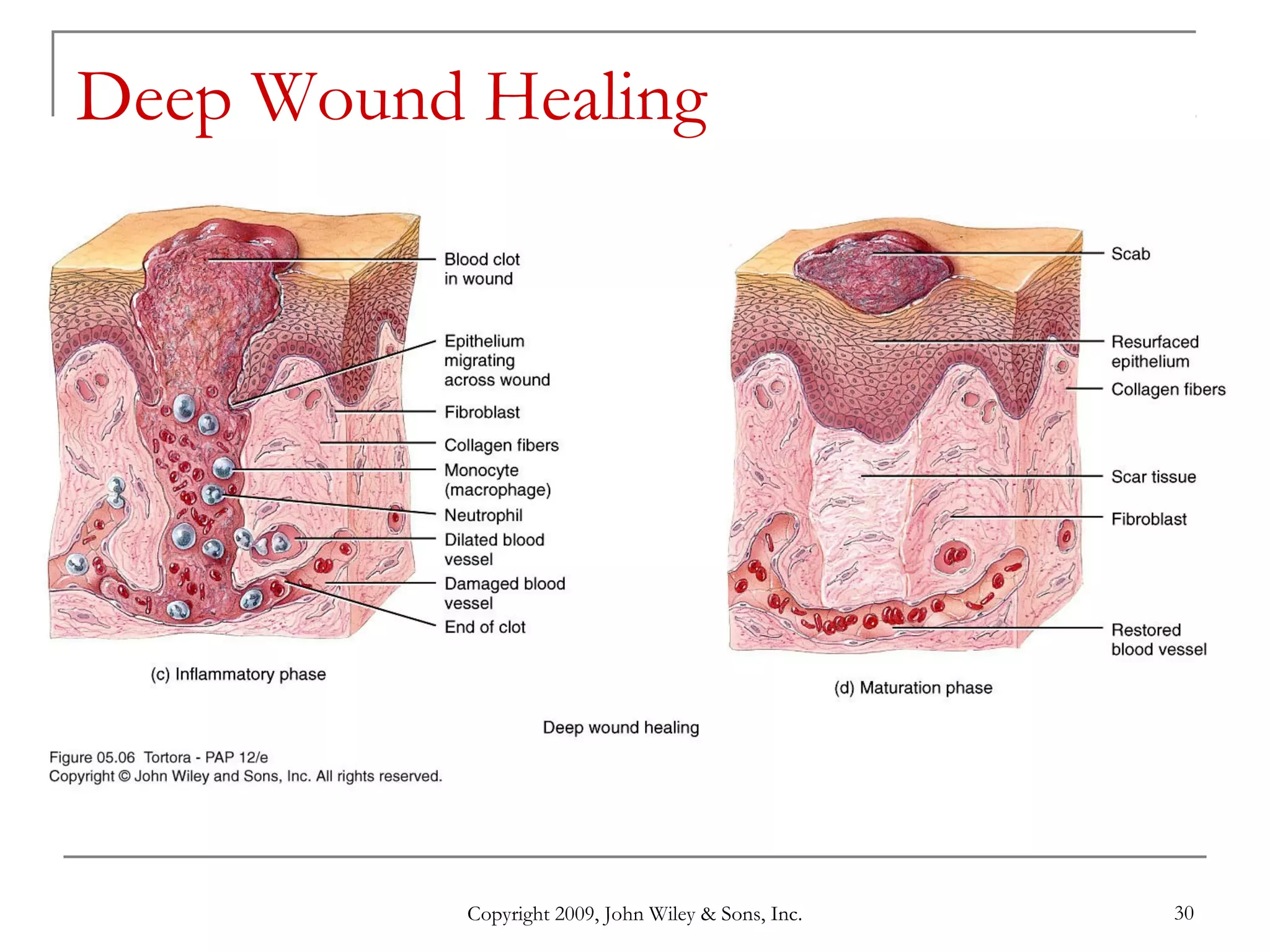
Copyright 2009, John Wiley & Sons, Inc. 1
Chapter 5
The Integumentary System
Copyright 2009, John Wiley & Sons, Inc. 2
Introduction
 The organs of the integumentary system
include the skin and its accessory structures
including hair, nails, and glands, as well as
blood vessels, muscles and nerves
 Dermatology is the medical specialty for the
diagnosis and treatment of disorders of the
integumentary system.
Copyright 2009, John Wiley & Sons, Inc. 3
Structure of the Skin
 The skin (cutaneous membrane) covers the
body and is the largest organ of the body by
surface area and weight
 Its area is about 2 square meters (22 square
feet) and weighs 4.5-5kg (10-11 lb), about
16% of body weight
 It is 0.5 – 4 mm thick, thinnest on the eyelids,
thickest on the heels; the average thickness
is 1 – 2 mm
Copyright 2009, John Wiley & Sons, Inc. 4
Structure of the Skin
 It consists of two major layers:
 outer, thinner layer called the epidermis,
consists of epithelial tissue
 inner, thicker layer called the dermis
 Beneath the dermis is a subcutaneous
(subQ) layer (also called hypodermis)
which attaches the skin to the underlying
tissues and organs.
Copyright 2009, John Wiley & Sons, Inc. 5
Components of the Integumentary System
Copyright 2009, John Wiley & Sons, Inc. 6
Structure of the Skin
 The epidermis has a number of important
characteristics:
 the epidermis is composed of keratinized
stratified squamous epithelium
 it contains four major types of cells:
 Keratinocytes (90% of the cells) produce
keratin which is a tough fibrous protein that
provides protection
Copyright 2009, John Wiley & Sons, Inc. 7
Structure of the Skin
 Melanocytes: which produce the pigment
melanin that protects against damage by
ultraviolet radiation
 Langerhans cells: involved in immune
responses, arise from red bone marrow
 Merkel cells: which function in the
sensation of touch along with the adjacent
tactile discs
Copyright 2009, John Wiley & Sons, Inc. 8
Types of Cells in the Epidermis
Copyright 2009, John Wiley & Sons, Inc. 9
Epidermis
 The epidermis contains four major layers (thin
skin) or five major layers (thick skin)
 Stratum basale (deepest layer) or stratum
germinativum, where continuous cell division
occurs which produces all the other layers
 Stratum spinosum, 8-10 layers of
keratinocytes
 Stratum granulosum, which includes
keratohyalin and lamellar granules
Copyright 2009, John Wiley & Sons, Inc. 10
Epidermis
 Stratum lucidum is present only in thick skin (the
skin of the fingertips, palms, and soles)
 Stratum corneum: composed of many sublayers of
flat, dead keratinocytes called corneocytes or
squames that are continuously shed and replaced by
cells from deeper strata; constant friction can stimulate
formation of a callus.
 Keratinization, the accumulation of more and more
protective keratin, occurs as cells move from the
deepest layer to the surface layer
 Dandruff - an excess of keratinized cells shed from
the scalp
Copyright 2009, John Wiley & Sons, Inc. 11
Layers of the Epidermis
Copyright 2009, John Wiley & Sons, Inc. 12
Dermis
 The dermis has several important
characteristics:
 is composed of connective tissue containing
collagen and elastic fibers
 contains two layers
 the outer papillary region consists of areolar
connective tissue containing thin collagen and
elastic fibers, dermal papillae (including
capillary loops), corpuscles of touch and
free nerve endings
Copyright 2009, John Wiley & Sons, Inc. 13
Dermis
 The deeper reticular region consists of
dense irregular connective tissue containing
collagen and elastic fibers adipose cells, hair
follicles, nerves, sebaceous (oil) glands, and
sudoriferous (sweat) glands
 Striae or stretch marks can appear if the
skin is stretched too much
Copyright 2009, John Wiley & Sons, Inc. 14
Dermis
 Lines of cleavage - “tension lines” in the
skin indicate the predominant direction of
underlying collagen fibers
 Epidermal ridges reflect contours of the
underlying dermal papillae and form the basis
for fingerprints (and footprints); their
function is to increase firmness of grip by
increasing friction.
 Dermatoglyphics - the study of the pattern
of epidermal ridges
Copyright 2009, John Wiley & Sons, Inc. 15
Structural Basis of Skin Color
 Variations in skin color arise from variations in
the amounts of three pigments: melanin,
carotene, and hemoglobin
 Melanin - a yellow-red or brown-black pigment
produced by melanocytes (located mostly in the
epidermis, where it absorbs UV radiation)
 The amount of melanin causes the skin’s color
to vary from pale yellow to red to tan to black
 The number of melanocytes are about the same
in all people; differences in skin color is due to
the amount of pigment produced
Copyright 2009, John Wiley & Sons, Inc. 16
Structural Basis of Skin Color
 A benign localized overgrowth of melanocytes is a
nevus or mole
 Albinism is an inherited inability to produce melanin
- vitiligo is a condition in which there is a partial or
complete loss of melanocytes from patches of skin
 Carotene - yellow-orange pigment (found in the
stratum corneum, dermis, and subcutaneous layer)
 Hemoglobin - red color (located in erythrocytes
flowing through dermal capillaries)
Copyright 2009, John Wiley & Sons, Inc. 17
Subcutaneous Layer
 Subcutaneous (subQ) layer (also called
hypodermis) is not part of the skin but,
among its functions, it attaches the skin to the
underlying tissues and organs; this layer (and
sometimes the dermis) contains lamellated
(pacinian) corpuscles which detect external
pressure applied to the skin.
Copyright 2009, John Wiley & Sons, Inc. 18
Accessory Structures of the Skin
 include hair, skin glands, and nails
 Hairs (pili) have a number of important
functions:
 protection
 reduction of heat loss
 sensing light touch
Copyright 2009, John Wiley & Sons, Inc. 19
Accessory Structures of the Skin - Hair
 Hair is composed of dead, keratinized
epidermal cells
 Hair consists of:
 shaft which mostly projects above the
surface of the skin
 root which penetrates into the dermis
 hair follicle
 epithelial root sheath
 dermal root sheath
Copyright 2009, John Wiley & Sons, Inc. 20
Copyright 2009, John Wiley & Sons, Inc. 21
Accessory Structures of the Skin
 There are different types of hairs including
lanugo, vellus hairs and terminal hairs
 Hair color is determined by the amount and
type of melanin
 Sebaceous (oil) glands are connected to
hair follicles
Copyright 2009, John Wiley & Sons, Inc. 22
Skin Glands
 Sebaceous glands secrete an oily substance
called sebum which prevents dehydration of
hair and skin, and inhibits growth of certain
bacteria
 Sudoriferous (sweat) glands-- 2 types:
 Eccrine sweat glands
 Apocrine sweat glands
Copyright 2009, John Wiley & Sons, Inc. 23
Sudoriferous (Sweat) Glands
 Numerous eccrine (or merocrine) sweat glands
helps to cool the body by evaporating, and also
eliminates small amounts of wastes
 Apocrine sweat glands, located mainly in the skin of
the axilla, groin, areolae, and bearded facial regions of
adult males.
 their excretory ducts open into hair follicles- this sweat is
secreted during emotional stress and sexual excitement.
Copyright 2009, John Wiley & Sons, Inc. 24
Ceruminous Glands
 Modified sweat glands located in the ear
canal
 Along with nearby sebaceous glands, they
are involved in producing a waxy secretion
called cerumen (earwax) which provides a
sticky barrier that prevents entry of foreign
bodies into the ear canal.
Copyright 2009, John Wiley & Sons, Inc. 25
Nails
 Nails are composed of hard, keratinized
epidermal cells located over the dorsal
surfaces of the ends of fingers and toes
 Each nail consists of:
 free edge
 transparent nail body (plate) with a whitish
lunula at its base
 nail root embedded in a fold of skin
Copyright 2009, John Wiley & Sons, Inc. 26
Nails
Copyright 2009, John Wiley & Sons, Inc. 27
Types of Skin
 There are two major types of skin:
 thin (hairy) skin covers all body regions
except the palms, palmar surfaces of digits,
and soles
 thick (hairless) skin covers the palms,
palmar surfaces of digits, and soles
Copyright 2009, John Wiley & Sons, Inc. 28
Functions of the Skin
 regulation of body temperature
 blood reservoir
 protection
 cutaneous sensations
 excretion and absorption
 synthesis of vitamin D
Copyright 2009, John Wiley & Sons, Inc. 29
Epidermal Wound Healing
Copyright 2009, John Wiley & Sons, Inc. 30
Deep Wound Healing
Copyright 2009, John Wiley & Sons, Inc. 31
Development of the Integumentary
System
 The epidermis develops from the ectoderm;
nails, hair, and skin glands are epidermal
derivatives - the epidermis of a fetus is
protected by a fatty substance called vernix
caseosa
 The dermis develops from the mesoderm
Copyright 2009, John Wiley & Sons, Inc. 32
Copyright 2009, John Wiley & Sons, Inc. 33
Aging and the Integumentary System
Effects:
• wrinkling
• decrease of skin’s immune responsiveness
• dehydration and cracking of the skin
• decreased sweat production
• decreased numbers of functional melanocytes
resulting in gray hair and atypical skin pigmentation
• loss of subcutaneous fat
• a general decrease in skin thickness
• an increased susceptibility to pathological conditions
 Growth of hair and nails decreases; nails may also
become more brittle with age.
Copyright 2009, John Wiley & Sons, Inc. 34
End of Chapter 5
Copyright 2009 John Wiley & Sons, Inc.
All rights reserved. Reproduction or translation of this work beyond
that permitted in section 117 of the 1976 United States Copyright
Act without express permission of the copyright owner is unlawful.
Request for further information should be addressed to the
Permission Department, John Wiley & Sons, Inc. The purchaser
may make back-up copies for his/her own use only and not for
distribution or resale. The Publishers assumes no responsibility for
errors, omissions, or damages caused by the use of theses
programs or from the use of the information herein.

integumentary system

  • 1.
    Copyright 2009, JohnWiley & Sons, Inc. 1 Chapter 5 The Integumentary System
  • 2.
    Copyright 2009, JohnWiley & Sons, Inc. 2 Introduction  The organs of the integumentary system include the skin and its accessory structures including hair, nails, and glands, as well as blood vessels, muscles and nerves  Dermatology is the medical specialty for the diagnosis and treatment of disorders of the integumentary system.
  • 3.
    Copyright 2009, JohnWiley & Sons, Inc. 3 Structure of the Skin  The skin (cutaneous membrane) covers the body and is the largest organ of the body by surface area and weight  Its area is about 2 square meters (22 square feet) and weighs 4.5-5kg (10-11 lb), about 16% of body weight  It is 0.5 – 4 mm thick, thinnest on the eyelids, thickest on the heels; the average thickness is 1 – 2 mm
  • 4.
    Copyright 2009, JohnWiley & Sons, Inc. 4 Structure of the Skin  It consists of two major layers:  outer, thinner layer called the epidermis, consists of epithelial tissue  inner, thicker layer called the dermis  Beneath the dermis is a subcutaneous (subQ) layer (also called hypodermis) which attaches the skin to the underlying tissues and organs.
  • 5.
    Copyright 2009, JohnWiley & Sons, Inc. 5 Components of the Integumentary System
  • 6.
    Copyright 2009, JohnWiley & Sons, Inc. 6 Structure of the Skin  The epidermis has a number of important characteristics:  the epidermis is composed of keratinized stratified squamous epithelium  it contains four major types of cells:  Keratinocytes (90% of the cells) produce keratin which is a tough fibrous protein that provides protection
  • 7.
    Copyright 2009, JohnWiley & Sons, Inc. 7 Structure of the Skin  Melanocytes: which produce the pigment melanin that protects against damage by ultraviolet radiation  Langerhans cells: involved in immune responses, arise from red bone marrow  Merkel cells: which function in the sensation of touch along with the adjacent tactile discs
  • 8.
    Copyright 2009, JohnWiley & Sons, Inc. 8 Types of Cells in the Epidermis
  • 9.
    Copyright 2009, JohnWiley & Sons, Inc. 9 Epidermis  The epidermis contains four major layers (thin skin) or five major layers (thick skin)  Stratum basale (deepest layer) or stratum germinativum, where continuous cell division occurs which produces all the other layers  Stratum spinosum, 8-10 layers of keratinocytes  Stratum granulosum, which includes keratohyalin and lamellar granules
  • 10.
    Copyright 2009, JohnWiley & Sons, Inc. 10 Epidermis  Stratum lucidum is present only in thick skin (the skin of the fingertips, palms, and soles)  Stratum corneum: composed of many sublayers of flat, dead keratinocytes called corneocytes or squames that are continuously shed and replaced by cells from deeper strata; constant friction can stimulate formation of a callus.  Keratinization, the accumulation of more and more protective keratin, occurs as cells move from the deepest layer to the surface layer  Dandruff - an excess of keratinized cells shed from the scalp
  • 11.
    Copyright 2009, JohnWiley & Sons, Inc. 11 Layers of the Epidermis
  • 12.
    Copyright 2009, JohnWiley & Sons, Inc. 12 Dermis  The dermis has several important characteristics:  is composed of connective tissue containing collagen and elastic fibers  contains two layers  the outer papillary region consists of areolar connective tissue containing thin collagen and elastic fibers, dermal papillae (including capillary loops), corpuscles of touch and free nerve endings
  • 13.
    Copyright 2009, JohnWiley & Sons, Inc. 13 Dermis  The deeper reticular region consists of dense irregular connective tissue containing collagen and elastic fibers adipose cells, hair follicles, nerves, sebaceous (oil) glands, and sudoriferous (sweat) glands  Striae or stretch marks can appear if the skin is stretched too much
  • 14.
    Copyright 2009, JohnWiley & Sons, Inc. 14 Dermis  Lines of cleavage - “tension lines” in the skin indicate the predominant direction of underlying collagen fibers  Epidermal ridges reflect contours of the underlying dermal papillae and form the basis for fingerprints (and footprints); their function is to increase firmness of grip by increasing friction.  Dermatoglyphics - the study of the pattern of epidermal ridges
  • 15.
    Copyright 2009, JohnWiley & Sons, Inc. 15 Structural Basis of Skin Color  Variations in skin color arise from variations in the amounts of three pigments: melanin, carotene, and hemoglobin  Melanin - a yellow-red or brown-black pigment produced by melanocytes (located mostly in the epidermis, where it absorbs UV radiation)  The amount of melanin causes the skin’s color to vary from pale yellow to red to tan to black  The number of melanocytes are about the same in all people; differences in skin color is due to the amount of pigment produced
  • 16.
    Copyright 2009, JohnWiley & Sons, Inc. 16 Structural Basis of Skin Color  A benign localized overgrowth of melanocytes is a nevus or mole  Albinism is an inherited inability to produce melanin - vitiligo is a condition in which there is a partial or complete loss of melanocytes from patches of skin  Carotene - yellow-orange pigment (found in the stratum corneum, dermis, and subcutaneous layer)  Hemoglobin - red color (located in erythrocytes flowing through dermal capillaries)
  • 17.
    Copyright 2009, JohnWiley & Sons, Inc. 17 Subcutaneous Layer  Subcutaneous (subQ) layer (also called hypodermis) is not part of the skin but, among its functions, it attaches the skin to the underlying tissues and organs; this layer (and sometimes the dermis) contains lamellated (pacinian) corpuscles which detect external pressure applied to the skin.
  • 18.
    Copyright 2009, JohnWiley & Sons, Inc. 18 Accessory Structures of the Skin  include hair, skin glands, and nails  Hairs (pili) have a number of important functions:  protection  reduction of heat loss  sensing light touch
  • 19.
    Copyright 2009, JohnWiley & Sons, Inc. 19 Accessory Structures of the Skin - Hair  Hair is composed of dead, keratinized epidermal cells  Hair consists of:  shaft which mostly projects above the surface of the skin  root which penetrates into the dermis  hair follicle  epithelial root sheath  dermal root sheath
  • 20.
    Copyright 2009, JohnWiley & Sons, Inc. 20
  • 21.
    Copyright 2009, JohnWiley & Sons, Inc. 21 Accessory Structures of the Skin  There are different types of hairs including lanugo, vellus hairs and terminal hairs  Hair color is determined by the amount and type of melanin  Sebaceous (oil) glands are connected to hair follicles
  • 22.
    Copyright 2009, JohnWiley & Sons, Inc. 22 Skin Glands  Sebaceous glands secrete an oily substance called sebum which prevents dehydration of hair and skin, and inhibits growth of certain bacteria  Sudoriferous (sweat) glands-- 2 types:  Eccrine sweat glands  Apocrine sweat glands
  • 23.
    Copyright 2009, JohnWiley & Sons, Inc. 23 Sudoriferous (Sweat) Glands  Numerous eccrine (or merocrine) sweat glands helps to cool the body by evaporating, and also eliminates small amounts of wastes  Apocrine sweat glands, located mainly in the skin of the axilla, groin, areolae, and bearded facial regions of adult males.  their excretory ducts open into hair follicles- this sweat is secreted during emotional stress and sexual excitement.
  • 24.
    Copyright 2009, JohnWiley & Sons, Inc. 24 Ceruminous Glands  Modified sweat glands located in the ear canal  Along with nearby sebaceous glands, they are involved in producing a waxy secretion called cerumen (earwax) which provides a sticky barrier that prevents entry of foreign bodies into the ear canal.
  • 25.
    Copyright 2009, JohnWiley & Sons, Inc. 25 Nails  Nails are composed of hard, keratinized epidermal cells located over the dorsal surfaces of the ends of fingers and toes  Each nail consists of:  free edge  transparent nail body (plate) with a whitish lunula at its base  nail root embedded in a fold of skin
  • 26.
    Copyright 2009, JohnWiley & Sons, Inc. 26 Nails
  • 27.
    Copyright 2009, JohnWiley & Sons, Inc. 27 Types of Skin  There are two major types of skin:  thin (hairy) skin covers all body regions except the palms, palmar surfaces of digits, and soles  thick (hairless) skin covers the palms, palmar surfaces of digits, and soles
  • 28.
    Copyright 2009, JohnWiley & Sons, Inc. 28 Functions of the Skin  regulation of body temperature  blood reservoir  protection  cutaneous sensations  excretion and absorption  synthesis of vitamin D
  • 29.
    Copyright 2009, JohnWiley & Sons, Inc. 29 Epidermal Wound Healing
  • 30.
    Copyright 2009, JohnWiley & Sons, Inc. 30 Deep Wound Healing
  • 31.
    Copyright 2009, JohnWiley & Sons, Inc. 31 Development of the Integumentary System  The epidermis develops from the ectoderm; nails, hair, and skin glands are epidermal derivatives - the epidermis of a fetus is protected by a fatty substance called vernix caseosa  The dermis develops from the mesoderm
  • 32.
    Copyright 2009, JohnWiley & Sons, Inc. 32
  • 33.
    Copyright 2009, JohnWiley & Sons, Inc. 33 Aging and the Integumentary System Effects: • wrinkling • decrease of skin’s immune responsiveness • dehydration and cracking of the skin • decreased sweat production • decreased numbers of functional melanocytes resulting in gray hair and atypical skin pigmentation • loss of subcutaneous fat • a general decrease in skin thickness • an increased susceptibility to pathological conditions  Growth of hair and nails decreases; nails may also become more brittle with age.
  • 34.
    Copyright 2009, JohnWiley & Sons, Inc. 34 End of Chapter 5 Copyright 2009 John Wiley & Sons, Inc. All rights reserved. Reproduction or translation of this work beyond that permitted in section 117 of the 1976 United States Copyright Act without express permission of the copyright owner is unlawful. Request for further information should be addressed to the Permission Department, John Wiley & Sons, Inc. The purchaser may make back-up copies for his/her own use only and not for distribution or resale. The Publishers assumes no responsibility for errors, omissions, or damages caused by the use of theses programs or from the use of the information herein.