Downloaded 50 times
![Research Article ISSN 2277-3657
Available online at www.ijpras.com
Volume 2, issue 1 (2013),31-35
International Journal of
Pharmaceutical Research &
Allied Sciences
31
Green Synthesis and Characterization of Zero Valent Silver
Nanoparticles from the Leaf Extract of Datura Metel
Akshaya Kumar Ojha , Jogeswari Rout , Shikha Behera and P.L.Nayak*
P.L.Nayak Research Foundation and Centre for Excellence in Nano Science and Technology, Synergy Institute of
Technology, Bhubaneswar, Odisha, India.
plnayak@rediffmail.com
Subject: Nanotechnology
Abstract
In the present work, nano scaled zero valent silver were synthesized from the plant extract of Datura metel under
atmospheric conditions through green synthesis. A systematic characterization of silver nanoparticles was performed
using UV, SEM,TEM and antimicrobial studies. The diameter of silver nanoparticles was predominantly found
within the range 50-100 nm.The novelty of this study is to comprehend a suitable biocompactible herbal reductant
for biosynthesis of zerovalent silver nanoparticles at a very cost effective level and the results are quite encouraging.
Keywords: Silver nanoparticles ; green Synthesis; UV, SEM, antimicrobial
1. Introduction
Nanotechnology is a reliable and enabling
environment friendly process for the synthesis
ofnanoscale particles. Nanosizeresults in specific
physicochemical characteristicssuch as high surface
area to volume ratio, whichpotentially results in high
reactivity [1]. Biosynthesis of nanoparticles is a kind
of bottom up approach where the main reaction
occurringis reduction/oxidation. With the antioxidant
or reducing properties of plant extracts, they are
usually responsible for the reduction of metal
compounds into theirrespective nanoparticles. Green
synthesis provides advancement over chemical
andphysical method as it is cost effective,
environment friendly, easily scaled up for largescale
synthesis and in this method there is no need to use
high pressure, energy, temperature and toxic
chemicals[2]. Green synthesis offer better
manipulation, control over crystal growth and their
stabilization. This has motivatedan upsurge in
research on the synthetic routes that allows better
control of shape and size forvarious
nanotechnological applications.
Silver has long been recognized as having inhibitory
effect on microbes present in medical and industrial
process [3, 4]. The most important application of
silver and silver nanoparticles is in medical industry
such as topical ointments to prevent infection against
burn and open wounds [5].
A number of approaches are available for the
synthesis of silver nanoparticles for example,
reduction in solutions[ 6] chemical and
photochemical reactions in reverse micelles [7],
thermal decomposition of silver compounds [8],
radiation assisted [9], electrochemical [10],
sonochemical[11], microwave assisted process [12]
and recently via green chemistry route [13,14,15].
Here in the current work we have reported the
synthesis of green silver nanoparticles using the leaf
extract of the plant – Datura metel (common name-
Kamkamawlaw). Aqueous silver nitrate solution,
after reacting with datura leaf extract, led to rapid
formation of highly stable, crystalline silver
nanoparticles. The rate of nanoparticle synthesis was
very high, which justifies use of plants over](https://image.slidesharecdn.com/silvernanoparticlesfromtheleafextractofdaturametel-150109035431-conversion-gate02/75/Silver-nanoparticles-from-the-leaf-extract-of-datura-metel-1-2048.jpg)
![Available online at www.ijpras.com
32
microorganisms in the biosynthesis of metal
nanoparticles through greener and safer methods. In
the subsequent sections we have described the
synthesis of silver nanoparticles based upon the
change in color, change in pH, change in absorbance
and the particle size formed after reduction.
2. Plant Description
Datura metel
Family : Solanaceae
Common name : Kamkamawlaw
It is a medicinal plant widely used in
phytomedicine to cure diseases such as asthma,
cough, convulsion and insanity. The leaves and seeds
are widely used in herbal medicine as anesthetic,
antispasmodic, bronchodilator and as hallucinogenic
.A variety of phytochemicals comprising of
alkaloids, flavonoids, phenols, tannins, saponins and
sterols have been found in it. The total alkaloid
content is 0.26 - 0.42 %. The plant and fruit are
spasmolytic, anticancerous and anthelmintic. Leaf is
antitumour, antirheumatic and vermicide. Flower is
antiasthamatic, anaesthetic and is employed in
swellings and eruptions on face [16].
Fig 1.a. Datura Plant (Datura metel )
3. Material and Experimental methods
3.1 Reagents and Chemicals
0.001 M Silver Nitrate was obtained from Sigma
Aldrich. Freshly prepared triple distilled water was
used throughout the experiment.
3.2 Collection of extracts
Datura leaves were collected from the local region.
They were washed and cleaned with triple distilled
water and dried with water absorbent paper. Then it
was cut into small pieces with an ethanol sterilized
knife and crushed with mortar and pestle dispensed in
10 ml of sterile distilled water and heated for 2-3
minutes at 70-80°C. The extract was then filtered
using Whatman No.1 filter paper. The filtrate was
collected in a clean and dried conical flask by
standard sterilized filtration method and was stored at
4°C.
3.3 Synthesis of Zero Valent Silver Nanoparticles
During the synthesis of Silver Nanoparticles both the
precursor and the reducing agent were mixed in a
clean sterilized flask in 1:1 proportion. For the
reduction of Ag ions, 5ml of filtered plant extract was
mixed to 5 ml of freshly prepared 0.001 M aqueous
of AgNO3 solution with constant stirring at 50-600
C.
The Silver Nanoparticles so prepared were stabilized
by adding 1% of chitosan and 1% of PVA.
3.4 UV-Vis Spectra analysis
The reduction of pure Ag ions to Ag0
was monitored
by measuring the UV-Vis spectrum by sampling of
aliquots (0.3 ml) of Ag Nanoparticle solution diluting
the sample in 3 ml distilled water. UV-Vis spectral
analysis was done by using UV-Vis
spectrophotometer Systronics 118 at the range
of 200-600 nm and observed the absorption peaks at
400-440 nm regions due to the excitation of surface
plasmon vibrations in the AgNPs solution, which are
identical to the characteristics UV-visible spectrum
of metallic Iron and it was recorded.
3.5 pH analysis
1 mM aqueous silver nitrate (AgNO3) solution shows
3.8 pH, there is concerned change in pH was
determined of silver nanoparticle synthesis using
extracts of plant and spices, which was determined
using Digital pH meter Systronics.
3.6 SEM analysis
Scanning Electron Microscopic (SEM) analysis was
done using Hitachi S-4500 SEM machine. Thin films
of the sample were prepared on a carbon coated
copper grid by just dropping a very small amount of
the sample on the grid, extra solution was removed
using a blotting paper and then the film on the SEM
grid were allowed to dry by putting it under a
mercury lamp for 5 min.
3.7 TEM analysis
After bioreduction,the mixtures were sampled for
TEM observation on H-600 Electron Microscope
(Hitachi) at a voltage of 120kV.
3.6 Antimicrobial Activity
3.6.1 Antibacterial assay
By disc diffusion method, the antibacterial activities
of the datura plant extract reduced AgNPs were](https://image.slidesharecdn.com/silvernanoparticlesfromtheleafextractofdaturametel-150109035431-conversion-gate02/75/Silver-nanoparticles-from-the-leaf-extract-of-datura-metel-2-2048.jpg)
![Available online at www.ijpras.com
35
Acknowledgement
The authors are greatly thankful to Shri Binod Dash,
Chairman, Synergy Institute of Technology for
providing facilities to carry out this piece of research
work at Centre of Excellence in Nanoscience and
Technology,Synergy Institute Of Technology
Bhubaneswar Odisha.
“Cite this article”
A. K. Ojha, J. Rout , S. Behera ,P. L. Nayak
“Green Synthesis and Characterization of Zero
Valent Silver Nanoparticles from the Leaf Extract
of Datura Metel” Int. J. of Pharm. Res. & All.
Sci.2013; Volume 2, Issue 1,31-35
References
[1] Peijenburg et al., “Nanosilver: A Review Of
Available Data And Knowledge Gaps in Human and
Environmental RiskAssessment”, J. Nanotoxicology,
(2009), Vol. 3, No. 2, pp. 109-113.
[2] Forough M and Farhad K :“Biological and Green
Synthesis of Silver Nanoparticles”, (2010), Vol. 34,
pp. 281-287
[3] R. M. Jose, L. E. Jose, C. Alejandra
Nanotechnology 16, 2346–2353. (2005)
[4] C. Lok, C. Ho, R. Chen, Q. He, W. Yu, H. Sun, P.
K. Tam, J. Chiu, C. Che J. Biol. Inorg.
Chem. 12, 527–534 (2007)
[5] M. Ip, S. L. Lui, V. K. M. Poon, I. Lung, A. Burd
J. Medical Microbiol. 55, 59–63 (2006)
[6] D.V. Goia, E. Matijevic N. J. Chem. 22, 1203
(1998)
[7] C. Taleb, M. Petit, P. Pileni Chem. Mater. 9, 950
(1997)
[8] K. Esumi, T. Tano, K. Torigoe, K. Meguro Chem.
Mater. 2, 564 (1990)
[9] A. Henglein Langmuir 17, 2329 (2001)
[10] L. Rodriguez-Sanchez, M. C. Blanco, M. A.
Lopez-Quintela J. Phys. Chem. B 104, 9683
(2000)
[11] J. J. Zhu, S. W. Liu, O. Palchik, Y. Koltypin, A.
Gedanken Langmuir 16, 6396 (2000)
[12] Pastoriza-Santos, L. M. Liz-Marzan Langmuir
18, 2888 (2002)
[13] N. A. Begum, S. Mondal, S. Basu, R. A. Laskar,
D. Mandal Colloids and Surfaces B:
Biointerfaces 71(1), 113-118 (2009)
[14] H. Bar, D. K. Bhui, G. P. Sahoo, P. Sarkar, S. P.
De, A. Misra. Colloids and Surfaces A:
Physicochemical and Engineering Aspects 339, 134–
139 (2009)
[15] J. Y. Song, B. S. Kim Bioprocess Biosyst. Eng.
32, 79–84 (2009) 489
[16] De Britto A J, Gracelin D H S. I. J.of Applied
Bio and Pharma Tech. 2011: Vol. 2(2); pp 429-433.](https://image.slidesharecdn.com/silvernanoparticlesfromtheleafextractofdaturametel-150109035431-conversion-gate02/75/Silver-nanoparticles-from-the-leaf-extract-of-datura-metel-5-2048.jpg)

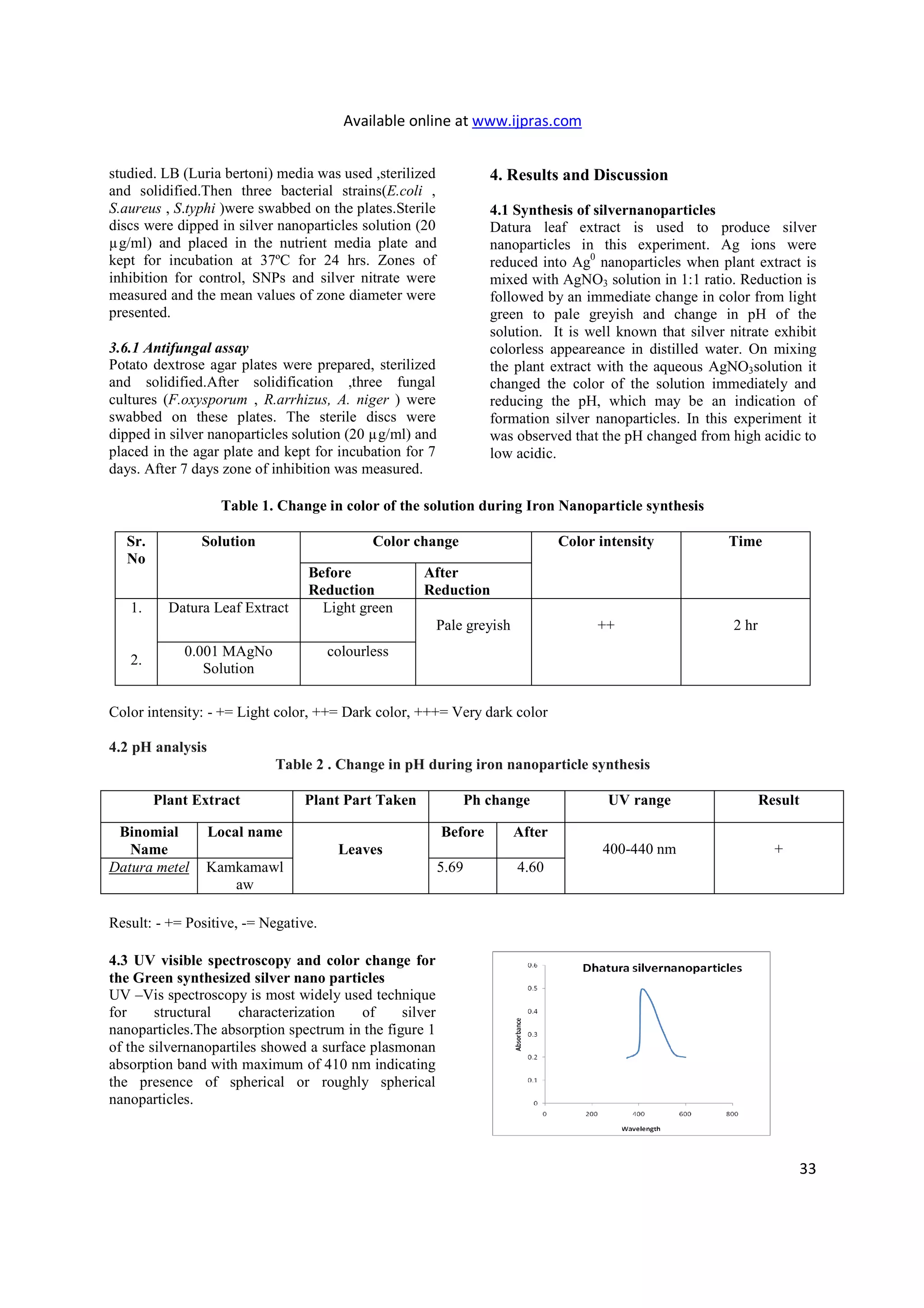
This research article describes the green synthesis and characterization of zero-valent silver nanoparticles using the leaf extract of the Datura metel plant. Silver nanoparticles were synthesized by mixing an aqueous solution of silver nitrate with an extract of D. metel leaves. Characterization using UV-Vis spectroscopy, SEM, and TEM showed the particles were predominantly between 50-100 nm in size. Antimicrobial testing demonstrated the silver nanoparticles had inhibitory effects against bacterial and fungal strains. The green synthesis method provides a low-cost and environmentally friendly approach for producing silver nanoparticles.