SlideShare a Scribd company logo
Nanoparticle Reactivity from Water Consumption throughout the Human Digestive
System
Brock E. Shilling1, Katherine Phetxumphou2, Andrea M. Dietrich2
1 NSF-REU Fellow, Virginia Polytechnic Institute and State University (Home Institution:
Biomolecular Engineering, Milwaukee School of Engineering)
2 Department of Civil and Environmental Engineering, Virginia Polytechnic Institute and State
University
Abstract
Although the amount of consumer products containing nanoparticles is increasing,
research data on the toxicity and reactivity of these nanoparticles is not readily available. The
objectives of this research were to: (1) to evaluate the amount of dissolution of copper and iron
nanoparticles in different digestive fluids: saliva, gastric, and intestinal; and (2) determine
reactivity of common nanoparticles: nano- iron, silver, copper, and silicon dioxide, in the human
digestive system by quantifying lipid oxidation using a TBARs assay. Dissolution results show
that gastric fluid most readily dissolves copper and iron nanoparticles as compared to other
digestive fluids. Since ferrous and cuprous solutions are known to induce lipid oxidation, they
were the controls in the TBARs experiments. It was found that all nanoparticles induced lipid
oxidation in at least one digestive fluid. Nanoiron caused the most lipid oxidation in all digestive
fluids. However, lipid oxidation results varied for copper, silver, and silicon dioxide
nanoparticles. Results show nanoparticles’ reactivity significantly differed among each other and
among each digestive fluid. These results suggest that nanoparticles exhibit a capability to react
in human digestive fluids, which may lead to health concerns and exposure limits.
Keywords: Nanoparticles, Lipid Oxidation, Reactivity, Digestive
1. Introduction
Every day, nanoparticles are becoming incorporated into more commercial and consumer
products due to their unique and beneficial properties. Nanomaterials are regarded as having a
measurement in at least one direction on the scale of 1-100 nanometers, and possessing a unique
property, which differs from the bulk material (Albanese et al., 2012). For instance, nanoscale
silver has been used in multitude of products for its antimicrobial property, while bulk silver
does not exhibit this ability. Due to characteristic properties like this, many industries are using
nanoparticles to improve current products and increase their efficiency. In 2009, it was reported
that there were already upwards of 600 products containing nanomaterials, and by now that
number has risen (Xia et al., 2009). However, these products may pose a health concern to
humans and other animals as they accumulate in the environment. The Food and Drug
Administration (FDA) does not require testing of nanoparticles when the bulk material is already
known to be safe (Trafton, 2014). Currently, this is the problem since many believe that if the
material of the nanoparticle at the bulk scale is safe, then so is the nanoparticle itself, which is
not always the case. Nanoparticles frequently exhibit enhanced reactivity as compared to their
bulk counterparts. It has been shown that nanoparticles can easily penetrate cells because of their
size and cause damage to DNA (Trafton, 2014). In depth research about nanoparticle toxicity and
exposure limits need to be conducted to ensure products are officially safe for use. As
nanotechnology and nanoparticles become more incorporated into every day products, it is
important to acknowledge the risks they may pose to the environment and human health. This
study focuses on several commonly used nanoparticles, such as iron, silver, copper, and silicon
dioxide, and their potential health concerns when introduced to the human digestive system.
1.1 Consumer nanoparticles
Several nanoparticle-containing products today pose a risk of being accidentally ingested.
Common sources are cosmetics, food, and clothing products (Fröhlich et al., 2012). Most
consumers do not even realize that nanoparticles are present in their life as sunscreen, toothpaste,
and food packaging. Silicon dioxide nanoparticles have been used as food additives, while silver
nanoparticles have been used in food packaging to improve shelf life (Fröhlich et al., 2012).
Table 1 below shows some of the most common nanoparticles being used in commercial
products, healthcare, and water treatment.
Table 1: Common nanoparticles currently used in different industries, including cosmetics,
food, water remediation, and healthcare (Shilling, 2015)
Nanoparticle Common Uses
Iron Groundwater remediation
Silver Food packaging, bandages, clothing, toothpaste
Copper Coatings on plastic
Silica Food additives, drug delivery techniques
Previously, the amount of nanoparticles being used was low enough that there were no
signs of impact on the environment or human health. However, with nanotechnology emerging
and nanoparticle use increasing, the levels of nanoparticles in the environment are starting to
become more significant (Nowack et al., 2007). Environmental nanoparticles levels need to be
monitored and exposure limits need to be researched and identified to ensure the correct
remediation protocols are being followed (Nowack et al., 2007).
1.2 Lipid oxidation
Lipids are ubiquitous in the human body and are one of the four basic organic compounds
which make up life. Lipids are insoluble in water and other polar solvents; they make up cell
membranes as well as provide long term energy storage. It has been shown that transition metals
degrade and oxidize lipids. The extent of lipid oxidation, also known as lipid peroxidation,
depends upon the nature of the lipid itself; it is dependent upon whether it is saturated or
unsaturated. Unsaturated lipids are oxidized because of the presence of carbon-carbon double
bonds. The lipid oxidation reaction is separated into three sections: initiation, propagation, and
termination (Sevanian et al., 1985). Fatty acids, a subsection of lipids, are common in biological
systems and take part in lipid oxidation. During initiation, an adjacently bonded hydrogen atom
to the double bond is removed by an initiator, such as a transition metal like iron (Sevanian et al.,
1985). This creates a fatty acid radical, which means it has unpaired electrons (Reaction 1). This
fatty acid radical proceeds to react with stable oxygen and creates a lipid peroxy radical; this is
known as propagation (Reaction 2, 3) (Sevanian et al., 1985). Radicals are known to be unstable
and reactive. When radicals react with stable molecules, another radical forms and a chain
reaction occurs. The termination portion occurs when one of these radicals reacts with another
radical to form a one of many possible byproducts which ends the chain reaction (Reaction 4, 5,
6) (Sevanian et al., 1985). A few of the prevalent byproducts of lipid oxidation are
malondialdehyde (MDA) and n-hexanal (Ömür-Özbek et al., 2012). The lipid peroxidation
stages are shown below, where R represents a carbon chain of the lipid and the symbol ∙
represents a free electron (Sevanian et al., 1985).
RH + Initiator  R∙ (1)
R∙ + O2  RO2∙ (2)
RO2∙ + RH  ROOH + R∙ (3)
RO2∙ + RO2∙  ROOR + O2 (4)
RO2∙ + R∙  ROOR (5)
R∙ + R∙  RR (6)
Lipid peroxidation is quantitatively measured by using the thiobarbituric acid reactive
substances (TBARs) assay to determine the amount of MDA produced. TBARs is a standard test
used in quantifying lipid oxidation in tissues, fluids, drugs, and food; although, it is tedious and
results vary due to lacking reactive aldehyde characterization (Phetxumphou, 2014). MDA alone
is colorless; however, with the presence of thiobarbituric acid (TBA), these two bind together to
form a product with a pink color. This color is analyzed by a UV-VIS spectrophotometer using a
wavelength of 532 nm. Figure 1 below shows the MDA and TBA reaction complex.
Figure 1: Reaction of the main byproduct of lipid peroxidation (MDA) and thiobarbituric
acid to form a pink colored complex which is quantitatively measured using UV-VIS
spectrophotometry.
Oxidative stress is damaging to human health and may contribute to disease pathogenesis
(Xia et al., 2009). With increase lipid peroxidation, cell membranes become damaged and the
cell becomes unable to regulate ion and water exchanges, eventually leading to cell death. Also,
byproducts of this reaction are able to cause mutations in DNA that may lead to cancer (Trafton,
2014). It has been proven that trace metal ions increase lipid peroxidation. Since nanoparticles’
reactivity is higher than their bulk counterparts, it raises the concern that nanoparticles have an
enhanced effect on lipid peroxidation, causing even low concentrations to trigger health
problems. The goal of this research was to evaluate the reactivity of iron, silver, copper, and
silicon dioxide nanoparticles throughout the human ingestion and digestion systems. Two
specific objectives were: (1) to evaluate the amount of dissolution of copper and iron
nanoparticles in artificial saliva, intestinal, and gastric digestive fluids; and (2) to quantitatively
determine lipid oxidation in artificial saliva, gastric fluid, and intestinal fluid induced by nano-
iron, silver, copper, and silicon dioxide using the TBARs assay.
2. ResearchMethods
2.1 Preparation of digestive fluids
Each artificial digestive fluid was prepared fresh before each day of experimentation
since proteins as well as lipids degrade with time. However, inorganic salt solutions were kept
for weeks at a time. Soybean oil was chosen as the lipid source for its oxidative stability, since
other lipids, like linoleic acid, oxidizes easily in air and would be difficult to work with
(Phetxumphou, 2014). Artificial saliva consisted of an inorganic salt solution, proteins, and
soybean oil; this solution is comparable to human saliva (Phetxumphou, 2014). The inorganic
saliva salt solution was based on saliva’s salt characteristics and included: NaCl, 0.126; KCl,
0.964; KSCN, 0.189; KH2PO4, 0.655; Na2SO4, 0.337; NH4Cl, 0.178; CaCl2, 0.155; NaHCO3,
0.568 grams, and dissolved in 1000 mL Nanopure® water (TDS=0 mg/L) (Phetxumphou, 2014).
The artificial saliva fluid consisted of: 30 mg soybean oil, 0.216 g mucin protein, 0.541 g α-
amylase protein, and 100 mL of the inorganic salt solution. The artificial intestinal fluid was
made up of 100 mL inorganic intestinal salt solution and 30 mg soybean oil. The inorganic
intestinal solution included: KH2PO4, 6.805; NaOH, 0.896 grams dissolved in 1000 mL
Nanopure® water, and the pH was 6.8 (Stippler et al., 2004). The pH of this fluid was adjusted
accordingly with NaOH and/or HCl. Artificial gastric fluid contained no salts or proteins. The
artificial gastric fluid imitated the conditions of fasted state simulated gastric fluid with a pH of
1.4-2.1 (Aburub et al., 2008). This fluid was prepared by adding 1 mL 99.9% HCl to 99 mL
Nanopure® water. The pH was adjusted with water or HCl depending on the initial reading.
Once desired pH was achieved, 30 mg of soybean oil was added to the fluid to act as the lipid
source.
2.2 Preparation of metallic and nanoparticle solutions
2.2.1 Preparation of metallic ions
The metallic ions ferrous (Fe+2) and cuprous (Cu+1) were used for this study. The targeted
total concentration of each metallic ion was 10 mg/L. The ferrous solution was prepared by
diluting 0.010 g FeSO4 ∙ 7H2O (Fisher Scientific, CAS #7782-63-0) into 200 mL Nanopure®
water. The cuprous solution was prepared by diluting 0.008 g CuCl (Sigma-Aldrich, CAS #7758-
89-6) into 500 mL Nanopure® water. Solutions were acid digested with 5% HNO3 by volume
and ran on the Atomic Absorbance Spectroscopy (AAS) (Perkin-Elmer, 5100PC AAS, Waltham,
MA, USA) to confirm the targeted concentrations of 10 mg/L.
2.2.2 Preparation of nanoparticle solutions
Only copper and iron nanoparticles were used for dissolution testing. The targeted
concentrations of copper and iron nanoparticles were 100 mg/L and 25 mg/L, respectively. The
copper solution was prepared by diluting 0.008 g Cu nanoparticles in 80 mL Nanopure® water.
The nZVI solution was prepared by adding 130 µL to 80 mL Nanopure® water.
For lipid oxidation experiments, nanoparticles copper, silver, silicon dioxide, and
nanoscale zero-valent iron (nZVI) were used. The targeted concentration of nanoparticles was 10
mg/L for lipid oxidation studies. Average human exposure to nanoparticles has been reported to
vary from 0-112 mg/individual/day, so an environmentally relevant value of 10 m/L was chosen
for the TBARs study (Fröhlich et al., 2012). Copper and silver nanoparticles exhibited poor
mixing abilities, so an increase in particles was necessary to achieve a AAS reading of
approximately 10 mg/L. Copper nanoparticles (Sigma-Aldrich, CAS #7440-50-8, Size 40-60
nm) were prepared at a concentration of 15 mg/L, and silver nanoparticles (Sigma-Aldrich, CAS
#7440-22-4, Size <100 nm) were prepared at a concentration of 50 mg/L. Silicon dioxide
nanoparticles (Sigma-Aldrich, CAS #7631-86-9, Size 10-20 nm) were prepared at a
concentration of 10 mg/L.
The nanoparticles: silver, copper, and silicon dioxide, arrived in the powder form, and
proper precautions and personal protective equipment were needed to ensure safety. In the
powder form, nanoparticles were weighed and handled in a fume hood while wearing a
laboratory coat, nitrile gloves, and safety glasses to minimize exposure. Nanopure® water was
used to suspend the nanoparticles in solution. Once in solution form, the nanoparticles were able
to be handled with gloves outside of the fume hood. Lastly, nZVI (provided by from Nanoiron
Ltd., Rajhrad, Czech Republic, EU, Size <50 nm), which was already in a slurry form, was
handled with gloves and safety glasses to minimize possible exposure. The targeted
concentration was 10 mg/L and was prepared by adding 50 µL to 60 mL of Nanopure® water.
All solutions were acid digested with 5% HNO3 by volume and ran on the AAS except silicon
dioxide nanoparticles, which were ran on the inductively coupled plasma mass spectrometry
(ICP-MS), to confirm concentrations of the ions and nanoparticle concentrations.
2.3 Measuring dissolution
Two sets of dissolutions tests were conducted. The first test exclusively used a copper
nanoparticle solution targeted at 100 mg/L. The second used an nZVI solution targeted at 25
mg/L. Artificial saliva, intestinal, and gastric fluids were prepared aforementioned. A one to one
ratio of digestive fluid and copper nanoparticle solution, 5 mL each, was prepared in a 15 mL
propylene conical tubes. A one to one ratio of Nanopure® and copper solution acted as the
control. Four time periods were selected at which AAS readings would be taken: 0, 15, 30, and
60 minutes. A total of 16 conical tubes were needed for each dissolution test, four per fluid and
the control. The 15, 30, and 60 minute tubes were placed in a 37ºC water bath to mimic body
conditions.
2.4 Measuring lipid oxidation
2.4.1 Thiobarbituric Acid Reactive Substances (TBARs) Assay
The Spanier TBARs method (1991) was modified to measure lipid oxidation in aqueous
samples by allowing low MDA concentrations to be read (Wang, 2002). MDA standards
(0.0313, 0.625, 0.125, 0.250, 0.500, 0.750, 1.00, 2.00, 4.00, and 10.0 µM) were prepared to
create a standard curve, Figure 2, using the UV-VIS spectrometer set to read at 532 nm. The best
fit line was determined and shown with Figure 2. The TBARs samples consisted of equal
volumes of digestive fluid and metallic or nanoparticle solutions, 5 mL each, in 15 mL propylene
conical tubes. Control samples were 1) Nanopure®; 2) Nanopure® and digestive fluid mixture;
3) digestive fluid only; and 4) Nanopure® and metallic/nanoparticle mixtures. All samples were
done in triplicates. Once prepared, the tubes were placed in 37ºC water bath for 15 minutes to
imitate body temperature. After incubation, samples were analyzed for lipid oxidation using the
TBARs method. This method measures the oxidative stress caused by each metallic and
nanoparticle solution. Once measured on the UV-VIS spectrometer, absorbance values were
averaged and converted to MDA concentration per mg of total metallic ion or nanoparticle using
the standard curve.
Figure 2: MDA standard curve.
3. Results and Discussion
3.1 Dissolution in digestive fluids
3.1.1 Dissolution of copper nanoparticles
For this dissolution, the copper nanoparticle solution was targeted at 100 mg/L. Since
samples were prepared with equal volume of digestive fluid and metal solution, the maximum
dissolution which could be achieved was 50 mg/L. The pH of the intestinal fluid was 6.8, and the
gastric fluid pH was 1.43. The amount of dissolution in gastric fluid increased with time. After
60 minutes of incubation, gastric fluid had the highest dissolution with 47.6%, then saliva with
14.2%, intestinal with 9.6 %, and lastly, Nanopure® with 9.0%. Gastric fluid considerably
differed from the other test fluids. Saliva, intestinal, and Nanopure® fluids yielded comparable
results with each other. Figure 3 is a graphical representation of the dissolution of copper
nanoparticles in digestive fluids over time.
Figure 3: The dissolution of copper nanoparticles in the artificial digestive fluids of saliva,
intestinal, and gastric. Nanopure® water was used as the control fluid.
The exhibited high dissolution of copper nanoparticles in gastric fluid was proposed to be
because of the fluid’s acidity. Nanopure®, saliva, and intestinal fluids all had pH values of
relatively neutral, while gastric was acidic. Hydrochloric acid has been reported to corrode and
react with metals (Zhang et al., 2004). However, metal copper does not readily react with
hydrochloric acid; it is a relatively slow reaction (Crundwell, 1992). Although bulk metal may
not quickly react with hydrochloric acid, copper nanoparticles may increase the rate of reaction
because of the increased surface area and reactivity, which nanoparticles are known for.
3.1.2 Dissolution of nZVI
The nZVI solution for this dissolution test was targeted at 25 mg/L, meaning the
maximum dissolution concentration would be 12.5 mg/L due to the samples diluted as an equal
volume mixture. The pH for the intestinal fluid was 6.8, and the gastric fluid pH was 1.51.
Similar to the copper nanoparticle dissolution, gastric fluid exhibited the greatest dissolution of
nZVI. After the 60 minute incubation period, gastric exhibited the highest dissolution of nZVI
with 94.4%, then intestinal with 57.5%, saliva was 54.7%, and lastly, the Nanopure® control
with 53.2%. All fluids showed a slight increase in dissolution over time as expected. By looking
at the graphical results in Figure 4 gastric results differ noticeably from the Nanopure® control
results, as compared to the other digestive fluids used.
Figure 4: The dissolution of nZVI in the artificial digestive fluids saliva, intestinal, and gastric.
Nanopure® water was used as the control fluid.
Similar to the copper dissolution, the proposed reason for gastric having increased
dissolution for nZVI is the acidity. In this dissolution test, results were closer to the projected
maximum. The nZVI particles were better suspended because they are in a stable slurry instead
of powder form, like the copper nanoparticles. The slurry was nearly a year old, and the iron
nanoparticles were capable of dissolving with time. This was verified by the results. Nanopure®
exhibited an over 50% dissolution result. If Nanopure® water were to dissolve nZVI this well,
the end dissolution should have been near the maximum 12.5 mg/L. However, since Nanopure®
dissolution neared 50%, the initial slurry was suggested to contain a mix of nZVI particles and
dissolved iron ions. Unlike copper, iron readily reacts with hydrochloric acid (Chetouani et al.,
2004). It is proposed that gastric fluid neared the maximum nZVI dissolution because of the
considerable amount of dissolved iron in the initial nZVI slurry and hydrochloric acid’s ability to
corrode and dissolve iron. These results indicated that if certain nanoparticles were ingested,
lipid oxidation may increase. The increased oxidation leads to more reactive oxygen
intermediates, which are associated with oxidative stress and cellular damage (Mittler, 2002).
3.2 Lipid oxidation using TBARs
With nanoparticles increasingly incorporated into consumer products, like food
packaging, it is essential to evaluate their reactivity in case of accidental consumption. In all
artificial fluids, nZVI samples did not significantly differ from the ferrous controls samples,
which were known to induce lipid oxidation. nZVI samples showed higher increases in lipid
oxidation than any other solution used, as seen in Figure 5. Cuprous, copper nanoparticles, and
silicon dioxide nanoparticles exhibited lower increases of lipid oxidation. Silver particles were
shown to have a similar increase in lipid oxidation as to the cuprous solution. In the silver
nanoparticle intestinal testing, an outlier was present, which was taken out. The outlier affected
the average increase of lipid oxidation and misconstrued the results. The silver nanoparticles
were difficult to suspend and readily aggregated on either the top or bottom of the solution. For
the outlier silver sample, it was believed that a clump was present. Therefore, it caused an
increase in the concentration of silver nanoparticles, which impacted the resulting average MDA
concentration. This silver sample outlier made the end result of MDA per milligram in intestinal
fluid much higher than expected. Copper nanoparticles did not readily dissolve in solution. This
made getting homogenous and repeatable samples for copper and silver nanoparticles
problematic. For further testing, it would be ideal to use stable slurries for all nanoparticles, like
the nZVI samples.
Figure 5: The increase in lipid oxidation in different artificial digestive fluids when
subjected to ferrous, cuprous, nZVI, copper nanoparticles, silver nanoparticles, or silicon
dioxide nanoparticles. *An outlier sample was removed.
Through the use of a two-way Anova statistical test, lipid oxidation between digestive
fluids significantly differed with p-value of 0.005, and lipid oxidation significantly differed
between tested metallic/nanoparticle solutions with a p-value of 0.011. It was proposed saliva
showed the highest lipid oxidation since the soybean oil was better dispersed due to the salts and
proteins that were present in saliva. In gastric fluid, the soybean oil stayed primarily at the top of
the fluid in one or two main clusters. The lipid source for saliva dispersed more evenly into
smaller clusters when mixed. When the lipids are more dispersed, there is more surface area to
react, which may be the cause of difference in lipid oxidation between the digestive fluids. For
future testing, an addition of an emulsifier would be encouraged to disperse the lipids more
consistently between fluids. Overall, it is important to realize that all nanoparticles increased
lipid oxidation, in at least one fluid. Lipid oxidation is harmful to the human health, and any
amount of increase may be concerning (Harris, 2013). Out of all of the nanoparticles tested,
nZVI exhibited consistently the highest reactivity, and is the highest concern for human health.
Further tests using stable slurries of these nanoparticles with a better dispersed lipid source is
encouraged to expand upon current findings.
4. Conclusion
For the dissolution tests of copper nanoparticles and nZVI, artificial gastric fluid was the
only digestive fluid which noticeably differed from the Nanopure® control. Gastric fluid had
dissolution results of 47.6% and 94.4% for copper nanoparticles and nZVI, respectively, while
the results of Nanopure® control were 9.0% and 53.2%, respectively. The gastric fluid’s
increased dissolution results were proposed to be due to the acidity of this fluid. The nZVI’s
increase in dissolution was because of the stable slurry it was in.
Ferrous and nZVI consistently yielded the highest increase in lipid oxidation throughout
the digestive fluids. The other solutions: cuprous, copper nanoparticles, silver nanoparticles, and
silicon dioxide nanoparticles, exhibited lower increases in lipid oxidation in comparison. Since
not all nanoparticles form stable solutions when prepared, stable and fresh slurries should be
used for future testing. Silver and copper nanoparticles readily aggregated at the top or bottom of
the solution, making precise, 10 mg/L samples difficult to attain. Nevertheless, lipid oxidation
between digestive fluids significantly differed. Lipid oxidation between nanoparticle samples
significantly differed. In conclusion, nanoparticles exhibited an increase in lipid oxidation in at
least one digestive fluid, if not all. Lipid oxidation can be a harmful reaction, which oxidizes
lipids that are present everywhere in the human body. With nanoparticle use in consumer
products increasing, it is crucial to monitor the increase of nanoparticles present in the
environment as well as further studying the effects of nanoparticles on human health.
5. Acknowledgements
First off, I want to thank Dr. Andrea Dietrich (Department of Civil and Environmental
Engineering) for being my faculty mentor and allowing me to conduct research in her lab. Also, I
want to thank my graduate mentor, Katherine Phetxumphou (Civil and Environmental
Engineering) for her help and support in this research process. I want to thank Dr. Lohani
(Environmental Education) for applying the funding, directing the Interdisciplinary Water and
Engineering REU, and allowing me this great opportunity. I want to thank my REU cohort for
the support during the research. We acknowledge the support of the National Science
Foundation through the NSF/REU Site Grant EEC-1359051. Any opinions, findings, and
conclusions or recommendations expressed in this paper are those of the author(s) and do not
necessarily reflect the views of the National Science Foundation.
References
Aburub, A., Risley, D. S., & Mishra, D. (2008). A critical evaluation of fasted state simulating
gastric fluid (FaSSGF) that contains sodium lauryl sulfate and proposal of a modified
recipe. International journal of pharmaceutics, 347(1), 16-22.
Albanese, A., Tang, P. S., & Chan, W. C. (2012). The effect of nanoparticle size, shape, and
surface chemistry on biological systems. Annual review of biomedical engineering, 14, 1-
16.
Chetouani, A., Hammouti, B., & Benkaddour, M. (2004). Corrosion inhibition of iron in
hydrochloric acid solution by jojoba oil. Pigment & resin technology, 33(1), 26-31.
Crundwell, F. K. (1992). The anodic dissolution of copper in hydrochloric acid solutions.
Electrochimica acta, 37(15), 2707-2714.
Fröhlich, E., & Roblegg, E. (2012). Models for oral uptake of nanoparticles in consumer
products. Toxicology, 291(1), 10-17.
Harris, L. J. (Ed.). (2013). Improving the safety and quality of nuts. Elsevier.
Jahnke, A., Holmbäck, J., Andersson, R. A., Kierkegaard, A., Mayer, P., & MacLeod, M. (2015).
Differences between lipids extracted from five species are not sufficient to explain
biomagnification of non-polar organic chemicals. Environmental Science & Technology Letters.
Lee, C., Kim, J. Y., Lee, W. I., Nelson, K. L., Yoon, J., & Sedlak, D. L. (2008). Bactericidal
effect of zero-valent iron nanoparticles on Escherichia coli. Environmental science &
technology, 42(13), 4927-4933.
Mittler, R. (2002). Oxidative stress, antioxidants and stress tolerance. Trends in plant science,
7(9), 405-410.
Nowack, B., & Bucheli, T. D. (2007). Occurrence, behavior and effects of nanoparticles in the
environment. Environmental pollution, 150(1), 5-22.
Ömür-Özbek, P., Dietrich, A. M., Duncan, S. E., & Lee, Y. (2012). Role of lipid oxidation,
chelating agents, and antioxidants in metallic flavor development in the oral cavity.
Journal of agricultural and food chemistry, 60(9), 2274-2280.
Phetxumphou, K. (2014). Consumers and their drinking water: communicating water quality
and assessing the reaction of zerovalent nanoiron (nZVI) with saliva (Doctoral
dissertation, Virginia Tech).
Romero, F. J., Bosch-Morell, F., Romero, M. J., Jareño, E. J., Romero, B., Marín, N., & Romá, J.
(1998). Lipid peroxidation products and antioxidants in human disease. Environmental Health
Perspectives, 106(Suppl 5), 1229.
Sevanian, A., & Hochstein, P. (1985). Mechanisms and consequences of lipid peroxidation in
biological systems. Annual review of nutrition, 5(1), 365-390.
Stippler, E., Kopp, S., & Dressman, J. B. (2004). Comparison of US pharmacopeia simulated
intestinal fluid TS (without pancreatin) and phosphate standard buffer pH 6.8, TS of the
international pharmacopoeia with respect to their use in in vitro dissolution testing. Dissolution
Technologies, 11(2), 6-11.
TaylorAlicia, A., MarcusIan, M., GuysiRisa, L., & WalkerSharon, L. (2015). Metal oxide
nanoparticles induce minimal phenotypic changes in a model colon gut microbiota.
Environmental Engineering Science.
Trafton, A. (2014). Tiny particles may pose big risk. Retrieved from
http://newsoffice.mit.edu/2014/tiny-particles-may-pose-big-risk.
Wang, T. (2002). Soybean oil. In F.D. Gunstone, Vegetable Oils in Food Technology, Oxford:
Blackwell Publishing, 18–58.
Xia, T., Li, N., & Nel, A. E. (2009). Potential health impact of nanoparticles. Annual review of
public health, 30, 137-150.
Zhang, D. Q., Gao, L. X., & Zhou, G. D. (2004). Inhibition of copper corrosion in aerated
hydrochloric acid solution by heterocyclic compounds containing a mercapto group.
Corrosion science, 46(12), 3031-3040.

More Related Content

What's hot

pH effects on the adsorption of saxitoxin by powdered activated carbon
pH effects on the adsorption of saxitoxin by powdered activated carbonpH effects on the adsorption of saxitoxin by powdered activated carbon
pH effects on the adsorption of saxitoxin by powdered activated carbon
hbuarque
 
Investigation of the effect of initial biomass on nitrate and phosphate remov...
Investigation of the effect of initial biomass on nitrate and phosphate remov...Investigation of the effect of initial biomass on nitrate and phosphate remov...
Investigation of the effect of initial biomass on nitrate and phosphate remov...
Alexander Decker
 
International Journal of Computational Engineering Research(IJCER)
International Journal of Computational Engineering Research(IJCER)International Journal of Computational Engineering Research(IJCER)
International Journal of Computational Engineering Research(IJCER)
ijceronline
 
Antioxidants
AntioxidantsAntioxidants
Antioxidants
Youssor mamdouh
 
Nanoparticles and production of reactive oxygen species
Nanoparticles and production of reactive oxygen speciesNanoparticles and production of reactive oxygen species
Nanoparticles and production of reactive oxygen speciesAdwaith P. Suratkar
 
Copper Nanoparticles and Antioxidant Stress: Problem Makers or Solvers?_ Cri...
 Copper Nanoparticles and Antioxidant Stress: Problem Makers or Solvers?_ Cri... Copper Nanoparticles and Antioxidant Stress: Problem Makers or Solvers?_ Cri...
Copper Nanoparticles and Antioxidant Stress: Problem Makers or Solvers?_ Cri...
CrimsonpublishersNTNF
 
A pilot study on effect of copper and cadmium toxicity in Tilapia Mossambicus
A pilot study on effect of copper and cadmium toxicity in Tilapia MossambicusA pilot study on effect of copper and cadmium toxicity in Tilapia Mossambicus
A pilot study on effect of copper and cadmium toxicity in Tilapia Mossambicus
researchanimalsciences
 
Antioxidants
AntioxidantsAntioxidants
Antioxidants
ssuserd509321
 
Free Radicals - Mandarin Version
Free Radicals - Mandarin VersionFree Radicals - Mandarin Version
Free Radicals - Mandarin Version
Sook Yen Wong
 
Versatile Importance of Schiff Bases
Versatile Importance of Schiff BasesVersatile Importance of Schiff Bases
Versatile Importance of Schiff Bases
YogeshIJTSRD
 
Isolation and Characterization of Nickel Tolerant Bacterial Strains from Elec...
Isolation and Characterization of Nickel Tolerant Bacterial Strains from Elec...Isolation and Characterization of Nickel Tolerant Bacterial Strains from Elec...
Isolation and Characterization of Nickel Tolerant Bacterial Strains from Elec...
Agriculture Journal IJOEAR
 
ROS ANTIOXIDENTS
ROS  ANTIOXIDENTSROS  ANTIOXIDENTS
ROS ANTIOXIDENTS
Dr Muhammad Mustansar
 
The Safety Use of Mercury Restoration in Dental Clinic (The Silent Hazard)
The Safety Use of Mercury Restoration in Dental Clinic  (The Silent Hazard)The Safety Use of Mercury Restoration in Dental Clinic  (The Silent Hazard)
The Safety Use of Mercury Restoration in Dental Clinic (The Silent Hazard)
Ameer Al-Ameedee
 
Reactive oxygen species and anti-oxidants
Reactive oxygen species and anti-oxidantsReactive oxygen species and anti-oxidants
Reactive oxygen species and anti-oxidants
Periowiki.com
 
MSc Project presentation
MSc Project presentationMSc Project presentation
MSc Project presentation
SameerHussain65
 
Co labats newsletter_02
Co labats newsletter_02Co labats newsletter_02
Co labats newsletter_02
Jokin Hidalgo
 

What's hot (20)

pH effects on the adsorption of saxitoxin by powdered activated carbon
pH effects on the adsorption of saxitoxin by powdered activated carbonpH effects on the adsorption of saxitoxin by powdered activated carbon
pH effects on the adsorption of saxitoxin by powdered activated carbon
 
Investigation of the effect of initial biomass on nitrate and phosphate remov...
Investigation of the effect of initial biomass on nitrate and phosphate remov...Investigation of the effect of initial biomass on nitrate and phosphate remov...
Investigation of the effect of initial biomass on nitrate and phosphate remov...
 
International Journal of Computational Engineering Research(IJCER)
International Journal of Computational Engineering Research(IJCER)International Journal of Computational Engineering Research(IJCER)
International Journal of Computational Engineering Research(IJCER)
 
I0445159
I0445159I0445159
I0445159
 
Antioxidants
AntioxidantsAntioxidants
Antioxidants
 
Nanoparticles and production of reactive oxygen species
Nanoparticles and production of reactive oxygen speciesNanoparticles and production of reactive oxygen species
Nanoparticles and production of reactive oxygen species
 
Zhu2020
Zhu2020Zhu2020
Zhu2020
 
Copper Nanoparticles and Antioxidant Stress: Problem Makers or Solvers?_ Cri...
 Copper Nanoparticles and Antioxidant Stress: Problem Makers or Solvers?_ Cri... Copper Nanoparticles and Antioxidant Stress: Problem Makers or Solvers?_ Cri...
Copper Nanoparticles and Antioxidant Stress: Problem Makers or Solvers?_ Cri...
 
A pilot study on effect of copper and cadmium toxicity in Tilapia Mossambicus
A pilot study on effect of copper and cadmium toxicity in Tilapia MossambicusA pilot study on effect of copper and cadmium toxicity in Tilapia Mossambicus
A pilot study on effect of copper and cadmium toxicity in Tilapia Mossambicus
 
Antioxidants
AntioxidantsAntioxidants
Antioxidants
 
Free Radicals - Mandarin Version
Free Radicals - Mandarin VersionFree Radicals - Mandarin Version
Free Radicals - Mandarin Version
 
Versatile Importance of Schiff Bases
Versatile Importance of Schiff BasesVersatile Importance of Schiff Bases
Versatile Importance of Schiff Bases
 
Isolation and Characterization of Nickel Tolerant Bacterial Strains from Elec...
Isolation and Characterization of Nickel Tolerant Bacterial Strains from Elec...Isolation and Characterization of Nickel Tolerant Bacterial Strains from Elec...
Isolation and Characterization of Nickel Tolerant Bacterial Strains from Elec...
 
Lipid peroxidation
Lipid peroxidation Lipid peroxidation
Lipid peroxidation
 
ROS ANTIOXIDENTS
ROS  ANTIOXIDENTSROS  ANTIOXIDENTS
ROS ANTIOXIDENTS
 
The Safety Use of Mercury Restoration in Dental Clinic (The Silent Hazard)
The Safety Use of Mercury Restoration in Dental Clinic  (The Silent Hazard)The Safety Use of Mercury Restoration in Dental Clinic  (The Silent Hazard)
The Safety Use of Mercury Restoration in Dental Clinic (The Silent Hazard)
 
Reactive oxygen species and anti-oxidants
Reactive oxygen species and anti-oxidantsReactive oxygen species and anti-oxidants
Reactive oxygen species and anti-oxidants
 
MSc Project presentation
MSc Project presentationMSc Project presentation
MSc Project presentation
 
Co labats newsletter_02
Co labats newsletter_02Co labats newsletter_02
Co labats newsletter_02
 
Cucurbita maxima
Cucurbita maximaCucurbita maxima
Cucurbita maxima
 

Viewers also liked

The effect of gold nanoparticle on renal function in rats
The effect of gold nanoparticle on renal function in ratsThe effect of gold nanoparticle on renal function in rats
The effect of gold nanoparticle on renal function in rats
Nanomedicine Journal (NMJ)
 
Silver nanoparticles from the leaf extract of datura metel
Silver nanoparticles from the leaf extract of datura metelSilver nanoparticles from the leaf extract of datura metel
Silver nanoparticles from the leaf extract of datura metel
Usman Arshad
 
Berbeco et al, Low Z target switching to increase tumor endothelial cell dose...
Berbeco et al, Low Z target switching to increase tumor endothelial cell dose...Berbeco et al, Low Z target switching to increase tumor endothelial cell dose...
Berbeco et al, Low Z target switching to increase tumor endothelial cell dose...David Parsons
 
GRAFTING OF NANOPARTICLES ON SURFACES OF SI
GRAFTING OF NANOPARTICLES ON SURFACES OF SIGRAFTING OF NANOPARTICLES ON SURFACES OF SI
GRAFTING OF NANOPARTICLES ON SURFACES OF SIDrix78
 
BME 626_Alzheimers_ Nanomedical Systems
BME 626_Alzheimers_ Nanomedical SystemsBME 626_Alzheimers_ Nanomedical Systems
BME 626_Alzheimers_ Nanomedical SystemsTejasvi Parupudi
 
Antimicrobial activity of trachyspermum ammi leaves mediated silver nanoparti...
Antimicrobial activity of trachyspermum ammi leaves mediated silver nanoparti...Antimicrobial activity of trachyspermum ammi leaves mediated silver nanoparti...
Antimicrobial activity of trachyspermum ammi leaves mediated silver nanoparti...
eSAT Journals
 
Making Graphene Holey. Gold-Nanoparticle-Mediated Hydroxyl Radical Attack on...
Making Graphene Holey.  Gold-Nanoparticle-Mediated Hydroxyl Radical Attack on...Making Graphene Holey.  Gold-Nanoparticle-Mediated Hydroxyl Radical Attack on...
Making Graphene Holey. Gold-Nanoparticle-Mediated Hydroxyl Radical Attack on...
kamatlab
 
Antimicrobial and cytotoxicity effect of silver nanoparticle synthesized by C...
Antimicrobial and cytotoxicity effect of silver nanoparticle synthesized by C...Antimicrobial and cytotoxicity effect of silver nanoparticle synthesized by C...
Antimicrobial and cytotoxicity effect of silver nanoparticle synthesized by C...
Nanomedicine Journal (NMJ)
 
Synthesis, characterization and role of zero valent iron nanoparticle in remo...
Synthesis, characterization and role of zero valent iron nanoparticle in remo...Synthesis, characterization and role of zero valent iron nanoparticle in remo...
Synthesis, characterization and role of zero valent iron nanoparticle in remo...
sadalltime
 
BBI Solutions - Nanoparticles
BBI Solutions - NanoparticlesBBI Solutions - Nanoparticles
BBI Solutions - Nanoparticles
BBISolutions
 
Nanoparticle use in pharmaceutical analysis
Nanoparticle use in pharmaceutical analysis Nanoparticle use in pharmaceutical analysis
Nanoparticle use in pharmaceutical analysis
Bhaumik Bavishi
 
Synthesis of Silver Nano Particles from Marine Bacteria Pseudomonas aerogenosa
Synthesis of Silver Nano Particles from Marine Bacteria Pseudomonas aerogenosaSynthesis of Silver Nano Particles from Marine Bacteria Pseudomonas aerogenosa
Synthesis of Silver Nano Particles from Marine Bacteria Pseudomonas aerogenosaKamalpreet Sarna
 
Gold nanoparticles: strange properties and applications
Gold nanoparticles: strange properties and applicationsGold nanoparticles: strange properties and applications
Gold nanoparticles: strange properties and applications
Expedeon
 
Nanoparticle Gold in therapy and cosmetology
 Nanoparticle Gold in therapy and cosmetology Nanoparticle Gold in therapy and cosmetology
Nanoparticle Gold in therapy and cosmetology
guest49edce
 
ferromagnetic nanomaterials
ferromagnetic nanomaterialsferromagnetic nanomaterials
ferromagnetic nanomaterials
University of Technology
 
Final Presentation on Iron Nanoparticles_Prajwal (1)
Final Presentation on Iron Nanoparticles_Prajwal (1)Final Presentation on Iron Nanoparticles_Prajwal (1)
Final Presentation on Iron Nanoparticles_Prajwal (1)Prajwal Bahukhandi
 
Biological methods for nanoparticle synthesis
Biological methods for nanoparticle synthesisBiological methods for nanoparticle synthesis
Biological methods for nanoparticle synthesis
Vaibhav Maurya
 
Gold nano particles
Gold nano particlesGold nano particles
Gold nano particles
shreni bandi
 
Final Report (Graphene supported platinum nanoparticles) (1)
Final Report (Graphene supported platinum nanoparticles) (1)Final Report (Graphene supported platinum nanoparticles) (1)
Final Report (Graphene supported platinum nanoparticles) (1)Sridharan Thirumalai
 

Viewers also liked (19)

The effect of gold nanoparticle on renal function in rats
The effect of gold nanoparticle on renal function in ratsThe effect of gold nanoparticle on renal function in rats
The effect of gold nanoparticle on renal function in rats
 
Silver nanoparticles from the leaf extract of datura metel
Silver nanoparticles from the leaf extract of datura metelSilver nanoparticles from the leaf extract of datura metel
Silver nanoparticles from the leaf extract of datura metel
 
Berbeco et al, Low Z target switching to increase tumor endothelial cell dose...
Berbeco et al, Low Z target switching to increase tumor endothelial cell dose...Berbeco et al, Low Z target switching to increase tumor endothelial cell dose...
Berbeco et al, Low Z target switching to increase tumor endothelial cell dose...
 
GRAFTING OF NANOPARTICLES ON SURFACES OF SI
GRAFTING OF NANOPARTICLES ON SURFACES OF SIGRAFTING OF NANOPARTICLES ON SURFACES OF SI
GRAFTING OF NANOPARTICLES ON SURFACES OF SI
 
BME 626_Alzheimers_ Nanomedical Systems
BME 626_Alzheimers_ Nanomedical SystemsBME 626_Alzheimers_ Nanomedical Systems
BME 626_Alzheimers_ Nanomedical Systems
 
Antimicrobial activity of trachyspermum ammi leaves mediated silver nanoparti...
Antimicrobial activity of trachyspermum ammi leaves mediated silver nanoparti...Antimicrobial activity of trachyspermum ammi leaves mediated silver nanoparti...
Antimicrobial activity of trachyspermum ammi leaves mediated silver nanoparti...
 
Making Graphene Holey. Gold-Nanoparticle-Mediated Hydroxyl Radical Attack on...
Making Graphene Holey.  Gold-Nanoparticle-Mediated Hydroxyl Radical Attack on...Making Graphene Holey.  Gold-Nanoparticle-Mediated Hydroxyl Radical Attack on...
Making Graphene Holey. Gold-Nanoparticle-Mediated Hydroxyl Radical Attack on...
 
Antimicrobial and cytotoxicity effect of silver nanoparticle synthesized by C...
Antimicrobial and cytotoxicity effect of silver nanoparticle synthesized by C...Antimicrobial and cytotoxicity effect of silver nanoparticle synthesized by C...
Antimicrobial and cytotoxicity effect of silver nanoparticle synthesized by C...
 
Synthesis, characterization and role of zero valent iron nanoparticle in remo...
Synthesis, characterization and role of zero valent iron nanoparticle in remo...Synthesis, characterization and role of zero valent iron nanoparticle in remo...
Synthesis, characterization and role of zero valent iron nanoparticle in remo...
 
BBI Solutions - Nanoparticles
BBI Solutions - NanoparticlesBBI Solutions - Nanoparticles
BBI Solutions - Nanoparticles
 
Nanoparticle use in pharmaceutical analysis
Nanoparticle use in pharmaceutical analysis Nanoparticle use in pharmaceutical analysis
Nanoparticle use in pharmaceutical analysis
 
Synthesis of Silver Nano Particles from Marine Bacteria Pseudomonas aerogenosa
Synthesis of Silver Nano Particles from Marine Bacteria Pseudomonas aerogenosaSynthesis of Silver Nano Particles from Marine Bacteria Pseudomonas aerogenosa
Synthesis of Silver Nano Particles from Marine Bacteria Pseudomonas aerogenosa
 
Gold nanoparticles: strange properties and applications
Gold nanoparticles: strange properties and applicationsGold nanoparticles: strange properties and applications
Gold nanoparticles: strange properties and applications
 
Nanoparticle Gold in therapy and cosmetology
 Nanoparticle Gold in therapy and cosmetology Nanoparticle Gold in therapy and cosmetology
Nanoparticle Gold in therapy and cosmetology
 
ferromagnetic nanomaterials
ferromagnetic nanomaterialsferromagnetic nanomaterials
ferromagnetic nanomaterials
 
Final Presentation on Iron Nanoparticles_Prajwal (1)
Final Presentation on Iron Nanoparticles_Prajwal (1)Final Presentation on Iron Nanoparticles_Prajwal (1)
Final Presentation on Iron Nanoparticles_Prajwal (1)
 
Biological methods for nanoparticle synthesis
Biological methods for nanoparticle synthesisBiological methods for nanoparticle synthesis
Biological methods for nanoparticle synthesis
 
Gold nano particles
Gold nano particlesGold nano particles
Gold nano particles
 
Final Report (Graphene supported platinum nanoparticles) (1)
Final Report (Graphene supported platinum nanoparticles) (1)Final Report (Graphene supported platinum nanoparticles) (1)
Final Report (Graphene supported platinum nanoparticles) (1)
 

Similar to Shilling_NanoparticleReactivity_2015_Final

Evaluation of the Concentration of Toxic Metals In Cosmetic Products In Nigeria
Evaluation of the Concentration of Toxic Metals In Cosmetic Products In Nigeria Evaluation of the Concentration of Toxic Metals In Cosmetic Products In Nigeria
Evaluation of the Concentration of Toxic Metals In Cosmetic Products In Nigeria
v2zq
 
F1804013742
F1804013742F1804013742
F1804013742
IOSR Journals
 
Evidence of heavy metals distribution in placenta in association with residua...
Evidence of heavy metals distribution in placenta in association with residua...Evidence of heavy metals distribution in placenta in association with residua...
Evidence of heavy metals distribution in placenta in association with residua...Alexander Decker
 
UGC Amit Proposal.docx
UGC Amit Proposal.docxUGC Amit Proposal.docx
UGC Amit Proposal.docx
NeerajOjha8
 
Antioxidant potentials of tannic acid on lipid peroxidation induced by severa...
Antioxidant potentials of tannic acid on lipid peroxidation induced by severa...Antioxidant potentials of tannic acid on lipid peroxidation induced by severa...
Antioxidant potentials of tannic acid on lipid peroxidation induced by severa...
Premier Publishers
 
Research proposal
Research proposalResearch proposal
Research proposal
Muhammad Salman
 
Dangers of bioaccumulation of some heavy metals consumed in sardine and macke...
Dangers of bioaccumulation of some heavy metals consumed in sardine and macke...Dangers of bioaccumulation of some heavy metals consumed in sardine and macke...
Dangers of bioaccumulation of some heavy metals consumed in sardine and macke...
Alexander Decker
 
Environmental Toxicity of Nanoparticles Environmental Toxicity of Nanoparticles
Environmental Toxicity of Nanoparticles Environmental Toxicity of NanoparticlesEnvironmental Toxicity of Nanoparticles Environmental Toxicity of Nanoparticles
Environmental Toxicity of Nanoparticles Environmental Toxicity of Nanoparticles
International Journal of Modern Research in Engineering and Technology
 
Nitrates In Water
Nitrates In WaterNitrates In Water
Nitrates In Water
Jennifer Wood
 
Charecteristics of wastewater
Charecteristics of wastewaterCharecteristics of wastewater
Charecteristics of wastewater
Venuri Lakshani
 
Risk in the use of silver nanoparticles on humain
Risk in the use of silver nanoparticles on humainRisk in the use of silver nanoparticles on humain
Risk in the use of silver nanoparticles on humain
Pierre Basmaji
 
Ijpr8471290198600
Ijpr8471290198600Ijpr8471290198600
Ijpr8471290198600
skedward
 
11.assessment of human health risk for arsenic, copper, nickel, mercury and z...
11.assessment of human health risk for arsenic, copper, nickel, mercury and z...11.assessment of human health risk for arsenic, copper, nickel, mercury and z...
11.assessment of human health risk for arsenic, copper, nickel, mercury and z...
Alexander Decker
 
Assessment of human health risk for arsenic, copper, nickel, mercury and zinc...
Assessment of human health risk for arsenic, copper, nickel, mercury and zinc...Assessment of human health risk for arsenic, copper, nickel, mercury and zinc...
Assessment of human health risk for arsenic, copper, nickel, mercury and zinc...Alexander Decker
 
Health risk assessment of consuming heavy metal contaminated benthic seafood,...
Health risk assessment of consuming heavy metal contaminated benthic seafood,...Health risk assessment of consuming heavy metal contaminated benthic seafood,...
Health risk assessment of consuming heavy metal contaminated benthic seafood,...
Premier Publishers
 
IRJET- Experimental Study on Impact of Leachate
IRJET- Experimental Study on Impact of LeachateIRJET- Experimental Study on Impact of Leachate
IRJET- Experimental Study on Impact of Leachate
IRJET Journal
 
IRJET- Experimental Study on Impact of Leachate
IRJET- Experimental Study on Impact of LeachateIRJET- Experimental Study on Impact of Leachate
IRJET- Experimental Study on Impact of Leachate
IRJET Journal
 
A Pilot Study on Effect of Copper and Cadmium Toxicity in Tilapia Mossambicus
A Pilot Study on Effect of Copper and Cadmium Toxicity in  Tilapia Mossambicus A Pilot Study on Effect of Copper and Cadmium Toxicity in  Tilapia Mossambicus
A Pilot Study on Effect of Copper and Cadmium Toxicity in Tilapia Mossambicus
researchanimalsciences
 
Characteristics of industrial waste
Characteristics of industrial wasteCharacteristics of industrial waste
Characteristics of industrial waste
tang mo
 
Toxicity of lead and mercury
Toxicity of lead and mercuryToxicity of lead and mercury
Toxicity of lead and mercury
SajawalNawaz2
 

Similar to Shilling_NanoparticleReactivity_2015_Final (20)

Evaluation of the Concentration of Toxic Metals In Cosmetic Products In Nigeria
Evaluation of the Concentration of Toxic Metals In Cosmetic Products In Nigeria Evaluation of the Concentration of Toxic Metals In Cosmetic Products In Nigeria
Evaluation of the Concentration of Toxic Metals In Cosmetic Products In Nigeria
 
F1804013742
F1804013742F1804013742
F1804013742
 
Evidence of heavy metals distribution in placenta in association with residua...
Evidence of heavy metals distribution in placenta in association with residua...Evidence of heavy metals distribution in placenta in association with residua...
Evidence of heavy metals distribution in placenta in association with residua...
 
UGC Amit Proposal.docx
UGC Amit Proposal.docxUGC Amit Proposal.docx
UGC Amit Proposal.docx
 
Antioxidant potentials of tannic acid on lipid peroxidation induced by severa...
Antioxidant potentials of tannic acid on lipid peroxidation induced by severa...Antioxidant potentials of tannic acid on lipid peroxidation induced by severa...
Antioxidant potentials of tannic acid on lipid peroxidation induced by severa...
 
Research proposal
Research proposalResearch proposal
Research proposal
 
Dangers of bioaccumulation of some heavy metals consumed in sardine and macke...
Dangers of bioaccumulation of some heavy metals consumed in sardine and macke...Dangers of bioaccumulation of some heavy metals consumed in sardine and macke...
Dangers of bioaccumulation of some heavy metals consumed in sardine and macke...
 
Environmental Toxicity of Nanoparticles Environmental Toxicity of Nanoparticles
Environmental Toxicity of Nanoparticles Environmental Toxicity of NanoparticlesEnvironmental Toxicity of Nanoparticles Environmental Toxicity of Nanoparticles
Environmental Toxicity of Nanoparticles Environmental Toxicity of Nanoparticles
 
Nitrates In Water
Nitrates In WaterNitrates In Water
Nitrates In Water
 
Charecteristics of wastewater
Charecteristics of wastewaterCharecteristics of wastewater
Charecteristics of wastewater
 
Risk in the use of silver nanoparticles on humain
Risk in the use of silver nanoparticles on humainRisk in the use of silver nanoparticles on humain
Risk in the use of silver nanoparticles on humain
 
Ijpr8471290198600
Ijpr8471290198600Ijpr8471290198600
Ijpr8471290198600
 
11.assessment of human health risk for arsenic, copper, nickel, mercury and z...
11.assessment of human health risk for arsenic, copper, nickel, mercury and z...11.assessment of human health risk for arsenic, copper, nickel, mercury and z...
11.assessment of human health risk for arsenic, copper, nickel, mercury and z...
 
Assessment of human health risk for arsenic, copper, nickel, mercury and zinc...
Assessment of human health risk for arsenic, copper, nickel, mercury and zinc...Assessment of human health risk for arsenic, copper, nickel, mercury and zinc...
Assessment of human health risk for arsenic, copper, nickel, mercury and zinc...
 
Health risk assessment of consuming heavy metal contaminated benthic seafood,...
Health risk assessment of consuming heavy metal contaminated benthic seafood,...Health risk assessment of consuming heavy metal contaminated benthic seafood,...
Health risk assessment of consuming heavy metal contaminated benthic seafood,...
 
IRJET- Experimental Study on Impact of Leachate
IRJET- Experimental Study on Impact of LeachateIRJET- Experimental Study on Impact of Leachate
IRJET- Experimental Study on Impact of Leachate
 
IRJET- Experimental Study on Impact of Leachate
IRJET- Experimental Study on Impact of LeachateIRJET- Experimental Study on Impact of Leachate
IRJET- Experimental Study on Impact of Leachate
 
A Pilot Study on Effect of Copper and Cadmium Toxicity in Tilapia Mossambicus
A Pilot Study on Effect of Copper and Cadmium Toxicity in  Tilapia Mossambicus A Pilot Study on Effect of Copper and Cadmium Toxicity in  Tilapia Mossambicus
A Pilot Study on Effect of Copper and Cadmium Toxicity in Tilapia Mossambicus
 
Characteristics of industrial waste
Characteristics of industrial wasteCharacteristics of industrial waste
Characteristics of industrial waste
 
Toxicity of lead and mercury
Toxicity of lead and mercuryToxicity of lead and mercury
Toxicity of lead and mercury
 

Shilling_NanoparticleReactivity_2015_Final

  • 1. Nanoparticle Reactivity from Water Consumption throughout the Human Digestive System Brock E. Shilling1, Katherine Phetxumphou2, Andrea M. Dietrich2 1 NSF-REU Fellow, Virginia Polytechnic Institute and State University (Home Institution: Biomolecular Engineering, Milwaukee School of Engineering) 2 Department of Civil and Environmental Engineering, Virginia Polytechnic Institute and State University Abstract Although the amount of consumer products containing nanoparticles is increasing, research data on the toxicity and reactivity of these nanoparticles is not readily available. The objectives of this research were to: (1) to evaluate the amount of dissolution of copper and iron nanoparticles in different digestive fluids: saliva, gastric, and intestinal; and (2) determine reactivity of common nanoparticles: nano- iron, silver, copper, and silicon dioxide, in the human digestive system by quantifying lipid oxidation using a TBARs assay. Dissolution results show that gastric fluid most readily dissolves copper and iron nanoparticles as compared to other digestive fluids. Since ferrous and cuprous solutions are known to induce lipid oxidation, they were the controls in the TBARs experiments. It was found that all nanoparticles induced lipid oxidation in at least one digestive fluid. Nanoiron caused the most lipid oxidation in all digestive fluids. However, lipid oxidation results varied for copper, silver, and silicon dioxide nanoparticles. Results show nanoparticles’ reactivity significantly differed among each other and among each digestive fluid. These results suggest that nanoparticles exhibit a capability to react in human digestive fluids, which may lead to health concerns and exposure limits. Keywords: Nanoparticles, Lipid Oxidation, Reactivity, Digestive 1. Introduction Every day, nanoparticles are becoming incorporated into more commercial and consumer products due to their unique and beneficial properties. Nanomaterials are regarded as having a measurement in at least one direction on the scale of 1-100 nanometers, and possessing a unique property, which differs from the bulk material (Albanese et al., 2012). For instance, nanoscale silver has been used in multitude of products for its antimicrobial property, while bulk silver does not exhibit this ability. Due to characteristic properties like this, many industries are using nanoparticles to improve current products and increase their efficiency. In 2009, it was reported that there were already upwards of 600 products containing nanomaterials, and by now that number has risen (Xia et al., 2009). However, these products may pose a health concern to humans and other animals as they accumulate in the environment. The Food and Drug Administration (FDA) does not require testing of nanoparticles when the bulk material is already known to be safe (Trafton, 2014). Currently, this is the problem since many believe that if the material of the nanoparticle at the bulk scale is safe, then so is the nanoparticle itself, which is not always the case. Nanoparticles frequently exhibit enhanced reactivity as compared to their bulk counterparts. It has been shown that nanoparticles can easily penetrate cells because of their size and cause damage to DNA (Trafton, 2014). In depth research about nanoparticle toxicity and
  • 2. exposure limits need to be conducted to ensure products are officially safe for use. As nanotechnology and nanoparticles become more incorporated into every day products, it is important to acknowledge the risks they may pose to the environment and human health. This study focuses on several commonly used nanoparticles, such as iron, silver, copper, and silicon dioxide, and their potential health concerns when introduced to the human digestive system. 1.1 Consumer nanoparticles Several nanoparticle-containing products today pose a risk of being accidentally ingested. Common sources are cosmetics, food, and clothing products (Fröhlich et al., 2012). Most consumers do not even realize that nanoparticles are present in their life as sunscreen, toothpaste, and food packaging. Silicon dioxide nanoparticles have been used as food additives, while silver nanoparticles have been used in food packaging to improve shelf life (Fröhlich et al., 2012). Table 1 below shows some of the most common nanoparticles being used in commercial products, healthcare, and water treatment. Table 1: Common nanoparticles currently used in different industries, including cosmetics, food, water remediation, and healthcare (Shilling, 2015) Nanoparticle Common Uses Iron Groundwater remediation Silver Food packaging, bandages, clothing, toothpaste Copper Coatings on plastic Silica Food additives, drug delivery techniques Previously, the amount of nanoparticles being used was low enough that there were no signs of impact on the environment or human health. However, with nanotechnology emerging and nanoparticle use increasing, the levels of nanoparticles in the environment are starting to become more significant (Nowack et al., 2007). Environmental nanoparticles levels need to be monitored and exposure limits need to be researched and identified to ensure the correct remediation protocols are being followed (Nowack et al., 2007). 1.2 Lipid oxidation Lipids are ubiquitous in the human body and are one of the four basic organic compounds which make up life. Lipids are insoluble in water and other polar solvents; they make up cell membranes as well as provide long term energy storage. It has been shown that transition metals degrade and oxidize lipids. The extent of lipid oxidation, also known as lipid peroxidation, depends upon the nature of the lipid itself; it is dependent upon whether it is saturated or unsaturated. Unsaturated lipids are oxidized because of the presence of carbon-carbon double bonds. The lipid oxidation reaction is separated into three sections: initiation, propagation, and termination (Sevanian et al., 1985). Fatty acids, a subsection of lipids, are common in biological systems and take part in lipid oxidation. During initiation, an adjacently bonded hydrogen atom to the double bond is removed by an initiator, such as a transition metal like iron (Sevanian et al., 1985). This creates a fatty acid radical, which means it has unpaired electrons (Reaction 1). This fatty acid radical proceeds to react with stable oxygen and creates a lipid peroxy radical; this is known as propagation (Reaction 2, 3) (Sevanian et al., 1985). Radicals are known to be unstable and reactive. When radicals react with stable molecules, another radical forms and a chain reaction occurs. The termination portion occurs when one of these radicals reacts with another
  • 3. radical to form a one of many possible byproducts which ends the chain reaction (Reaction 4, 5, 6) (Sevanian et al., 1985). A few of the prevalent byproducts of lipid oxidation are malondialdehyde (MDA) and n-hexanal (Ömür-Özbek et al., 2012). The lipid peroxidation stages are shown below, where R represents a carbon chain of the lipid and the symbol ∙ represents a free electron (Sevanian et al., 1985). RH + Initiator  R∙ (1) R∙ + O2  RO2∙ (2) RO2∙ + RH  ROOH + R∙ (3) RO2∙ + RO2∙  ROOR + O2 (4) RO2∙ + R∙  ROOR (5) R∙ + R∙  RR (6) Lipid peroxidation is quantitatively measured by using the thiobarbituric acid reactive substances (TBARs) assay to determine the amount of MDA produced. TBARs is a standard test used in quantifying lipid oxidation in tissues, fluids, drugs, and food; although, it is tedious and results vary due to lacking reactive aldehyde characterization (Phetxumphou, 2014). MDA alone is colorless; however, with the presence of thiobarbituric acid (TBA), these two bind together to form a product with a pink color. This color is analyzed by a UV-VIS spectrophotometer using a wavelength of 532 nm. Figure 1 below shows the MDA and TBA reaction complex. Figure 1: Reaction of the main byproduct of lipid peroxidation (MDA) and thiobarbituric acid to form a pink colored complex which is quantitatively measured using UV-VIS spectrophotometry. Oxidative stress is damaging to human health and may contribute to disease pathogenesis (Xia et al., 2009). With increase lipid peroxidation, cell membranes become damaged and the cell becomes unable to regulate ion and water exchanges, eventually leading to cell death. Also, byproducts of this reaction are able to cause mutations in DNA that may lead to cancer (Trafton, 2014). It has been proven that trace metal ions increase lipid peroxidation. Since nanoparticles’ reactivity is higher than their bulk counterparts, it raises the concern that nanoparticles have an enhanced effect on lipid peroxidation, causing even low concentrations to trigger health problems. The goal of this research was to evaluate the reactivity of iron, silver, copper, and silicon dioxide nanoparticles throughout the human ingestion and digestion systems. Two specific objectives were: (1) to evaluate the amount of dissolution of copper and iron nanoparticles in artificial saliva, intestinal, and gastric digestive fluids; and (2) to quantitatively determine lipid oxidation in artificial saliva, gastric fluid, and intestinal fluid induced by nano- iron, silver, copper, and silicon dioxide using the TBARs assay.
  • 4. 2. ResearchMethods 2.1 Preparation of digestive fluids Each artificial digestive fluid was prepared fresh before each day of experimentation since proteins as well as lipids degrade with time. However, inorganic salt solutions were kept for weeks at a time. Soybean oil was chosen as the lipid source for its oxidative stability, since other lipids, like linoleic acid, oxidizes easily in air and would be difficult to work with (Phetxumphou, 2014). Artificial saliva consisted of an inorganic salt solution, proteins, and soybean oil; this solution is comparable to human saliva (Phetxumphou, 2014). The inorganic saliva salt solution was based on saliva’s salt characteristics and included: NaCl, 0.126; KCl, 0.964; KSCN, 0.189; KH2PO4, 0.655; Na2SO4, 0.337; NH4Cl, 0.178; CaCl2, 0.155; NaHCO3, 0.568 grams, and dissolved in 1000 mL Nanopure® water (TDS=0 mg/L) (Phetxumphou, 2014). The artificial saliva fluid consisted of: 30 mg soybean oil, 0.216 g mucin protein, 0.541 g α- amylase protein, and 100 mL of the inorganic salt solution. The artificial intestinal fluid was made up of 100 mL inorganic intestinal salt solution and 30 mg soybean oil. The inorganic intestinal solution included: KH2PO4, 6.805; NaOH, 0.896 grams dissolved in 1000 mL Nanopure® water, and the pH was 6.8 (Stippler et al., 2004). The pH of this fluid was adjusted accordingly with NaOH and/or HCl. Artificial gastric fluid contained no salts or proteins. The artificial gastric fluid imitated the conditions of fasted state simulated gastric fluid with a pH of 1.4-2.1 (Aburub et al., 2008). This fluid was prepared by adding 1 mL 99.9% HCl to 99 mL Nanopure® water. The pH was adjusted with water or HCl depending on the initial reading. Once desired pH was achieved, 30 mg of soybean oil was added to the fluid to act as the lipid source. 2.2 Preparation of metallic and nanoparticle solutions 2.2.1 Preparation of metallic ions The metallic ions ferrous (Fe+2) and cuprous (Cu+1) were used for this study. The targeted total concentration of each metallic ion was 10 mg/L. The ferrous solution was prepared by diluting 0.010 g FeSO4 ∙ 7H2O (Fisher Scientific, CAS #7782-63-0) into 200 mL Nanopure® water. The cuprous solution was prepared by diluting 0.008 g CuCl (Sigma-Aldrich, CAS #7758- 89-6) into 500 mL Nanopure® water. Solutions were acid digested with 5% HNO3 by volume and ran on the Atomic Absorbance Spectroscopy (AAS) (Perkin-Elmer, 5100PC AAS, Waltham, MA, USA) to confirm the targeted concentrations of 10 mg/L. 2.2.2 Preparation of nanoparticle solutions Only copper and iron nanoparticles were used for dissolution testing. The targeted concentrations of copper and iron nanoparticles were 100 mg/L and 25 mg/L, respectively. The copper solution was prepared by diluting 0.008 g Cu nanoparticles in 80 mL Nanopure® water. The nZVI solution was prepared by adding 130 µL to 80 mL Nanopure® water. For lipid oxidation experiments, nanoparticles copper, silver, silicon dioxide, and nanoscale zero-valent iron (nZVI) were used. The targeted concentration of nanoparticles was 10 mg/L for lipid oxidation studies. Average human exposure to nanoparticles has been reported to vary from 0-112 mg/individual/day, so an environmentally relevant value of 10 m/L was chosen for the TBARs study (Fröhlich et al., 2012). Copper and silver nanoparticles exhibited poor mixing abilities, so an increase in particles was necessary to achieve a AAS reading of
  • 5. approximately 10 mg/L. Copper nanoparticles (Sigma-Aldrich, CAS #7440-50-8, Size 40-60 nm) were prepared at a concentration of 15 mg/L, and silver nanoparticles (Sigma-Aldrich, CAS #7440-22-4, Size <100 nm) were prepared at a concentration of 50 mg/L. Silicon dioxide nanoparticles (Sigma-Aldrich, CAS #7631-86-9, Size 10-20 nm) were prepared at a concentration of 10 mg/L. The nanoparticles: silver, copper, and silicon dioxide, arrived in the powder form, and proper precautions and personal protective equipment were needed to ensure safety. In the powder form, nanoparticles were weighed and handled in a fume hood while wearing a laboratory coat, nitrile gloves, and safety glasses to minimize exposure. Nanopure® water was used to suspend the nanoparticles in solution. Once in solution form, the nanoparticles were able to be handled with gloves outside of the fume hood. Lastly, nZVI (provided by from Nanoiron Ltd., Rajhrad, Czech Republic, EU, Size <50 nm), which was already in a slurry form, was handled with gloves and safety glasses to minimize possible exposure. The targeted concentration was 10 mg/L and was prepared by adding 50 µL to 60 mL of Nanopure® water. All solutions were acid digested with 5% HNO3 by volume and ran on the AAS except silicon dioxide nanoparticles, which were ran on the inductively coupled plasma mass spectrometry (ICP-MS), to confirm concentrations of the ions and nanoparticle concentrations. 2.3 Measuring dissolution Two sets of dissolutions tests were conducted. The first test exclusively used a copper nanoparticle solution targeted at 100 mg/L. The second used an nZVI solution targeted at 25 mg/L. Artificial saliva, intestinal, and gastric fluids were prepared aforementioned. A one to one ratio of digestive fluid and copper nanoparticle solution, 5 mL each, was prepared in a 15 mL propylene conical tubes. A one to one ratio of Nanopure® and copper solution acted as the control. Four time periods were selected at which AAS readings would be taken: 0, 15, 30, and 60 minutes. A total of 16 conical tubes were needed for each dissolution test, four per fluid and the control. The 15, 30, and 60 minute tubes were placed in a 37ºC water bath to mimic body conditions. 2.4 Measuring lipid oxidation 2.4.1 Thiobarbituric Acid Reactive Substances (TBARs) Assay The Spanier TBARs method (1991) was modified to measure lipid oxidation in aqueous samples by allowing low MDA concentrations to be read (Wang, 2002). MDA standards (0.0313, 0.625, 0.125, 0.250, 0.500, 0.750, 1.00, 2.00, 4.00, and 10.0 µM) were prepared to create a standard curve, Figure 2, using the UV-VIS spectrometer set to read at 532 nm. The best fit line was determined and shown with Figure 2. The TBARs samples consisted of equal volumes of digestive fluid and metallic or nanoparticle solutions, 5 mL each, in 15 mL propylene conical tubes. Control samples were 1) Nanopure®; 2) Nanopure® and digestive fluid mixture; 3) digestive fluid only; and 4) Nanopure® and metallic/nanoparticle mixtures. All samples were done in triplicates. Once prepared, the tubes were placed in 37ºC water bath for 15 minutes to imitate body temperature. After incubation, samples were analyzed for lipid oxidation using the TBARs method. This method measures the oxidative stress caused by each metallic and nanoparticle solution. Once measured on the UV-VIS spectrometer, absorbance values were averaged and converted to MDA concentration per mg of total metallic ion or nanoparticle using the standard curve.
  • 6. Figure 2: MDA standard curve. 3. Results and Discussion 3.1 Dissolution in digestive fluids 3.1.1 Dissolution of copper nanoparticles For this dissolution, the copper nanoparticle solution was targeted at 100 mg/L. Since samples were prepared with equal volume of digestive fluid and metal solution, the maximum dissolution which could be achieved was 50 mg/L. The pH of the intestinal fluid was 6.8, and the gastric fluid pH was 1.43. The amount of dissolution in gastric fluid increased with time. After 60 minutes of incubation, gastric fluid had the highest dissolution with 47.6%, then saliva with 14.2%, intestinal with 9.6 %, and lastly, Nanopure® with 9.0%. Gastric fluid considerably differed from the other test fluids. Saliva, intestinal, and Nanopure® fluids yielded comparable results with each other. Figure 3 is a graphical representation of the dissolution of copper nanoparticles in digestive fluids over time.
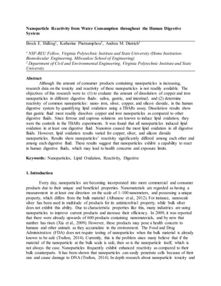
  • 7. Figure 3: The dissolution of copper nanoparticles in the artificial digestive fluids of saliva, intestinal, and gastric. Nanopure® water was used as the control fluid. The exhibited high dissolution of copper nanoparticles in gastric fluid was proposed to be because of the fluid’s acidity. Nanopure®, saliva, and intestinal fluids all had pH values of relatively neutral, while gastric was acidic. Hydrochloric acid has been reported to corrode and react with metals (Zhang et al., 2004). However, metal copper does not readily react with hydrochloric acid; it is a relatively slow reaction (Crundwell, 1992). Although bulk metal may not quickly react with hydrochloric acid, copper nanoparticles may increase the rate of reaction because of the increased surface area and reactivity, which nanoparticles are known for. 3.1.2 Dissolution of nZVI The nZVI solution for this dissolution test was targeted at 25 mg/L, meaning the maximum dissolution concentration would be 12.5 mg/L due to the samples diluted as an equal volume mixture. The pH for the intestinal fluid was 6.8, and the gastric fluid pH was 1.51. Similar to the copper nanoparticle dissolution, gastric fluid exhibited the greatest dissolution of nZVI. After the 60 minute incubation period, gastric exhibited the highest dissolution of nZVI with 94.4%, then intestinal with 57.5%, saliva was 54.7%, and lastly, the Nanopure® control with 53.2%. All fluids showed a slight increase in dissolution over time as expected. By looking at the graphical results in Figure 4 gastric results differ noticeably from the Nanopure® control results, as compared to the other digestive fluids used.
  • 8. Figure 4: The dissolution of nZVI in the artificial digestive fluids saliva, intestinal, and gastric. Nanopure® water was used as the control fluid. Similar to the copper dissolution, the proposed reason for gastric having increased dissolution for nZVI is the acidity. In this dissolution test, results were closer to the projected maximum. The nZVI particles were better suspended because they are in a stable slurry instead of powder form, like the copper nanoparticles. The slurry was nearly a year old, and the iron nanoparticles were capable of dissolving with time. This was verified by the results. Nanopure® exhibited an over 50% dissolution result. If Nanopure® water were to dissolve nZVI this well, the end dissolution should have been near the maximum 12.5 mg/L. However, since Nanopure® dissolution neared 50%, the initial slurry was suggested to contain a mix of nZVI particles and dissolved iron ions. Unlike copper, iron readily reacts with hydrochloric acid (Chetouani et al., 2004). It is proposed that gastric fluid neared the maximum nZVI dissolution because of the considerable amount of dissolved iron in the initial nZVI slurry and hydrochloric acid’s ability to corrode and dissolve iron. These results indicated that if certain nanoparticles were ingested, lipid oxidation may increase. The increased oxidation leads to more reactive oxygen intermediates, which are associated with oxidative stress and cellular damage (Mittler, 2002). 3.2 Lipid oxidation using TBARs With nanoparticles increasingly incorporated into consumer products, like food packaging, it is essential to evaluate their reactivity in case of accidental consumption. In all artificial fluids, nZVI samples did not significantly differ from the ferrous controls samples, which were known to induce lipid oxidation. nZVI samples showed higher increases in lipid oxidation than any other solution used, as seen in Figure 5. Cuprous, copper nanoparticles, and silicon dioxide nanoparticles exhibited lower increases of lipid oxidation. Silver particles were shown to have a similar increase in lipid oxidation as to the cuprous solution. In the silver nanoparticle intestinal testing, an outlier was present, which was taken out. The outlier affected the average increase of lipid oxidation and misconstrued the results. The silver nanoparticles were difficult to suspend and readily aggregated on either the top or bottom of the solution. For the outlier silver sample, it was believed that a clump was present. Therefore, it caused an increase in the concentration of silver nanoparticles, which impacted the resulting average MDA
  • 9. concentration. This silver sample outlier made the end result of MDA per milligram in intestinal fluid much higher than expected. Copper nanoparticles did not readily dissolve in solution. This made getting homogenous and repeatable samples for copper and silver nanoparticles problematic. For further testing, it would be ideal to use stable slurries for all nanoparticles, like the nZVI samples. Figure 5: The increase in lipid oxidation in different artificial digestive fluids when subjected to ferrous, cuprous, nZVI, copper nanoparticles, silver nanoparticles, or silicon dioxide nanoparticles. *An outlier sample was removed. Through the use of a two-way Anova statistical test, lipid oxidation between digestive fluids significantly differed with p-value of 0.005, and lipid oxidation significantly differed between tested metallic/nanoparticle solutions with a p-value of 0.011. It was proposed saliva showed the highest lipid oxidation since the soybean oil was better dispersed due to the salts and proteins that were present in saliva. In gastric fluid, the soybean oil stayed primarily at the top of the fluid in one or two main clusters. The lipid source for saliva dispersed more evenly into smaller clusters when mixed. When the lipids are more dispersed, there is more surface area to react, which may be the cause of difference in lipid oxidation between the digestive fluids. For future testing, an addition of an emulsifier would be encouraged to disperse the lipids more consistently between fluids. Overall, it is important to realize that all nanoparticles increased lipid oxidation, in at least one fluid. Lipid oxidation is harmful to the human health, and any amount of increase may be concerning (Harris, 2013). Out of all of the nanoparticles tested, nZVI exhibited consistently the highest reactivity, and is the highest concern for human health. Further tests using stable slurries of these nanoparticles with a better dispersed lipid source is encouraged to expand upon current findings. 4. Conclusion For the dissolution tests of copper nanoparticles and nZVI, artificial gastric fluid was the only digestive fluid which noticeably differed from the Nanopure® control. Gastric fluid had
  • 10. dissolution results of 47.6% and 94.4% for copper nanoparticles and nZVI, respectively, while the results of Nanopure® control were 9.0% and 53.2%, respectively. The gastric fluid’s increased dissolution results were proposed to be due to the acidity of this fluid. The nZVI’s increase in dissolution was because of the stable slurry it was in. Ferrous and nZVI consistently yielded the highest increase in lipid oxidation throughout the digestive fluids. The other solutions: cuprous, copper nanoparticles, silver nanoparticles, and silicon dioxide nanoparticles, exhibited lower increases in lipid oxidation in comparison. Since not all nanoparticles form stable solutions when prepared, stable and fresh slurries should be used for future testing. Silver and copper nanoparticles readily aggregated at the top or bottom of the solution, making precise, 10 mg/L samples difficult to attain. Nevertheless, lipid oxidation between digestive fluids significantly differed. Lipid oxidation between nanoparticle samples significantly differed. In conclusion, nanoparticles exhibited an increase in lipid oxidation in at least one digestive fluid, if not all. Lipid oxidation can be a harmful reaction, which oxidizes lipids that are present everywhere in the human body. With nanoparticle use in consumer products increasing, it is crucial to monitor the increase of nanoparticles present in the environment as well as further studying the effects of nanoparticles on human health. 5. Acknowledgements First off, I want to thank Dr. Andrea Dietrich (Department of Civil and Environmental Engineering) for being my faculty mentor and allowing me to conduct research in her lab. Also, I want to thank my graduate mentor, Katherine Phetxumphou (Civil and Environmental Engineering) for her help and support in this research process. I want to thank Dr. Lohani (Environmental Education) for applying the funding, directing the Interdisciplinary Water and Engineering REU, and allowing me this great opportunity. I want to thank my REU cohort for the support during the research. We acknowledge the support of the National Science Foundation through the NSF/REU Site Grant EEC-1359051. Any opinions, findings, and conclusions or recommendations expressed in this paper are those of the author(s) and do not necessarily reflect the views of the National Science Foundation. References Aburub, A., Risley, D. S., & Mishra, D. (2008). A critical evaluation of fasted state simulating gastric fluid (FaSSGF) that contains sodium lauryl sulfate and proposal of a modified recipe. International journal of pharmaceutics, 347(1), 16-22. Albanese, A., Tang, P. S., & Chan, W. C. (2012). The effect of nanoparticle size, shape, and surface chemistry on biological systems. Annual review of biomedical engineering, 14, 1- 16. Chetouani, A., Hammouti, B., & Benkaddour, M. (2004). Corrosion inhibition of iron in hydrochloric acid solution by jojoba oil. Pigment & resin technology, 33(1), 26-31. Crundwell, F. K. (1992). The anodic dissolution of copper in hydrochloric acid solutions.
  • 11. Electrochimica acta, 37(15), 2707-2714. Fröhlich, E., & Roblegg, E. (2012). Models for oral uptake of nanoparticles in consumer products. Toxicology, 291(1), 10-17. Harris, L. J. (Ed.). (2013). Improving the safety and quality of nuts. Elsevier. Jahnke, A., Holmbäck, J., Andersson, R. A., Kierkegaard, A., Mayer, P., & MacLeod, M. (2015). Differences between lipids extracted from five species are not sufficient to explain biomagnification of non-polar organic chemicals. Environmental Science & Technology Letters. Lee, C., Kim, J. Y., Lee, W. I., Nelson, K. L., Yoon, J., & Sedlak, D. L. (2008). Bactericidal effect of zero-valent iron nanoparticles on Escherichia coli. Environmental science & technology, 42(13), 4927-4933. Mittler, R. (2002). Oxidative stress, antioxidants and stress tolerance. Trends in plant science, 7(9), 405-410. Nowack, B., & Bucheli, T. D. (2007). Occurrence, behavior and effects of nanoparticles in the environment. Environmental pollution, 150(1), 5-22. Ömür-Özbek, P., Dietrich, A. M., Duncan, S. E., & Lee, Y. (2012). Role of lipid oxidation, chelating agents, and antioxidants in metallic flavor development in the oral cavity. Journal of agricultural and food chemistry, 60(9), 2274-2280. Phetxumphou, K. (2014). Consumers and their drinking water: communicating water quality and assessing the reaction of zerovalent nanoiron (nZVI) with saliva (Doctoral dissertation, Virginia Tech). Romero, F. J., Bosch-Morell, F., Romero, M. J., Jareño, E. J., Romero, B., Marín, N., & Romá, J. (1998). Lipid peroxidation products and antioxidants in human disease. Environmental Health Perspectives, 106(Suppl 5), 1229. Sevanian, A., & Hochstein, P. (1985). Mechanisms and consequences of lipid peroxidation in biological systems. Annual review of nutrition, 5(1), 365-390. Stippler, E., Kopp, S., & Dressman, J. B. (2004). Comparison of US pharmacopeia simulated intestinal fluid TS (without pancreatin) and phosphate standard buffer pH 6.8, TS of the international pharmacopoeia with respect to their use in in vitro dissolution testing. Dissolution Technologies, 11(2), 6-11. TaylorAlicia, A., MarcusIan, M., GuysiRisa, L., & WalkerSharon, L. (2015). Metal oxide nanoparticles induce minimal phenotypic changes in a model colon gut microbiota. Environmental Engineering Science.
  • 12. Trafton, A. (2014). Tiny particles may pose big risk. Retrieved from http://newsoffice.mit.edu/2014/tiny-particles-may-pose-big-risk. Wang, T. (2002). Soybean oil. In F.D. Gunstone, Vegetable Oils in Food Technology, Oxford: Blackwell Publishing, 18–58. Xia, T., Li, N., & Nel, A. E. (2009). Potential health impact of nanoparticles. Annual review of public health, 30, 137-150. Zhang, D. Q., Gao, L. X., & Zhou, G. D. (2004). Inhibition of copper corrosion in aerated hydrochloric acid solution by heterocyclic compounds containing a mercapto group. Corrosion science, 46(12), 3031-3040.