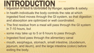
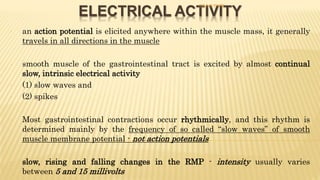
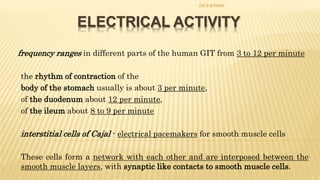
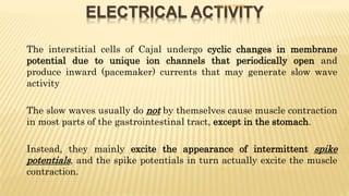
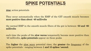
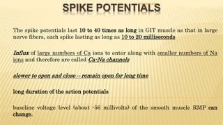
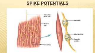
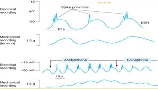
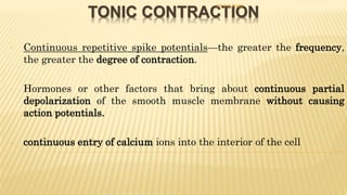
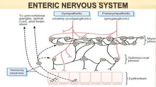
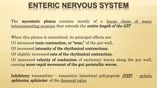
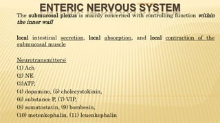
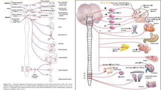
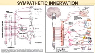
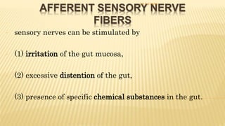
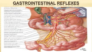
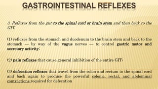
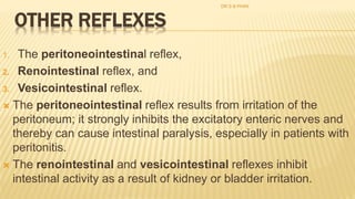
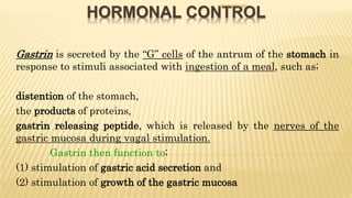
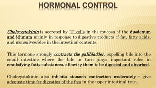
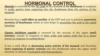
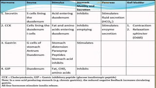
Call Girls Coimbatore Just Call 9907093804 Top Class Call Girl Service Available
3. git lecture 3
1. GENERAL CONTROL OF THE
GASTROINTESTINAL TRACT
INTRODUCTION
ELECTRICAL ACTIVITY OF GIT WALL
ENTERIC NERVOUS SYSTEM (Intrinsic)
AUTONOMIC CONTROL (Extrinsic)
AFFERENT SENSORY NERVE FIBERS
GASTROINTESTINAL REFLEXES
HORMONAL CONTROL
CONTENTS
DR S B PHIRI
2. INTRODUCTION Ingestion of food is controlled by hunger, appetite & satiety.
In health individuals the body controls the rate at which
ingested food moves through the GI system, so that digestion
and absorption are optimized or well coordinated.
The first residue from a meal will pass through the GI system
in 7-10 hours, but
some may take up to 5 or 6 hours to pass through.
Ingested food pass through the alimentary canal
mouth, esophagus, stomach, small intestine, (duodenum,
jejunum, and ileum), and the large intestine (colon) before
exiting the body.
DR S B PHIRI
3. THE GI WALL HAS FOUR LAYERS
The basic structure of the GIT wall is
similar in the stomach and intestine
though with some variations between
different sections of the GIT.
An inner layer—mucosa
A middle layer--- submucosa
An outer layer---muscle
Cover-----------serosaDR S B PHIRI
4. ELECTRICAL ACTIVITY
- an action potential is elicited anywhere within the muscle mass, it generally
travels in all directions in the muscle
- smooth muscle of the gastrointestinal tract is excited by almost continual
slow, intrinsic electrical activity
- (1) slow waves and
- (2) spikes
- Most gastrointestinal contractions occur rhythmically, and this rhythm is
determined mainly by the frequency of so called “slow waves” of smooth
muscle membrane potential - not action potentials
- slow, rising and falling changes in the RMP - intensity usually varies
between 5 and 15 millivolts
DR S B PHIRI
5. ELECTRICAL ACTIVITY
frequency ranges in different parts of the human GIT from 3 to 12 per minute
- the rhythm of contraction of the
- body of the stomach usually is about 3 per minute,
- of the duodenum about 12 per minute,
- of the ileum about 8 to 9 per minute
- interstitial cells of Cajal - electrical pacemakers for smooth muscle cells
- These cells form a network with each other and are interposed between the
smooth muscle layers, with synaptic like contacts to smooth muscle cells.
DR S B PHIRI
6. ELECTRICAL ACTIVITY
- The interstitial cells of Cajal undergo cyclic changes in membrane
potential due to unique ion channels that periodically open and
produce inward (pacemaker) currents that may generate slow wave
activity
- The slow waves usually do not by themselves cause muscle contraction
in most parts of the gastrointestinal tract, except in the stomach.
- Instead, they mainly excite the appearance of intermittent spike
potentials, and the spike potentials in turn actually excite the muscle
contraction.
DR S B PHIRI
7. SPIKE POTENTIALS
- true action potentials
- They occur automatically when the RMP of the GIT smooth muscle becomes
more positive than about -40 millivolts
- the normal RMP in the smooth muscle fibers of the gut is between -50 and -60
millivolts
- each time the peaks of the slow waves temporarily become more positive than
-40 millivolts, spike potentials appear on these peaks
- The higher the slow wave potential rises, the greater the frequency of the
spike potentials - ranging between 1 and 10 spikes / second.
DR S B PHIRI
8. SPIKE POTENTIALS
- The spike potentials last 10 to 40 times as long in GIT muscle as that in large
nerve fibers, each spike lasting as long as 10 to 20 milliseconds
- Influx of large numbers of Ca ions to enter along with smaller numbers of Na
ions and therefore are called Ca-Na channels
- slower to open and close – remain open for long time
- long duration of the action potentials
- baseline voltage level (about -56 millivolts) of the smooth muscle RMP can
change.
DR S B PHIRI
9. SPIKE POTENTIALS
- When the potential becomes less negative - depolarization of the membrane, the
muscle fibers become more excitable.
- When the potential becomes more negative - hyperpolarization, the fibers become
less excitable
- Factors that depolarize the membrane
- (1) stretching of the muscle,
- (2) stimulation by acetylcholine,
- (3) stimulation by parasympathetic nerves that secrete acetylcholine at their
endings,
- (4) stimulation by several specific gastrointestinal hormones.
- Important factors that hyperpolarize the membrane
- (1) the effect of norepinephrine or epinephrine on the fiber membrane
- (2) stimulation of the sympathetic nerves that secrete mainly norepinephrine at
their endings.
DR S B PHIRI
11. TONIC CONTRACTION
- Continuous repetitive spike potentials—the greater the frequency,
the greater the degree of contraction.
- Hormones or other factors that bring about continuous partial
depolarization of the smooth muscle membrane without causing
action potentials.
- continuous entry of calcium ions into the interior of the cell
DR S B PHIRI
12. ENTERIC NERVOUS SYSTEM
- Lies in the wall of the gut, beginning in the esophagus and
- extending all the way to the anus
- controlling gastrointestinal movements and secretion.
- (1) an outer plexus lying between the longitudinal and circular muscle layers,
called the myenteric plexus or Auerbach’s plexus,
- controls mainly the gastrointestinal movements
- (2) an inner plexus, called the submucosal plexus or Meissner’s plexus, that
lies in the submucosa.
- controls mainly gastrointestinal secretion and local blood flow
DR S B PHIRI
13. ENTERIC NERVOUS SYSTEM
- the extrinsic sympathetic and parasympathetic fibers that connect to both the
myenteric and submucosal plexuses.
- the enteric nervous system can function on its own, independently of these
extrinsic nerves,
- stimulation by the parasympathetic and sympathetic systems can greatly enhance
or inhibit gastrointestinal functions
- sensory nerve endings that originate in the gastrointestinal epithelium or gut wall
and send afferent fibers to both plexuses of the enteric system,
- as well as (1) to the prevertebral ganglia of the sympathetic nervous system, (2) to
the spinal cord, and (3) in the vagus nerves all the way to the brain stem.
- These sensory nerves can elicit local reflexes within the gut wall
DR S B PHIRI
14. ENTERIC NERVOUS SYSTEM
- The myenteric plexus consists mostly of a linear chain of many
interconnecting neurons that extends the entire length of the GIT
- When this plexus is stimulated, its principal effects are
- (1) increased tonic contraction, or “tone,” of the gut wall,
- (2) increased intensity of the rhythmical contractions,
- (3) slightly increased rate of the rhythmical contraction,
- (4) increased velocity of conduction of excitatory waves along the gut wall,
causing more rapid movement of the gut peristaltic waves.
- Inhibitory transmitter - vasoactive intestinal polypeptide (VIP) - pyloric
sphincter, sphincter of the ileocecal valve
DR S B PHIRI
15. ENTERIC NERVOUS SYSTEM
- The submucosal plexus is mainly concerned with controlling function within
the inner wall
- local intestinal secretion, local absorption, and local contraction of the
submucosal muscle
- Neurotransmitters:
- (1) Ach
- (2) NE
- (3)ATP,
- (4) dopamine, (5) cholecystokinin,
- (6) substance P, (7) VIP,
- (8) somatostatin, (9) bombesin,
- (10) metenkephalin, (11) leuenkephalin
DR S B PHIRI
16. AUTONOMIC CONTROL
- Parasympathetic
- the cranial parasympathetic nerve fibers - mouth and pharyngeal regions of
the alimentary tract, esophagus, stomach, and pancreas and somewhat less
to the intestines down through the first half of the large intestine.
- The sacral parasympathetics originate in the 2nd, 3rd & 4th sacral segments of
the spinal cord and pass through the pelvic nerves to the distal half of the
large intestine and all the way to the anus.
- The sigmoidal, rectal, and anal regions are considerably better supplied with
parasympathetic fibers than are the other intestinal areas - defecation
reflexes
DR S B PHIRI
17. SYMPATHETIC INNERVATION
- spinal cord between segments T-5 and L-2.
- Pre ganglionic - sympathetic chains - celiac ganglion and various mesenteric
ganglia – post ganglionic
- innervate essentially all of the gastrointestinal tract – inhibitory
- (1) to a slight extent by direct effect of secreted NE to inhibit intestinal tract
smooth muscle
- (2) to a major extent by an inhibitory effect of NE on the neurons of the
entire enteric nervous system
DR S B PHIRI
18. AFFERENT SENSORY NERVE
FIBERS
- sensory nerves can be stimulated by
- (1) irritation of the gut mucosa,
- (2) excessive distention of the gut,
- (3) presence of specific chemical substances in the gut.
DR S B PHIRI
19. GASTROINTESTINAL REFLEXES
- A reflex is an involuntary movement or exercise of function in a part, excited in
response to a stimulus applied to the periphery and transmitted to the brain or
spinal cord
-
- 1. Reflexes that are integrated entirely within the gut wall enteric nervous
system – secretion, peristalsis, mixing contractions
- 2. Reflexes from the gut to the prevertebral sympathetic ganglia and then back
to the GIT
- signals from the stomach to cause evacuation of the colon (the gastrocolic reflex),
- signals from the colon and small intestine to inhibit stomach motility and
stomach secretion (the enterogastric reflexes),
- reflexes from the colon to inhibit emptying of ileal contents into the colon (the
colonoileal reflex).
DR S B PHIRI
20. GASTROINTESTINAL REFLEXES
- 3. Reflexes from the gut to the spinal cord or brain stem and then back to the
GIT.
- (1) reflexes from the stomach and duodenum to the brain stem and back to the
stomach — by way of the vagus nerves — to control gastric motor and
secretory activity;
- (2) pain reflexes that cause general inhibition of the entire GIT;
- (3) defecation reflexes that travel from the colon and rectum to the spinal cord
and back again to produce the powerful colonic, rectal, and abdominal
contractions required for defecation
DR S B PHIRI
21. OTHER REFLEXES
1. The peritoneointestinal reflex,
2. Renointestinal reflex, and
3. Vesicointestinal reflex.
The peritoneointestinal reflex results from irritation of the
peritoneum; it strongly inhibits the excitatory enteric nerves and
thereby can cause intestinal paralysis, especially in patients with
peritonitis.
The renointestinal and vesicointestinal reflexes inhibit
intestinal activity as a result of kidney or bladder irritation.
DR S B PHIRI
22. HORMONAL CONTROL
- Gastrin is secreted by the “G” cells of the antrum of the stomach in
response to stimuli associated with ingestion of a meal, such as;
- distention of the stomach,
- the products of proteins,
- gastrin releasing peptide, which is released by the nerves of the
gastric mucosa during vagal stimulation.
- Gastrin then function to;
- (1) stimulation of gastric acid secretion and
- (2) stimulation of growth of the gastric mucosa
DR S B PHIRI
23. HORMONAL CONTROL
- Cholecystokinin is secreted by “I” cells in the mucosa of the duodenum
and jejunum mainly in response to digestive products of fat, fatty acids,
and monoglycerides in the intestinal contents.
- This hormone strongly contracts the gallbladder, expelling bile into the
small intestine where the bile in turn plays important roles in
emulsifying fatty substances, allowing them to be digested and absorbed.
- Cholecystokinin also inhibits stomach contraction moderately - give
adequate time for digestion of the fats in the upper intestinal tract.
DR S B PHIRI
24. HORMONAL CONTROL- Secretin is secreted by the “S” cells in the mucosa of the duodenum in response
to acidic gastric juice emptying into the duodenum from the pylorus of the
stomach.
- Secretin has a mild effect on motility of the GIT and acts to promote pancreatic
secretion of bicarbonate which in turn helps to neutralize the acid in the small
intestine
- Gastric inhibitory peptide is secreted by the mucosa of the upper small
intestine, mainly in response to fatty acids and amino acids but to a lesser
extent in response to carbohydrate.
- It has a mild effect in decreasing motor activity of the stomach and therefore
slows emptying of gastric contents into the duodenum when the upper small
intestine is already overloaded with food products.
DR S B PHIRI
25. HORMONAL CONTROL
- Motilin is secreted by the upper duodenum during fasting, and the
only known function of this hormone is to increase gastrointestinal
motility.
- Motilin is released cyclically and stimulates waves of
gastrointestinal motility called interdigestive myoelectric
complexes;
- that move through the stomach and small intestine every 90
minutes in a fasted person.
- They also called motor migrating complexes
DR S B PHIRI