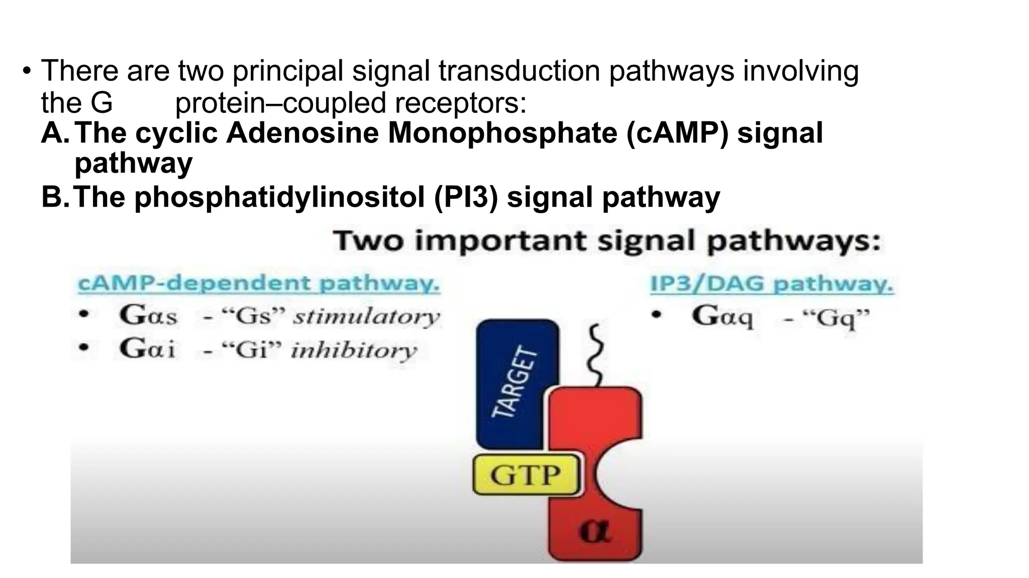
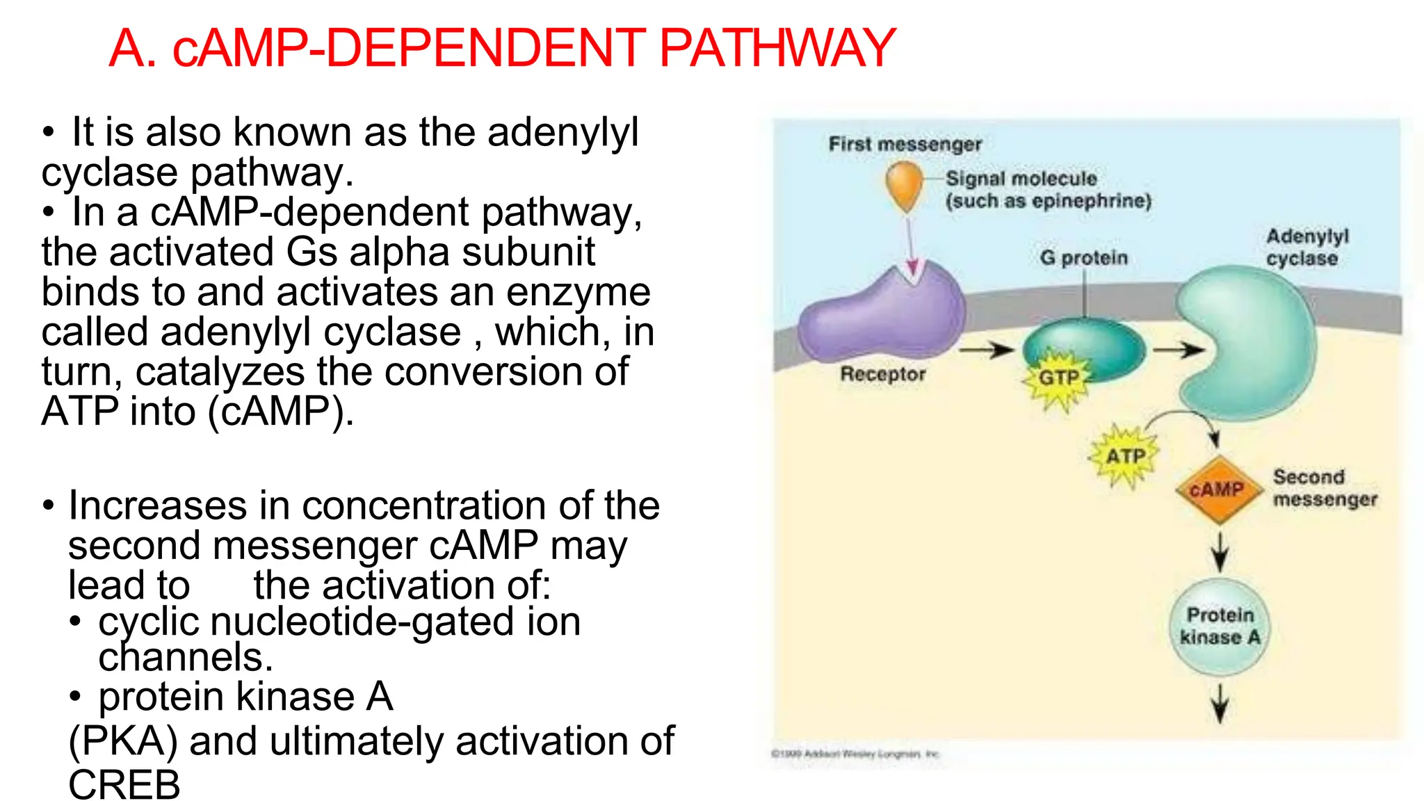
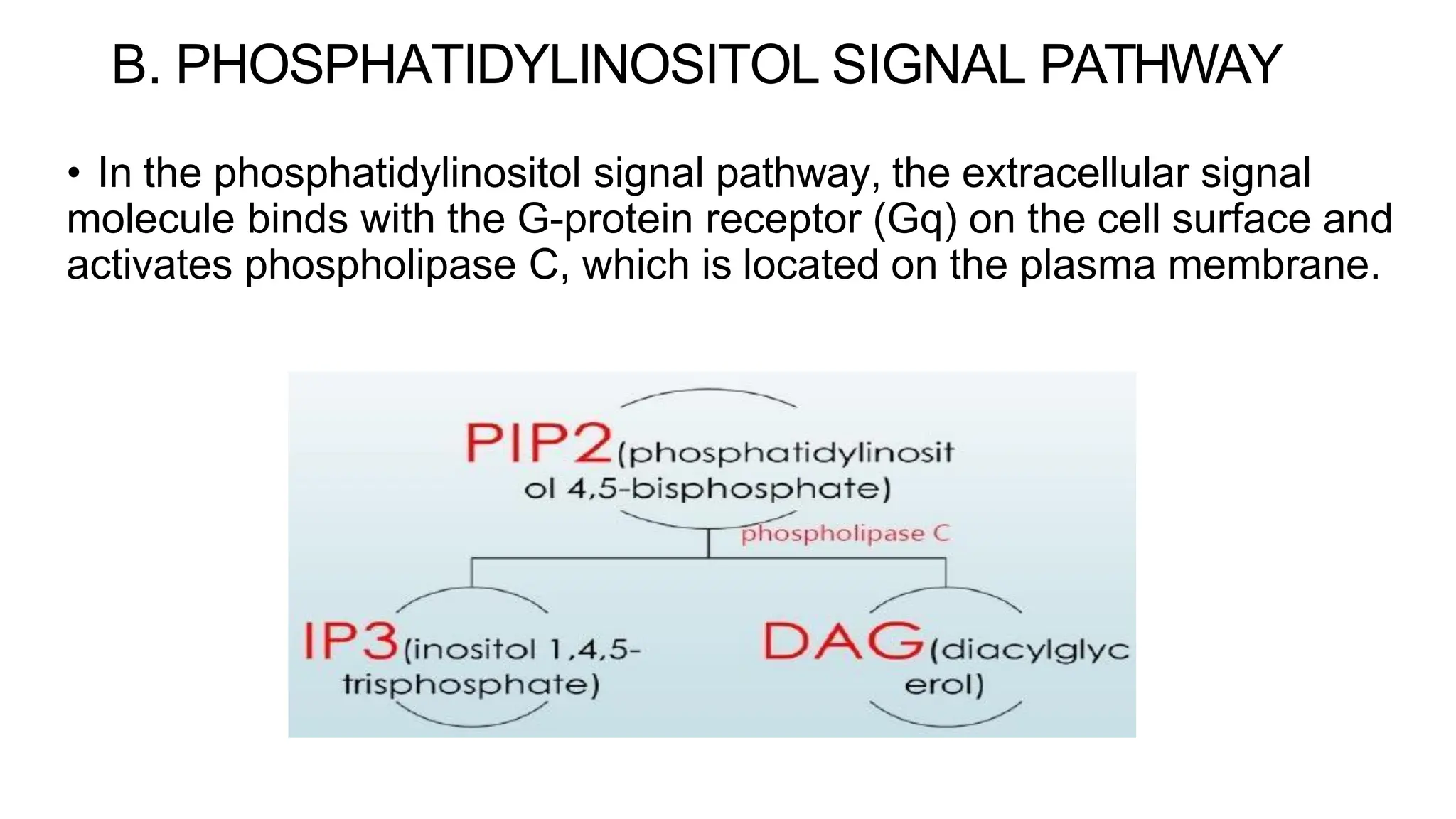
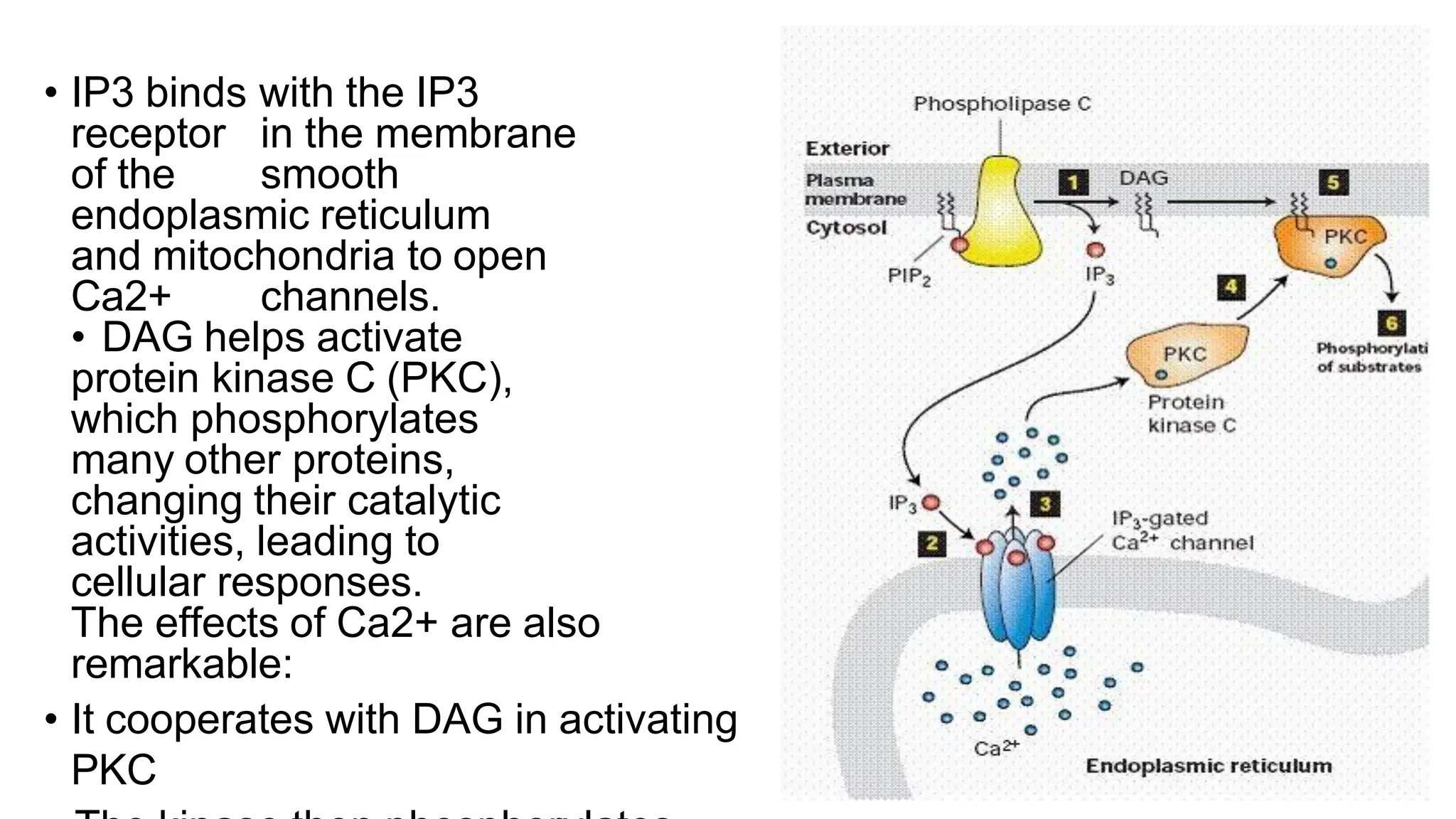
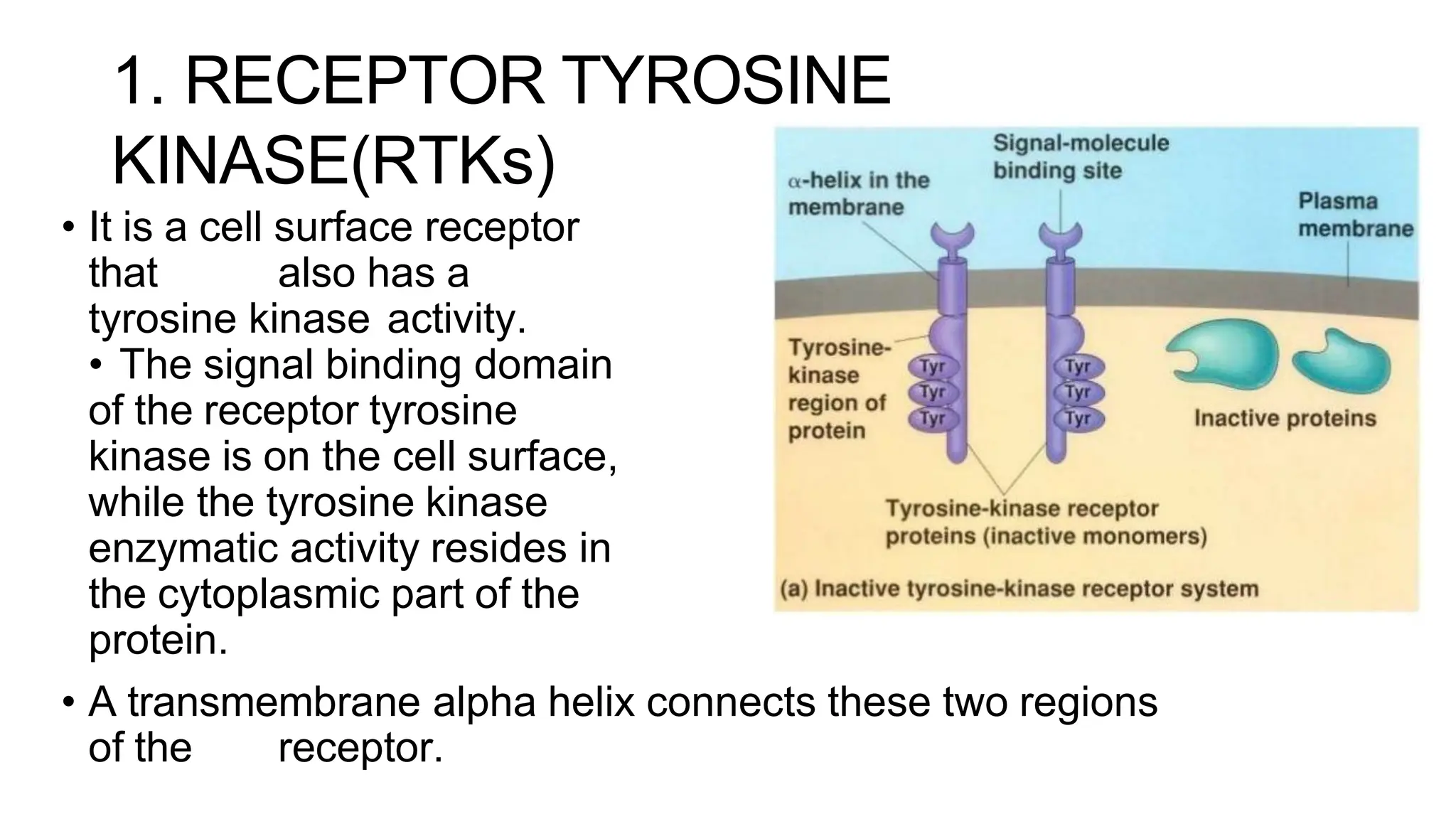
Cell communication and
signal transduction
Dr. Rubia Anwer
Senior Lecturer
Introduction
• In human body, numerous processes are required for coordinating
individual cells to support the body.
• At the cellular level, sensing of environments and cell communication for
coordination relies on signal transduction; modeling signal transduction
systems as self-organizing allows one to explain how equilibria are
maintained.
• Many disease processes, such as diabetes, cancer, inflammation and
heart disease arise from defects or dysregulations in these pathways,
highlighting the importance of these processes in human biology and
medicine
Cell communication
• Cell communication occurs through chemical signals and cellular
receptors by either;
1)Direct contact of molecules on two cells surfaces
2) Or the release of a "chemical signal" recognized by another cell
(near or far).
• Hormones are carried by the circulatory systems to many sites.
• Growth factors are released to act on nearby tissues.
• Ligands are signals that bind cell surface receptors (as
observed with insulin (a ligand) and the insulin receptor) or that
can pass into the cell and bind an internal receptor (such as the
steroid hormones).
SIGNAL TRANSDUCTION
• Any process occurring within cells that convert one kind of
signal/stimulus into another type.
• It also known as cell signaling in which the transmission of
molecular signals from a cell's exterior to its interior.
• Signals received by cells must be transmitted effectively into the cell
to ensure an appropriate response.
• This step is initiated by cell-surface receptors which triggers a
biochemical chain of events inside the cell, creating a response.
Cell Signaling
• Signaling is also defined as “the ability of a cell to change behavior in
response to a receptor-ligand interaction”.
• The ligand is the primary messenger.
• As the result of binding the receptor, other molecules or second
messengers are produced within the target cell.
• Second messengers communicate the signal from one location to
another (such as from plasma membrane to nucleus) leading to
cascade of events/changes within a cell
Components of cell signaling
1. Signaling molecules: Bacteria,
Viruses, UV
2. Signaling receptors: GPCR
3. Signaling conveyers: G-protein
4. Signaling transducer: cAMP
5. Transcription factors: CREB, NF-kB
Ligands
The ligands that bind and activate receptors include;
• Light-sensitive compounds
• Hormones
• Neurotransmitters, and vary in size from small molecules to peptides to
large proteins.
• Messenger molecules may be amino acids, peptides, proteins, fatty
acids, lipids, nucleosides or nucleotides.
• Hydrophilic messengers bind to cell membrane receptors.
• Hydrophobic messengers bind to intracellular receptors which
regulate expression of specific genes.
• A ligand binds its receptor through a number of specific weak non-
covalent bonds by fitting into a specific binding site or "pocket ".
RECEPTORS
• Receptors can be roughly divided into two major classes:
intracellular receptors and extracellular receptors.
EXTRACELLULAR RECEPTORS
• Extracellular receptors are integral transmembrane proteins and
make up most receptors.
• They span the plasma membrane of the cell, with one part of the
receptor on the outside of the cell and the other on the inside.
• Signal transduction occurs as a result of a ligand binding to the
outside region of the receptor (the ligand does not pass through
the membrane).
Extracellular Receptors
• G-protein-coupled receptors
(GPCR)
• Receptors with Kinase activity
• Ligand-gated ion channel
receptors
A. G-PROTEIN–COUPLED RECEPTORS (GPCRs)
• Also known as seven-transmembrane domain receptors, 7TM
receptors, heptahelical receptors, and G protein–linked receptors
(GPLR).
• These constitute a large protein family of receptors that sense
molecules outside the cell and activate inside signal transduction
pathways and, ultimately, cellular responses.
• Coupling with G proteins, they are called seven-transmembrane
receptors because they pass through the cell membrane seven times.
• G-protein–coupled receptors are involved in many diseases, and are
also the target of approximately 40% of all modern medicinal drugs.
• G proteins, also known as guanine nucleotide-binding proteins, are a family
of proteins that act as molecular switches inside cells, and are involved in
transmitting signals from a variety of stimuli outside a cell to its interior.
• When they are bound to GTP, they are 'on', and, when they are bound to
GDP, they are 'off'.
• G proteins belong to the larger group of enzymes called GTPases
• There are two classes of G proteins;
1.The first function as monomeric
small GTPases
2.The second form and function
as heterotrimeric G protein
complexes.
• Heterotrimeric class of
complexes is made up of alpha
(α), beta (β) and gamma (γ)
subunits. The beta and gamma
subunits can form a stable
dimeric complex referred to as
MECHANISM
1. In the inactive state, the GPCR is bound to a heterotrimeric G
protein complex.
2. Binding of an agonist to the GPCR results in a conformation change
in the receptor that is transmitted to the bound Gα subunit of the
heterotrimeric G
protein.
3. The activated G α subunit exchanges GTP in place of GDP
which in turn triggers the dissociation of Gα subunit from the Gβγ
dimer and from the receptor.
4. The dissociated G α and Gβγ subunits interact with other
intracellular proteins to continue the signal transduction cascade.
5. While the freed GPCR is able to rebind to another heterotrimeric G
protein to form a new complex that is ready to initiate another round of
signal transduction.
• There are two principal signal transduction pathways involving
the G protein–coupled receptors:
A.The cyclic Adenosine Monophosphate (cAMP) signal
pathway
B.The phosphatidylinositol (PI3) signal pathway
A. cAMP-DEPENDENT PATHWAY
• It is also known as the adenylyl
cyclase pathway.
• In a cAMP-dependent pathway,
the activated Gs alpha subunit
binds to and activates an enzyme
called adenylyl cyclase , which, in
turn, catalyzes the conversion of
ATP into (cAMP).
• Increases in concentration of the
second messenger cAMP may
lead to the activation of:
• cyclic nucleotide-gated ion
channels.
• protein kinase A
(PKA) and ultimately activation of
CREB
The PKA enzyme is also known as cAMP-dependent enzyme
because it gets activated only if cAMP is present.
• Once PKA is activated, it phosphorylates a number of other proteins
including:
1. enzymes that convert glycogen into glucose
2. enzymes that promote muscle contraction in the heart leading
to an increase in heart rate
3. transcription factors, which regulate gene expression
B. PHOSPHATIDYLINOSITOL SIGNAL PATHWAY
• In the phosphatidylinositol signal pathway, the extracellular signal
molecule binds with the G-protein receptor (Gq) on the cell surface and
activates phospholipase C, which is located on the plasma membrane.
• IP3 binds with the IP3
receptor in the membrane
of the smooth
endoplasmic reticulum
and mitochondria to open
Ca2+ channels.
• DAG helps activate
protein kinase C (PKC),
which phosphorylates
many other proteins,
changing their catalytic
activities, leading to
cellular responses.
The effects of Ca2+ are also
remarkable:
• It cooperates with DAG in activating
PKC
B. RECEPTORS WITH KINASE ACTIVITY
• A kinase is a type of enzyme that transfers phosphate groups
from high- energy donor molecules, such as ATP to specific
target molecules (substrates); the process is termed
phosphorylation.
• Kinase enzymes that specifically phosphorylate tyrosine amino
acids are termed tyrosine kinases .
1. RECEPTOR TYROSINE
KINASE(RTKs)
• It is a cell surface receptor
that also has a
tyrosine kinase activity.
• The signal binding domain
of the receptor tyrosine
kinase is on the cell surface,
while the tyrosine kinase
enzymatic activity resides in
the cytoplasmic part of the
protein.
• A transmembrane alpha helix connects these two regions
of the receptor.
• The most important groups of signals that bind to receptor tyrosine
kinases are:
- Peptide growth factors like nerve growth factor (NGF) and
epidermal growth factor (EGF)
- Peptide hormones, like insulin.
- Binding of signal molecules to the extracellular domains of
receptor tyrosine kinase molecules causes two receptor
molecules to dimerize.
- This brings the cytoplasmic tails of the receptors close to each
other and causes the tyrosine kinase activity of these tails to be
turned on.
• The activated tails then phosphorylate each other on
several tyrosine residues. This is called
autophosphorylation.
• The phosphorylation of tyrosines on the receptor tails
triggers the assembly of an intracellular signaling complex on
the tails.
• The newly phosphorylated tyrosines serve as binding
sites for a variety of signaling proteins that then pass the
message on to yet other proteins.
2. NON RECPETOR TYROSINE KINASE
(nRTKs)
• Non-receptor tyrosine kinases are a subgroup of protein
family tyrosine kinases, enzymes that can transfer the
phosphate group from ATP to a tyrosine residue of a
protein (phosphorylation).
• Unlike the receptor tyrosine kinases (RTKs), the second
subgroup of tyrosine kinases, the non-receptor tyrosine kinases
are cytoplasmic enzymes.
• nRTKs regulate cell's growth, proliferation, differentiation,
adhesion,
migration and apoptosis and they are critical components
in the regulation of the immune system.
LIGAND-GATED ION CHANNEL
• Ligand-gated ion channels (LGICs) are a group of transmembrane ion
channel proteins which open to allow ions such as Na+, K+,Ca2+, or Cl-
to pass through the membrane in response to the binding of a chemical
messenger (i.e. a ligand), such as a neurotransmitter.
• A ligand-gated ion channel, upon binding with a ligand, change
conformation to open a channel in the cell membrane through which
ions relaying signals can pass.
• An example of this mechanism is found in the receiving cell of a
neural synapse.
INTRACELLULAR RECEPTORS
• Intracellular receptors are receptors located inside the cell
rather than on its cell membrane.
• Classic hormones that use intracellular receptors include
thyroid and steroid hormones.
Examples are:
- Class of nuclear receptors located in the cell nucleus and
cytoplasm
- IP3 receptor located on the endoplasmic reticulum.
• The ligands that bind to them are usually:
- Intracellular second messengers like inositol trisphosphate
(IP3)
- Extracellular lipophilic hormones like steroid hormones.
- Activated nuclear receptors attach to the DNA
- Due to their enabling gene transcription, they are
alternatively called inductors of gene expression.

Lecture 6 cell comunication and signal transduction.pdf

  • 1.
    Cell communication and signaltransduction Dr. Rubia Anwer Senior Lecturer
  • 2.
    Introduction • In humanbody, numerous processes are required for coordinating individual cells to support the body. • At the cellular level, sensing of environments and cell communication for coordination relies on signal transduction; modeling signal transduction systems as self-organizing allows one to explain how equilibria are maintained. • Many disease processes, such as diabetes, cancer, inflammation and heart disease arise from defects or dysregulations in these pathways, highlighting the importance of these processes in human biology and medicine
  • 3.
    Cell communication • Cellcommunication occurs through chemical signals and cellular receptors by either; 1)Direct contact of molecules on two cells surfaces 2) Or the release of a "chemical signal" recognized by another cell (near or far). • Hormones are carried by the circulatory systems to many sites. • Growth factors are released to act on nearby tissues. • Ligands are signals that bind cell surface receptors (as observed with insulin (a ligand) and the insulin receptor) or that can pass into the cell and bind an internal receptor (such as the steroid hormones).
  • 4.
    SIGNAL TRANSDUCTION • Anyprocess occurring within cells that convert one kind of signal/stimulus into another type. • It also known as cell signaling in which the transmission of molecular signals from a cell's exterior to its interior. • Signals received by cells must be transmitted effectively into the cell to ensure an appropriate response. • This step is initiated by cell-surface receptors which triggers a biochemical chain of events inside the cell, creating a response.
  • 5.
    Cell Signaling • Signalingis also defined as “the ability of a cell to change behavior in response to a receptor-ligand interaction”. • The ligand is the primary messenger. • As the result of binding the receptor, other molecules or second messengers are produced within the target cell. • Second messengers communicate the signal from one location to another (such as from plasma membrane to nucleus) leading to cascade of events/changes within a cell
  • 7.
    Components of cellsignaling 1. Signaling molecules: Bacteria, Viruses, UV 2. Signaling receptors: GPCR 3. Signaling conveyers: G-protein 4. Signaling transducer: cAMP 5. Transcription factors: CREB, NF-kB
  • 8.
    Ligands The ligands thatbind and activate receptors include; • Light-sensitive compounds • Hormones • Neurotransmitters, and vary in size from small molecules to peptides to large proteins. • Messenger molecules may be amino acids, peptides, proteins, fatty acids, lipids, nucleosides or nucleotides. • Hydrophilic messengers bind to cell membrane receptors. • Hydrophobic messengers bind to intracellular receptors which regulate expression of specific genes. • A ligand binds its receptor through a number of specific weak non- covalent bonds by fitting into a specific binding site or "pocket ".
  • 10.
    RECEPTORS • Receptors canbe roughly divided into two major classes: intracellular receptors and extracellular receptors. EXTRACELLULAR RECEPTORS • Extracellular receptors are integral transmembrane proteins and make up most receptors. • They span the plasma membrane of the cell, with one part of the receptor on the outside of the cell and the other on the inside. • Signal transduction occurs as a result of a ligand binding to the outside region of the receptor (the ligand does not pass through the membrane).
  • 11.
    Extracellular Receptors • G-protein-coupledreceptors (GPCR) • Receptors with Kinase activity • Ligand-gated ion channel receptors
  • 12.
    A. G-PROTEIN–COUPLED RECEPTORS(GPCRs) • Also known as seven-transmembrane domain receptors, 7TM receptors, heptahelical receptors, and G protein–linked receptors (GPLR). • These constitute a large protein family of receptors that sense molecules outside the cell and activate inside signal transduction pathways and, ultimately, cellular responses. • Coupling with G proteins, they are called seven-transmembrane receptors because they pass through the cell membrane seven times. • G-protein–coupled receptors are involved in many diseases, and are also the target of approximately 40% of all modern medicinal drugs.
  • 14.
    • G proteins,also known as guanine nucleotide-binding proteins, are a family of proteins that act as molecular switches inside cells, and are involved in transmitting signals from a variety of stimuli outside a cell to its interior. • When they are bound to GTP, they are 'on', and, when they are bound to GDP, they are 'off'. • G proteins belong to the larger group of enzymes called GTPases • There are two classes of G proteins; 1.The first function as monomeric small GTPases 2.The second form and function as heterotrimeric G protein complexes. • Heterotrimeric class of complexes is made up of alpha (α), beta (β) and gamma (γ) subunits. The beta and gamma subunits can form a stable dimeric complex referred to as
  • 15.
    MECHANISM 1. In theinactive state, the GPCR is bound to a heterotrimeric G protein complex. 2. Binding of an agonist to the GPCR results in a conformation change in the receptor that is transmitted to the bound Gα subunit of the heterotrimeric G protein. 3. The activated G α subunit exchanges GTP in place of GDP which in turn triggers the dissociation of Gα subunit from the Gβγ dimer and from the receptor. 4. The dissociated G α and Gβγ subunits interact with other intracellular proteins to continue the signal transduction cascade. 5. While the freed GPCR is able to rebind to another heterotrimeric G protein to form a new complex that is ready to initiate another round of signal transduction.
  • 17.
    • There aretwo principal signal transduction pathways involving the G protein–coupled receptors: A.The cyclic Adenosine Monophosphate (cAMP) signal pathway B.The phosphatidylinositol (PI3) signal pathway
  • 18.
    A. cAMP-DEPENDENT PATHWAY •It is also known as the adenylyl cyclase pathway. • In a cAMP-dependent pathway, the activated Gs alpha subunit binds to and activates an enzyme called adenylyl cyclase , which, in turn, catalyzes the conversion of ATP into (cAMP). • Increases in concentration of the second messenger cAMP may lead to the activation of: • cyclic nucleotide-gated ion channels. • protein kinase A (PKA) and ultimately activation of CREB
  • 19.
    The PKA enzymeis also known as cAMP-dependent enzyme because it gets activated only if cAMP is present. • Once PKA is activated, it phosphorylates a number of other proteins including: 1. enzymes that convert glycogen into glucose 2. enzymes that promote muscle contraction in the heart leading to an increase in heart rate 3. transcription factors, which regulate gene expression
  • 21.
    B. PHOSPHATIDYLINOSITOL SIGNALPATHWAY • In the phosphatidylinositol signal pathway, the extracellular signal molecule binds with the G-protein receptor (Gq) on the cell surface and activates phospholipase C, which is located on the plasma membrane.
  • 22.
    • IP3 bindswith the IP3 receptor in the membrane of the smooth endoplasmic reticulum and mitochondria to open Ca2+ channels. • DAG helps activate protein kinase C (PKC), which phosphorylates many other proteins, changing their catalytic activities, leading to cellular responses. The effects of Ca2+ are also remarkable: • It cooperates with DAG in activating PKC
  • 23.
    B. RECEPTORS WITHKINASE ACTIVITY • A kinase is a type of enzyme that transfers phosphate groups from high- energy donor molecules, such as ATP to specific target molecules (substrates); the process is termed phosphorylation. • Kinase enzymes that specifically phosphorylate tyrosine amino acids are termed tyrosine kinases .
  • 24.
    1. RECEPTOR TYROSINE KINASE(RTKs) •It is a cell surface receptor that also has a tyrosine kinase activity. • The signal binding domain of the receptor tyrosine kinase is on the cell surface, while the tyrosine kinase enzymatic activity resides in the cytoplasmic part of the protein. • A transmembrane alpha helix connects these two regions of the receptor.
  • 25.
    • The mostimportant groups of signals that bind to receptor tyrosine kinases are: - Peptide growth factors like nerve growth factor (NGF) and epidermal growth factor (EGF) - Peptide hormones, like insulin. - Binding of signal molecules to the extracellular domains of receptor tyrosine kinase molecules causes two receptor molecules to dimerize. - This brings the cytoplasmic tails of the receptors close to each other and causes the tyrosine kinase activity of these tails to be turned on.
  • 26.
    • The activatedtails then phosphorylate each other on several tyrosine residues. This is called autophosphorylation. • The phosphorylation of tyrosines on the receptor tails triggers the assembly of an intracellular signaling complex on the tails. • The newly phosphorylated tyrosines serve as binding sites for a variety of signaling proteins that then pass the message on to yet other proteins.
  • 28.
    2. NON RECPETORTYROSINE KINASE (nRTKs) • Non-receptor tyrosine kinases are a subgroup of protein family tyrosine kinases, enzymes that can transfer the phosphate group from ATP to a tyrosine residue of a protein (phosphorylation). • Unlike the receptor tyrosine kinases (RTKs), the second subgroup of tyrosine kinases, the non-receptor tyrosine kinases are cytoplasmic enzymes. • nRTKs regulate cell's growth, proliferation, differentiation, adhesion, migration and apoptosis and they are critical components in the regulation of the immune system.
  • 29.
    LIGAND-GATED ION CHANNEL •Ligand-gated ion channels (LGICs) are a group of transmembrane ion channel proteins which open to allow ions such as Na+, K+,Ca2+, or Cl- to pass through the membrane in response to the binding of a chemical messenger (i.e. a ligand), such as a neurotransmitter. • A ligand-gated ion channel, upon binding with a ligand, change conformation to open a channel in the cell membrane through which ions relaying signals can pass. • An example of this mechanism is found in the receiving cell of a neural synapse.
  • 31.
    INTRACELLULAR RECEPTORS • Intracellularreceptors are receptors located inside the cell rather than on its cell membrane. • Classic hormones that use intracellular receptors include thyroid and steroid hormones. Examples are: - Class of nuclear receptors located in the cell nucleus and cytoplasm - IP3 receptor located on the endoplasmic reticulum.
  • 32.
    • The ligandsthat bind to them are usually: - Intracellular second messengers like inositol trisphosphate (IP3) - Extracellular lipophilic hormones like steroid hormones. - Activated nuclear receptors attach to the DNA - Due to their enabling gene transcription, they are alternatively called inductors of gene expression.