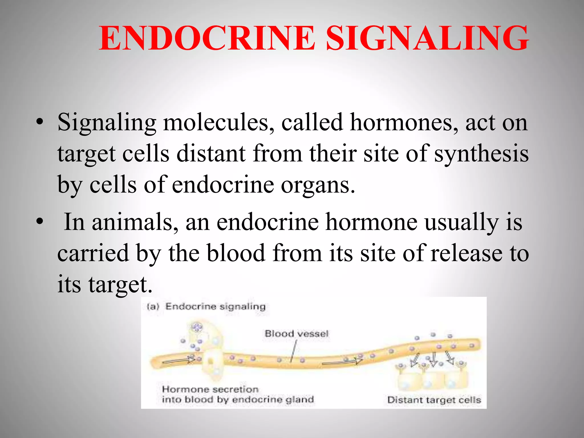
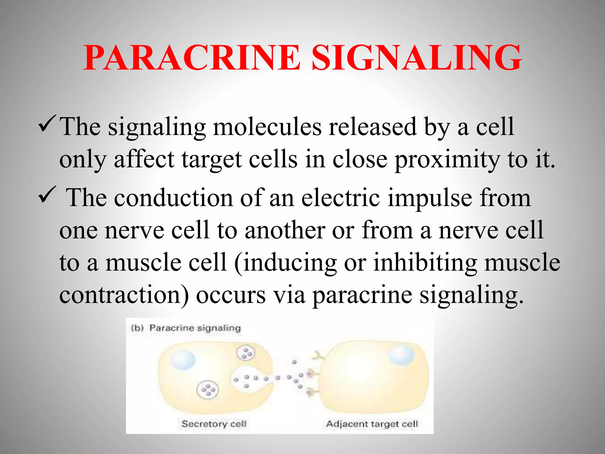
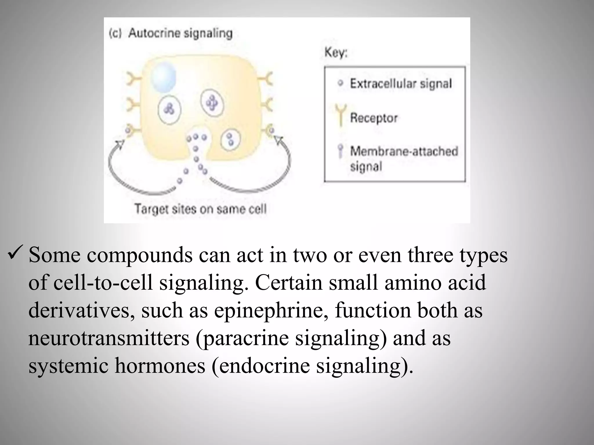
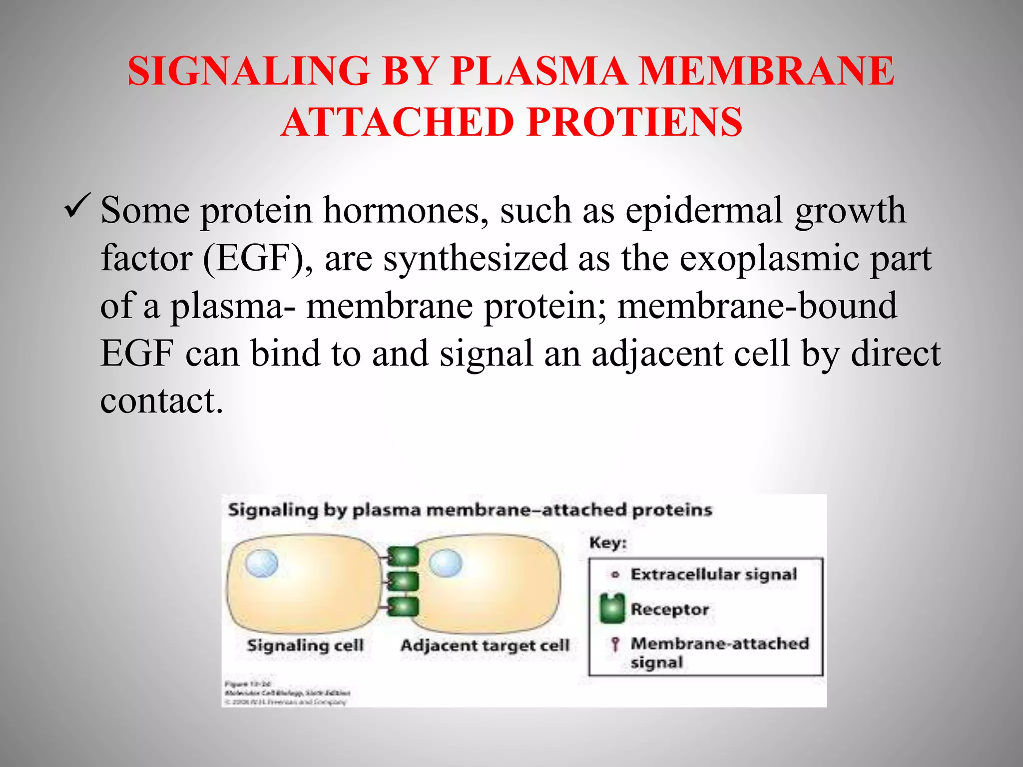
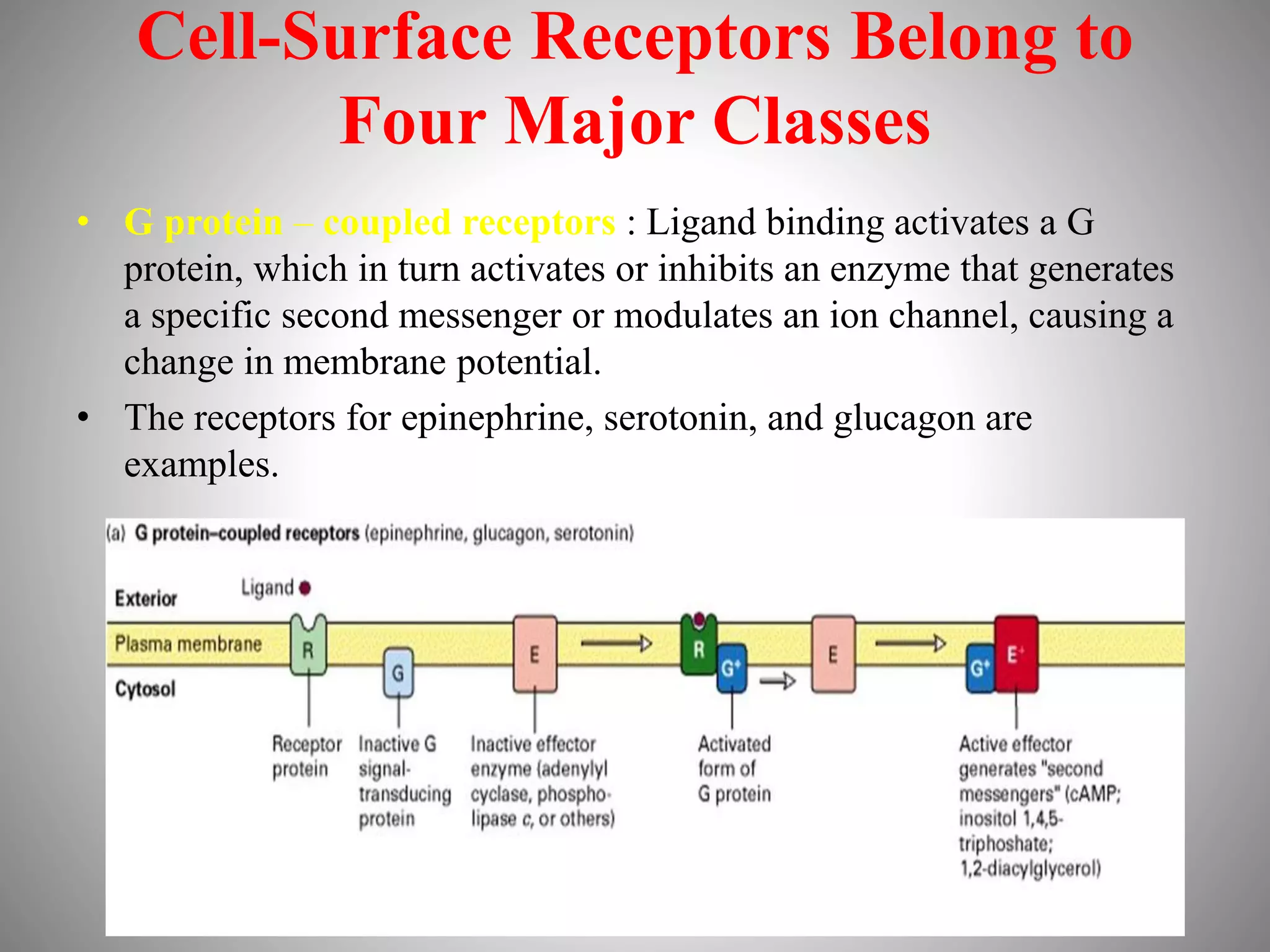
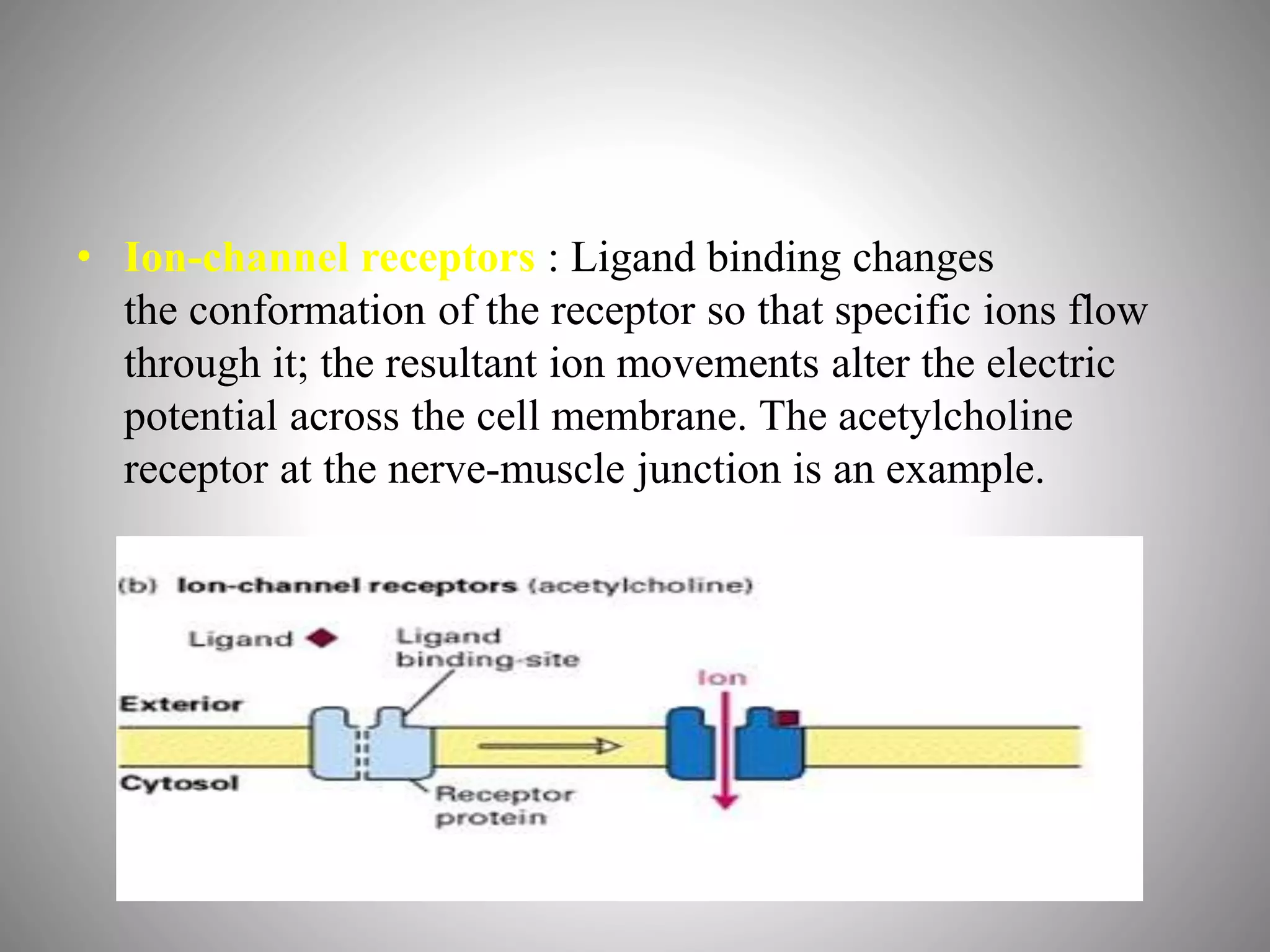
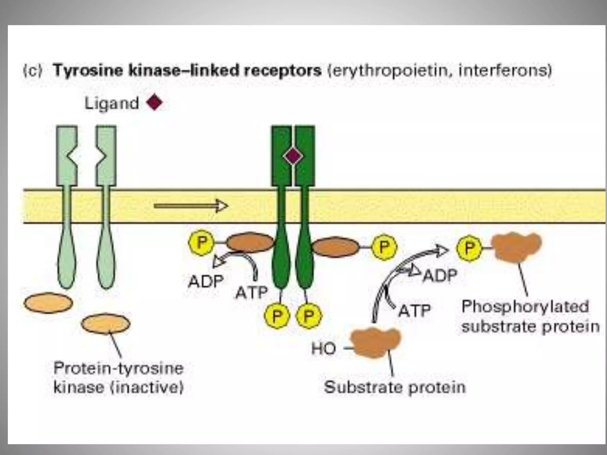
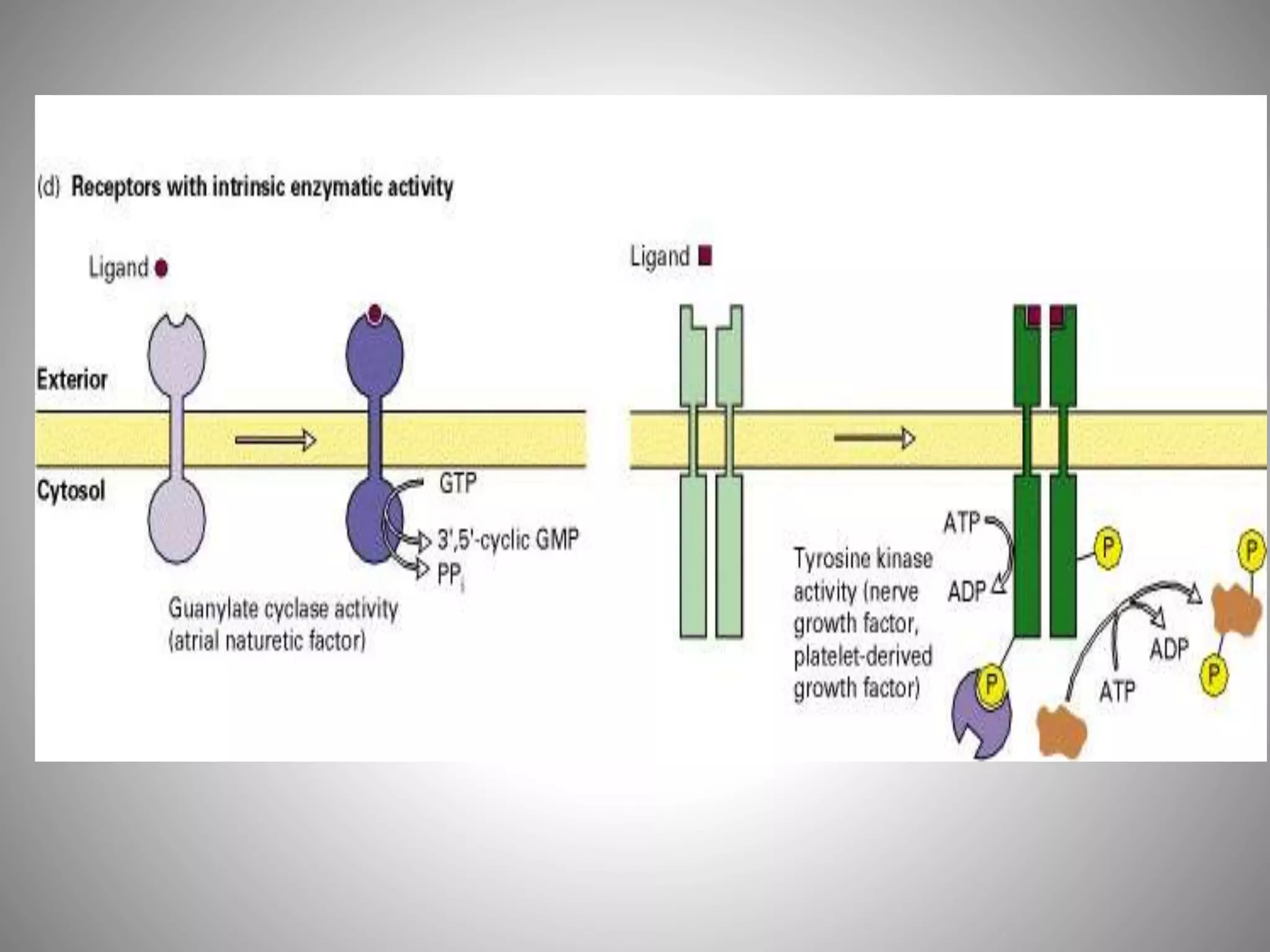
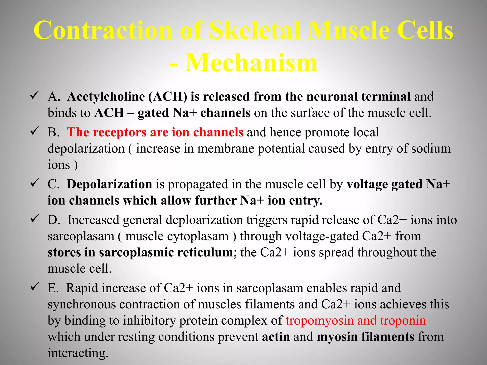
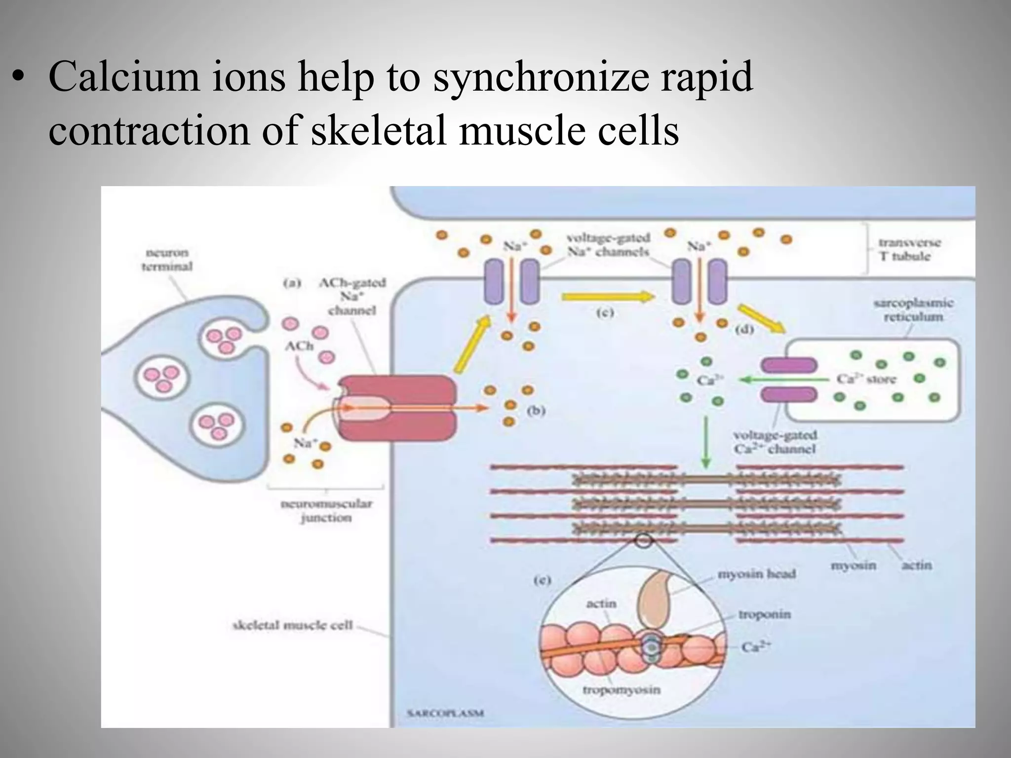
BIO-MEMBRANE, BIO-ENERGY CELL
TRAFFICKING
TOPIC: MOLECULAR INTERACTION,
REGULATION & SIGNALLING RECEPTORS &
VESICLES
ANANTHAKUMAR.T
BMS14312
Overview of Extracellular Signaling
Communication by extracellular signals usually involves six
steps:
1. Synthesis
2. Release of the signaling molecule by the signaling cell;
3. Transport of the signal to the target cell;
4. Detection of the signal by a specific receptor protein;
5. A change in cellular metabolism, function, or development
triggered by the receptor-signal complex;
6. Removal of the signal, which often terminates the cellular
response.
Signaling Molecules Operate over Various
Distances in Animals
• In animals, signaling by extracellular, secreted
molecules can be classified into three types ;
1. Endocrine,
2. Paracrine,
3. Autocrine
• Based on the distance over which the signal acts.
• In addition, certain membrane-bound proteins on
one cell can directly signal an adjacent cell
ENDOCRINE SIGNALING
• Signaling molecules, called hormones, act on
target cells distant from their site of synthesis
by cells of endocrine organs.
• In animals, an endocrine hormone usually is
carried by the blood from its site of release to
its target.
PARACRINE SIGNALING
The signaling molecules released by a cell
only affect target cells in close proximity to it.
 The conduction of an electric impulse from
one nerve cell to another or from a nerve cell
to a muscle cell (inducing or inhibiting muscle
contraction) occurs via paracrine signaling.
AUTOCRINE SIGNALING
Cells respond to substances that they themselves
release.
Many growth factors act in this fashion, and
cultured cells often secrete growth factors that
stimulate their own growth and proliferation.
 This type of signaling is particularly common
in tumor cells, many of which overproduce and
release growth factors that stimulate
inappropriate, unregulated proliferation of
themselves as well as adjacent non tumor cells;
this process may lead to formation of tumor mass.
 Some compounds can act in two or even three types
of cell-to-cell signaling. Certain small amino acid
derivatives, such as epinephrine, function both as
neurotransmitters (paracrine signaling) and as
systemic hormones (endocrine signaling).
SIGNALING BY PLASMA MEMBRANE
ATTACHED PROTIENS
 Some protein hormones, such as epidermal growth
factor (EGF), are synthesized as the exoplasmic part
of a plasma- membrane protein; membrane-bound
EGF can bind to and signal an adjacent cell by direct
contact.
RECEPTORS
• Most receptors are on cell surface --- Water soluble
signaling molecules cannot cross membrane lipid
bilayer but are capable of binding to specific
receptors embedded in the plasma membrane.
• Receptors have an a) Extracellular domain that binds
signaling molecule b) hydrophobic transmembrane
domain and c) intracellular domain.
• On binding a ligand, there induces a conformational
change in the receptor, in particular of the
intracellular region which in turn activates a relay of
intracellular signaling molecules… thus bringing
about appropriate cellular responses such as altered
metabolism, altered cytoskeleton or altered gene
expression or cell division.
PROPERTIES OF RECEPTORS
• Specificity – Signaling molecule or ligand recognizes
and binds to its specific receptor just as an enzyme
binds to its substrate and this binding is independent
from competition from other signaling molecules
• High Affinity – Receptors usually bind to their
signaling molecules with high affinity as determined
by their affinity constants Ki and also the interaction
shows low dissociation constants Kd indicating high
affinity binding.
• Saturation Kinetics – Number of receptors on the surface
of a cell is limited and the number varies from less than a
dozen to several thousand depending on the receptor type.
Increasing the signal ligand concentration would eventually
result in receptor saturation meaning all sites being
occupied by ligands.
• Reversibility – Normally the binding of a signaling ligand
to a receptor is reversible and this is necessary property to
prevent permanent activation of the receptor
• Physiological Response – Ligand receptor binding triggers
a physiological response characteristic for its interaction
such as binding of insulin to insulin receptor (INSR)
stimulates glucose transport into target cells. The
magnitude of the response is directly related to the number
of receptors to which the ligands bind.
Cell-Surface Receptors Belong to
Four Major Classes
• G protein – coupled receptors : Ligand binding activates a G
protein, which in turn activates or inhibits an enzyme that generates
a specific second messenger or modulates an ion channel, causing a
change in membrane potential.
• The receptors for epinephrine, serotonin, and glucagon are
examples.
• Ion-channel receptors : Ligand binding changes
the conformation of the receptor so that specific ions flow
through it; the resultant ion movements alter the electric
potential across the cell membrane. The acetylcholine
receptor at the nerve-muscle junction is an example.
Tyrosine kinase – linked receptors: These
receptors lack intrinsic catalytic activity,
but ligand binding stimulates formation of a
dimeric receptor, which then interacts with and
activates one or more cytosolic protein-tyrosine
kinases.
The receptors for many cytokines, the interferons,
and human growth factor are of this type.
These tyrosine kinase – linked receptors
sometimes are referred to as the cytokine-receptor
superfamily.
 Receptors with intrinsic enzymatic activity : Several
types of receptors have intrinsic catalytic activity,
which is activated by binding of ligand.
 For instance, some activated receptors catalyze
conversion of GTP to cGMP; others act
as protein phosphatases, removing phosphate groups
from phosphotyrosine residues in substrate proteins,
thereby modifying their activity.
 The receptors for insulin and many growth factors are
ligand-triggered protein kinases; in most cases, the
ligand binds as a dimer, leading to dimerization of
the receptor and activation of its kinase activity.
 These receptors — often referred to as receptor
serine/threonine kinases or receptor tyrosine kinases —
auto phosphorylate residues in their own
cytosolic domain and also can phosphorylate various
substrate proteins.
SIGNAL TRANSDUCTION
MECHANISMS
 Signaling information has to be transmitted from the receptor in the
plasma membrane across the cytoplasam to the nucleus ( If gene
transcription is the response required), the cytoskeleton ( If cell
movement or another change to cell morphology is the response) or
various other subcellular compartments.
 These responses are expected to occur within a suitable time frame so
as to have some synchronicity with regarding transmission or signal
and cellular response.
 In a typical model of signaling a chain of intracellular mediators
successively activates the next target in the chain, as in a branching
network of activation, diversification and modulation of response.
 Thus the branched molecular network of activation ( or deactivation )
of signaling molecules linking receptor activation to the intracellular
targets is referred to as a signal transduction pathway ( or cascade ).
SECOND MESSENGERS
 Intracellular signaling molecules have properties that allow control
of the speed, duration and target of the signal and be categorized
accordingly.
 Broadly they can be divided into 2 groups on the basis of their
molecular characteristics, Second Messengers and Signaling
Proteins.
 1. Second Messengers are small readily diffusible intracellular
mediators regulating the activity of other target signaling molecules.
 The calcium ions Ca2+ is a classic example of a second messenger
being released in large quantities in response to a signal (amplifying
the signal ) and diffusing rapidly through the cytosol and help in
broadcasting the signal quickly to several distant parts of the cell.
 Eg. Ca2+ ions mediate and coordinate contraction of skeletal
muscle cells. Thus if rapid response is needed second messengers
will definitely be present in the signalling pathway.
 2. Second messengers are water soluble such as cyclic
nucleotides cAMP and cGMP and act similarly to Ca2+ ions
by diffusing through the cytosol, whereas second
messengers such as diacylglycerol ( DAG ) are lipid
soluble and diffuse along the inside of the plasma membrane
where other key signaling proteins are anchored.
 3. In the G protien coupled receptor (GPCR) system the
effector enzyme adenylyl cyclase synthesizes the second
messenger cAMP from ATP and guanylyl cyclase
synthesizes cGMP from GTP.
 4. Resultant rise in cAMP and cGMP affects various cell
types in different ways. Diverse effects of cAMP are
mediated by enzymes called protein kinases A (PKAs) and
does not function in signalling pathways mediated by
receptor tyrosine kinases (RTKs).
Contraction of Skeletal Muscle Cells
- Mechanism
 A. Acetylcholine (ACH) is released from the neuronal terminal and
binds to ACH – gated Na+ channels on the surface of the muscle cell.
 B. The receptors are ion channels and hence promote local
depolarization ( increase in membrane potential caused by entry of sodium
ions )
 C. Depolarization is propagated in the muscle cell by voltage gated Na+
ion channels which allow further Na+ ion entry.
 D. Increased general deploarization triggers rapid release of Ca2+ ions into
sarcoplasam ( muscle cytoplasam ) through voltage-gated Ca2+ from
stores in sarcoplasmic reticulum; the Ca2+ ions spread throughout the
muscle cell.
 E. Rapid increase of Ca2+ ions in sarcoplasam enables rapid and
synchronous contraction of muscles filaments and Ca2+ ions achieves this
by binding to inhibitory protein complex of tropomyosin and troponin
which under resting conditions prevent actin and myosin filaments from
interacting.
• Calcium ions help to synchronize rapid
contraction of skeletal muscle cells
THANK YOU

Molecular interaction, Regulation and Signalling receptors and vesicles

  • 1.
    BIO-MEMBRANE, BIO-ENERGY CELL TRAFFICKING TOPIC:MOLECULAR INTERACTION, REGULATION & SIGNALLING RECEPTORS & VESICLES ANANTHAKUMAR.T BMS14312
  • 2.
    Overview of ExtracellularSignaling Communication by extracellular signals usually involves six steps: 1. Synthesis 2. Release of the signaling molecule by the signaling cell; 3. Transport of the signal to the target cell; 4. Detection of the signal by a specific receptor protein; 5. A change in cellular metabolism, function, or development triggered by the receptor-signal complex; 6. Removal of the signal, which often terminates the cellular response.
  • 3.
    Signaling Molecules Operateover Various Distances in Animals • In animals, signaling by extracellular, secreted molecules can be classified into three types ; 1. Endocrine, 2. Paracrine, 3. Autocrine • Based on the distance over which the signal acts. • In addition, certain membrane-bound proteins on one cell can directly signal an adjacent cell
  • 4.
    ENDOCRINE SIGNALING • Signalingmolecules, called hormones, act on target cells distant from their site of synthesis by cells of endocrine organs. • In animals, an endocrine hormone usually is carried by the blood from its site of release to its target.
  • 5.
    PARACRINE SIGNALING The signalingmolecules released by a cell only affect target cells in close proximity to it.  The conduction of an electric impulse from one nerve cell to another or from a nerve cell to a muscle cell (inducing or inhibiting muscle contraction) occurs via paracrine signaling.
  • 6.
    AUTOCRINE SIGNALING Cells respondto substances that they themselves release. Many growth factors act in this fashion, and cultured cells often secrete growth factors that stimulate their own growth and proliferation.  This type of signaling is particularly common in tumor cells, many of which overproduce and release growth factors that stimulate inappropriate, unregulated proliferation of themselves as well as adjacent non tumor cells; this process may lead to formation of tumor mass.
  • 7.
     Some compoundscan act in two or even three types of cell-to-cell signaling. Certain small amino acid derivatives, such as epinephrine, function both as neurotransmitters (paracrine signaling) and as systemic hormones (endocrine signaling).
  • 8.
    SIGNALING BY PLASMAMEMBRANE ATTACHED PROTIENS  Some protein hormones, such as epidermal growth factor (EGF), are synthesized as the exoplasmic part of a plasma- membrane protein; membrane-bound EGF can bind to and signal an adjacent cell by direct contact.
  • 9.
    RECEPTORS • Most receptorsare on cell surface --- Water soluble signaling molecules cannot cross membrane lipid bilayer but are capable of binding to specific receptors embedded in the plasma membrane. • Receptors have an a) Extracellular domain that binds signaling molecule b) hydrophobic transmembrane domain and c) intracellular domain. • On binding a ligand, there induces a conformational change in the receptor, in particular of the intracellular region which in turn activates a relay of intracellular signaling molecules… thus bringing about appropriate cellular responses such as altered metabolism, altered cytoskeleton or altered gene expression or cell division.
  • 10.
    PROPERTIES OF RECEPTORS •Specificity – Signaling molecule or ligand recognizes and binds to its specific receptor just as an enzyme binds to its substrate and this binding is independent from competition from other signaling molecules • High Affinity – Receptors usually bind to their signaling molecules with high affinity as determined by their affinity constants Ki and also the interaction shows low dissociation constants Kd indicating high affinity binding.
  • 11.
    • Saturation Kinetics– Number of receptors on the surface of a cell is limited and the number varies from less than a dozen to several thousand depending on the receptor type. Increasing the signal ligand concentration would eventually result in receptor saturation meaning all sites being occupied by ligands. • Reversibility – Normally the binding of a signaling ligand to a receptor is reversible and this is necessary property to prevent permanent activation of the receptor • Physiological Response – Ligand receptor binding triggers a physiological response characteristic for its interaction such as binding of insulin to insulin receptor (INSR) stimulates glucose transport into target cells. The magnitude of the response is directly related to the number of receptors to which the ligands bind.
  • 12.
    Cell-Surface Receptors Belongto Four Major Classes • G protein – coupled receptors : Ligand binding activates a G protein, which in turn activates or inhibits an enzyme that generates a specific second messenger or modulates an ion channel, causing a change in membrane potential. • The receptors for epinephrine, serotonin, and glucagon are examples.
  • 13.
    • Ion-channel receptors: Ligand binding changes the conformation of the receptor so that specific ions flow through it; the resultant ion movements alter the electric potential across the cell membrane. The acetylcholine receptor at the nerve-muscle junction is an example.
  • 14.
    Tyrosine kinase – linked receptors:These receptors lack intrinsic catalytic activity, but ligand binding stimulates formation of a dimeric receptor, which then interacts with and activates one or more cytosolic protein-tyrosine kinases. The receptors for many cytokines, the interferons, and human growth factor are of this type. These tyrosine kinase – linked receptors sometimes are referred to as the cytokine-receptor superfamily.
  • 16.
     Receptors withintrinsic enzymatic activity : Several types of receptors have intrinsic catalytic activity, which is activated by binding of ligand.  For instance, some activated receptors catalyze conversion of GTP to cGMP; others act as protein phosphatases, removing phosphate groups from phosphotyrosine residues in substrate proteins, thereby modifying their activity.  The receptors for insulin and many growth factors are ligand-triggered protein kinases; in most cases, the ligand binds as a dimer, leading to dimerization of the receptor and activation of its kinase activity.  These receptors — often referred to as receptor serine/threonine kinases or receptor tyrosine kinases — auto phosphorylate residues in their own cytosolic domain and also can phosphorylate various substrate proteins.
  • 18.
    SIGNAL TRANSDUCTION MECHANISMS  Signalinginformation has to be transmitted from the receptor in the plasma membrane across the cytoplasam to the nucleus ( If gene transcription is the response required), the cytoskeleton ( If cell movement or another change to cell morphology is the response) or various other subcellular compartments.  These responses are expected to occur within a suitable time frame so as to have some synchronicity with regarding transmission or signal and cellular response.  In a typical model of signaling a chain of intracellular mediators successively activates the next target in the chain, as in a branching network of activation, diversification and modulation of response.  Thus the branched molecular network of activation ( or deactivation ) of signaling molecules linking receptor activation to the intracellular targets is referred to as a signal transduction pathway ( or cascade ).
  • 19.
    SECOND MESSENGERS  Intracellularsignaling molecules have properties that allow control of the speed, duration and target of the signal and be categorized accordingly.  Broadly they can be divided into 2 groups on the basis of their molecular characteristics, Second Messengers and Signaling Proteins.  1. Second Messengers are small readily diffusible intracellular mediators regulating the activity of other target signaling molecules.  The calcium ions Ca2+ is a classic example of a second messenger being released in large quantities in response to a signal (amplifying the signal ) and diffusing rapidly through the cytosol and help in broadcasting the signal quickly to several distant parts of the cell.  Eg. Ca2+ ions mediate and coordinate contraction of skeletal muscle cells. Thus if rapid response is needed second messengers will definitely be present in the signalling pathway.
  • 20.
     2. Secondmessengers are water soluble such as cyclic nucleotides cAMP and cGMP and act similarly to Ca2+ ions by diffusing through the cytosol, whereas second messengers such as diacylglycerol ( DAG ) are lipid soluble and diffuse along the inside of the plasma membrane where other key signaling proteins are anchored.  3. In the G protien coupled receptor (GPCR) system the effector enzyme adenylyl cyclase synthesizes the second messenger cAMP from ATP and guanylyl cyclase synthesizes cGMP from GTP.  4. Resultant rise in cAMP and cGMP affects various cell types in different ways. Diverse effects of cAMP are mediated by enzymes called protein kinases A (PKAs) and does not function in signalling pathways mediated by receptor tyrosine kinases (RTKs).
  • 21.
    Contraction of SkeletalMuscle Cells - Mechanism  A. Acetylcholine (ACH) is released from the neuronal terminal and binds to ACH – gated Na+ channels on the surface of the muscle cell.  B. The receptors are ion channels and hence promote local depolarization ( increase in membrane potential caused by entry of sodium ions )  C. Depolarization is propagated in the muscle cell by voltage gated Na+ ion channels which allow further Na+ ion entry.  D. Increased general deploarization triggers rapid release of Ca2+ ions into sarcoplasam ( muscle cytoplasam ) through voltage-gated Ca2+ from stores in sarcoplasmic reticulum; the Ca2+ ions spread throughout the muscle cell.  E. Rapid increase of Ca2+ ions in sarcoplasam enables rapid and synchronous contraction of muscles filaments and Ca2+ ions achieves this by binding to inhibitory protein complex of tropomyosin and troponin which under resting conditions prevent actin and myosin filaments from interacting.
  • 22.
    • Calcium ionshelp to synchronize rapid contraction of skeletal muscle cells
  • 23.