Learning Objectives
The studentswill learn:
• Intracellular signaling pathway
• Intercellular signaling pathways
• Extracellular signaling molecules and
receptors
• Intracellular signaling molecules and
receptors
3.
CELL SIGNALING
Cell signalingis part of a complex system of communication
that governs basic cellular activities and coordinates cell
actions.
The ability of cells to perceive and correctly respond to their
microenvironment is the basis of development, tissue repair,
and immunity as well as normal tissue homeostasis
Errors in cellular information processing are responsible for
diseases such as cancer, autoimmunity, and diabetes.
By understanding cell signaling, diseases may be
treated effectively and, theoretically, artificial tissues may be
created.
3
4.
Definition
s
• Signaling: Cell-cellcommunication via signals.
• Signal transduction: Process of converting
extracellular signals into intra-cellular
responses.
• Ligand: The signaling molecule.
• Receptors: Bind specific ligands and in turn
activate one or more intracellular pathways.
These pathways depend on intracellular
signaling proteins which process the signal and
transmit the signal to appropriate intracellular
targets. The targets at the end of signaling
pathways are called effector proteins.
4
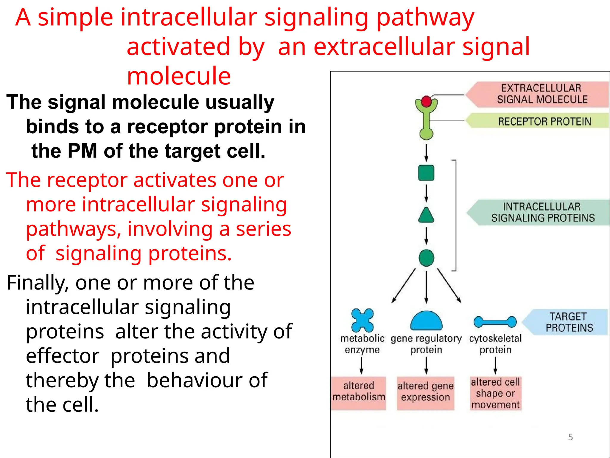
5.
5
A simple intracellularsignaling pathway
activated by an extracellular signal
molecule
The signal molecule usually
binds to a receptor protein in
the PM of the target cell.
The receptor activates one or
more intracellular signaling
pathways, involving a series
of signaling proteins.
Finally, one or more of the
intracellular signaling
proteins alter the activity of
effector proteins and
thereby the behaviour of
the cell.
6.
Four forms ofintercellular signaling
6
• Cells usually communicate with each other
through extracellular messenger molecules.
1. Juxtacrine Signaling
This involves direct cell-to-cell contact, where
signaling molecules remain bound to the
surface of one cell and interact with receptors
on an adjacent cell.
Function: It plays a key role in tissue
development and immune system function.
Example: Notch signaling, crucial for cell
differentiation during embryonic
development.
7.
Four forms ofintercellular signaling
• 2. paracrine signaling depends
on local mediators that are
released into the extracellular
space and act on neighbouring
cells. E.g. nerve-muscle
• synaptic signaling is
performed by neurons that
transmit signals electrically
along their axons and release
neurotransmitters at synapses.
8.
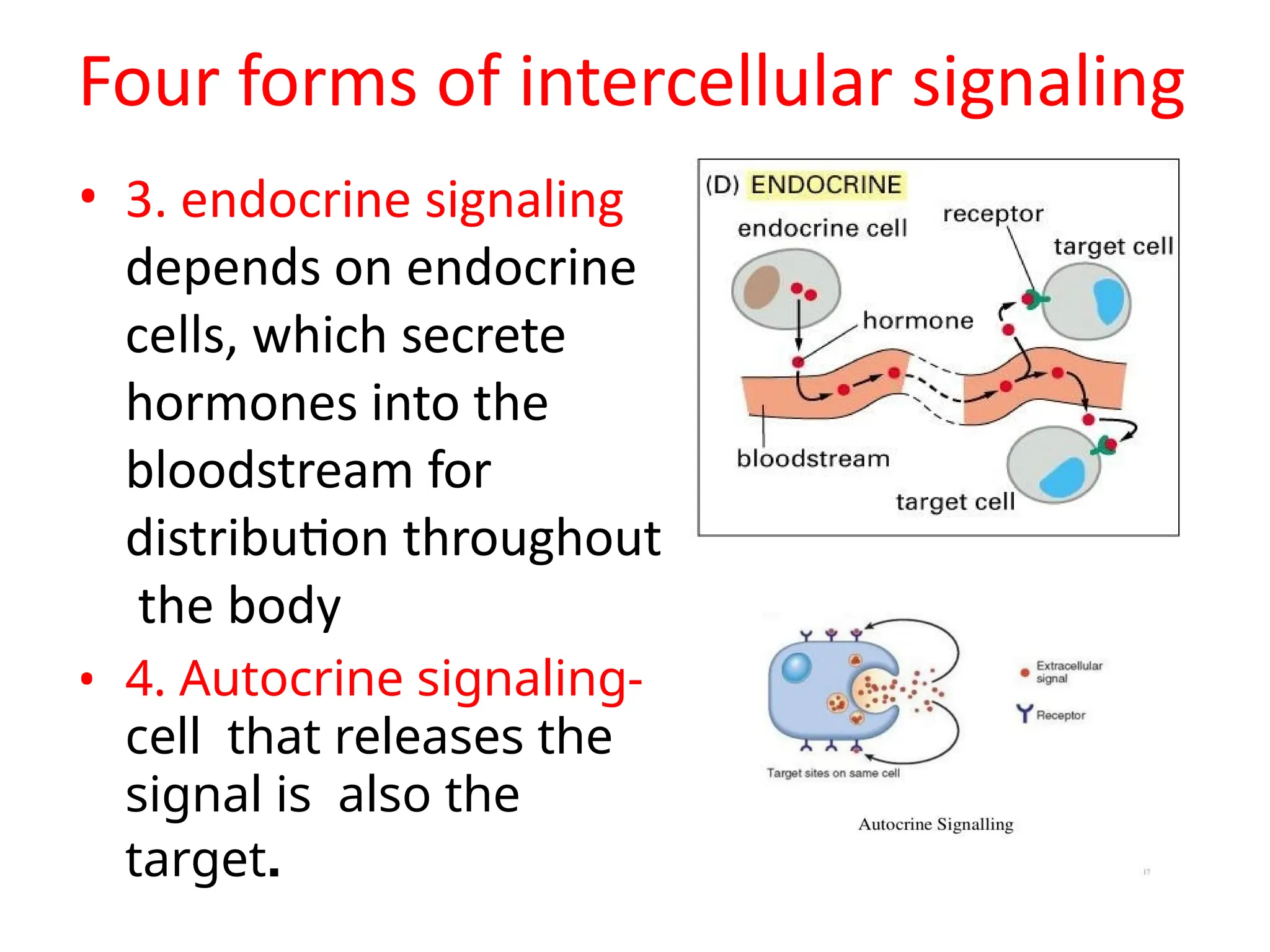
Four forms ofintercellular signaling
• 3. endocrine signaling
depends on endocrine
cells, which secrete
hormones into the
bloodstream for
distribution throughout
the body
• 4. Autocrine signaling-
cell that releases the
signal is also the
target.
9.
9
Each cell isprogrammed
to respond to specific
combinations of
extracellular signals
A cell may require multiple signals (A,B,C) to survive. Additional
signals to grow and divide (D,E) or differentiate (F,G). If appropriate
survival signals are deprived off, the cell undergoes apoptosis.
10.
Key Components ofCell Signaling
A cell signaling event involves several key players that work together to create a
response:
1. Signaling Molecule (Ligand):
These are the messengers that carry the signal. They can be proteins,
hormones, neurotransmitters, or other molecules.
2. Receptor:
Proteins located on the cell surface or inside the cell that specifically bind the
ligand to initiate the signaling process.
3. Intracellular Signaling Pathway:
Once the ligand binds to the receptor, it triggers a series of chemical changes
or protein interactions inside the cell, leading to signal amplification and
processing.
4. Effector Molecules:
These are the molecules or enzymes that directly carry out the cell's response
to the signal.
11.
Extracellular signal moleculesbind to
specific receptors
proteins, small peptides, amino
• Extracellular signal molecules
include
acids,
nucleotides, steroids, retinoids, fatty
acid derivatives, NO, CO.
• Target cells respond by means of receptors.
• Receptors are of two types:
12.
11
Receptors
1.
mostly,
receptors
transmembrane proteins on
Arethe
target-cell surface. When they bind
to Extra cellular molecule (a ligand),
and act as signal transducer, they
become
various
activated and
generate intracellular
signals that
alter the behaviour of cell.
2. Intracellular receptors-the signal
molecule has to be small to diffuse
across the PM and bind to receptor
proteins inside the target cell-either
in the cytosol or nucleus.
13.
Types of Signalingligands
Produced by signaling cells, bind to receptors
in target cells, act as chemical signals
1. Small Hydrophobic Ligands
2. Other Ligands
3. Water-Soluble Ligands
14.
Small hydrophobic ligands
•Small hydrophobic ligands can directly diffuse
through the plasma membrane and interact
with internal receptors. Important members
of this class of ligands are
the steroid hormones.
15.
Other Ligands
• Nitricoxide (NO) is a gas that also acts as a
ligand. It is able to diffuse directly across the
plasma membrane; one of its roles is to
interact with receptors in smooth muscle and
induce relaxation of the tissue. NO has a very
short half-life; therefore, it only functions over
short distances.
16.
Water-soluble ligands
• Water-solubleligands are polar and, therefore, cannot
pass through the plasma membrane unaided. Instead,
most water-soluble ligands bind to the extracellular
receptors.
• Cell-surface receptors include: ion-channel, G-protein,
and enzyme-linked protein receptors. The binding of
these ligands to these receptors results in a series of
cellular changes. These water soluble ligands are quite
diverse and include small molecules, peptides, and
proteins.
17.
16
Types of SignalingLigands:
A. Ligands that bind to cell-surface receptors:
1.Neurotransmitters (NT), i.e. norepinephrine,
histamine - hydrophilic (charged, polar)
2.Peptide hormones (P), i.e. insulin - can't cross
membrane
3. Growth factors (GF),
4. Lipophilic signaling molecules, i.e.
prostaglandins
B.Ligands that bind to intracellular receptors:
lipid soluble hormones that diffuse across the
plasma membrane and interact with receptors in the
cytosol or nucleus. i.e. steroids, thyroxine,
retinoic acid, nitric oxide.
Signaling molecules
18.
Cell surface receptors
•Ion -channel-linked receptors bind a ligand and open
a channel through the membrane that allows specific
ions to pass through.
• G-protein-linked receptors bind a ligand and activate
a membrane protein called a G-protein, which then
interacts with either an ion channel or an enzyme in
the membrane.
• Enzyme-linked receptors are cell-surface receptors
with intracellular domains that are associated with
an enzyme.
19.
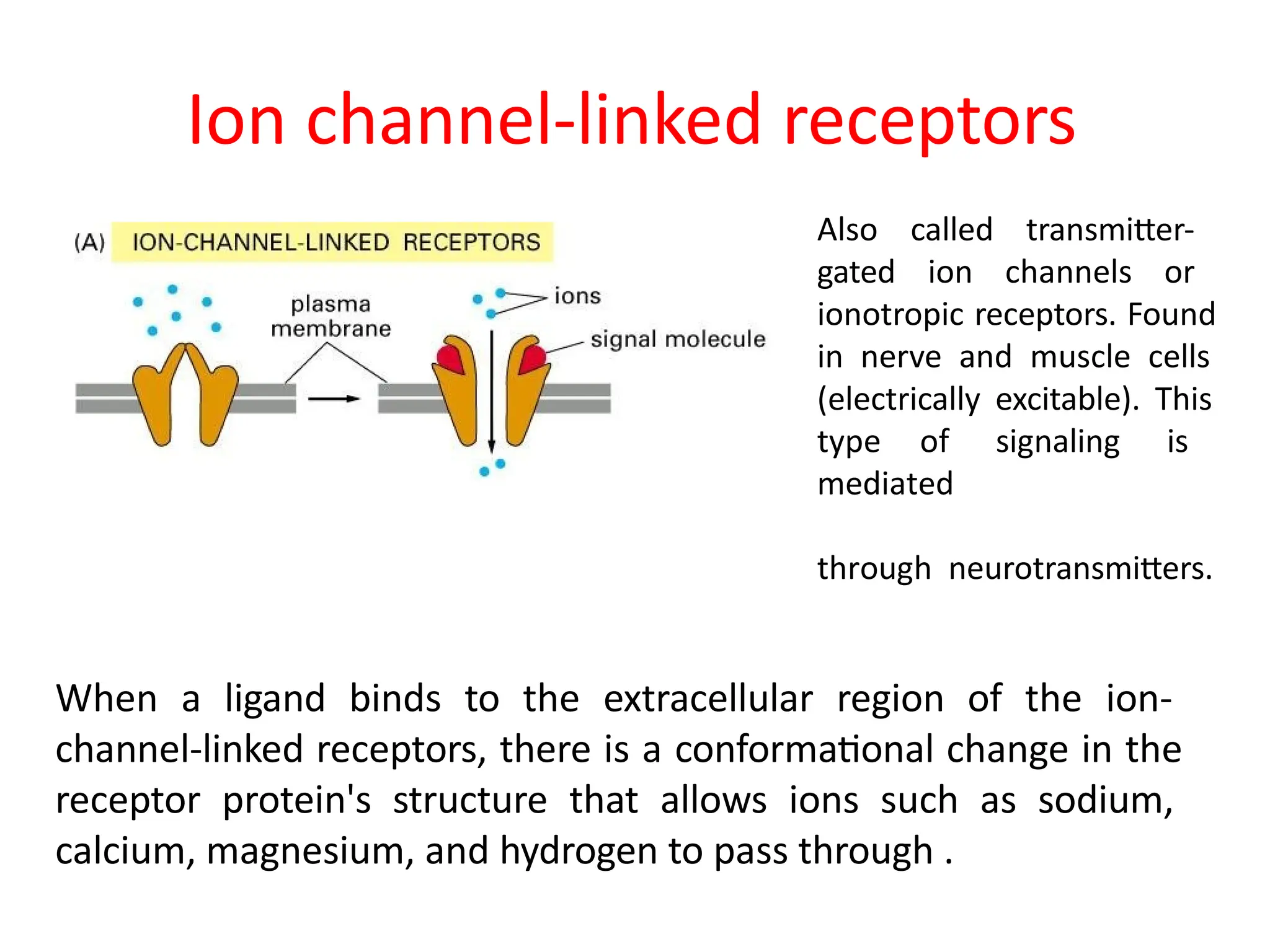
Ion channel-linked receptors
Alsocalled transmitter-
gated ion channels or
ionotropic receptors. Found
in nerve and muscle cells
(electrically excitable). This
type of signaling is
mediated
through neurotransmitters.
When a ligand binds to the extracellular region of the ion-
channel-linked receptors, there is a conformational change in the
receptor protein's structure that allows ions such as sodium,
calcium, magnesium, and hydrogen to pass through .
Signal molecules
20
Biogenic amines:Adrenaline, noradrenaline, dopamine,,
histamine, acetylcholine
Amino acids and ions: Glutamate, Ca2+, GABA
Lipids : prostaglandins, leukotrienes ( produced in
leukocytes by the oxidation of arachidonic acid )
Peptides / proteins :
GnRH, angiotensin, bradykinin, thrombin, bombesin, glucagon,
calcitonin, vasoactive intestinal peptides, PTH, FSH, LH, TSH
Nucleotides : Adenosine nucleotides, adenine nucleotides,
uridine nucleotides
Others : Light, odorants, pheromones, opiates
22.
• Robert Lefkowitzand Brian Kobilka: the 2012
Nobel Prize in Chemistry for groundbreaking
discoveries that revealed the inner workings of
G-protein–coupled receptors.
21
Historical background
23.
G-protein-linked receptors
• Thebinding of ligands to the extracellular
domain of these receptors induces a
conformational change in the receptor and
exposes a binding site for a G protein (bound to
the inner face of the plasma membrane). G
protein consists of α,β,γ subunits.
24.
Mammalian Gprotein complexes are made up of
• 20 alpha (α)
• 6 beta (β)
• 12 gamma (γ) subunits.
Beta and gamma subunits
can form a stable dimeric
complex referred to as the
beta-gamma complex.
α subunit
β subunit
γ subunit
23
heterotrimeric G proteins
25.
G-protein-linked receptors
• Inthe resting state, α is bound to GDP.
• Ligand binding induces a conformational change in the receptor,
such that the cytosolic domain of the receptor interacts with the G
protein and stimulates the release of bound GDP from α
subunit and its exchange for GTP.
• The activated GTP-bound α subunit then dissociates from β and γ,
which remain together and function as a βγ complex.
• α subunit moves along the inner membrane and interact with
another membrane bound protein ‘primary effector’
adenyl cyclase to elicit an intracellular response.
• The activity of the α subunit is terminated by hydrolysis of the
bound GTP, and the inactive α subunit (now with GDP bound) then
reassociates with the βγ complex, ready for the cycle to start
anew.
26.
• ‘primary effector’creates a second messenger
which may activate a ‘secondary effector’-
protein kinase
• the G protein associated with the epinephrine
receptor is called Gs because its α subunit
stimulates adenyl cyclase.
• In additionto regulating target enzymes, both the
α and βγ subunits of some G proteins directly
regulate ion channels. Example: action of the
neurotransmitter acetylcholine on heart muscle.
• Heart muscle cells have a different acetylcholine
receptor, which is G protein-coupled.
• This G protein is designated Gi because its α
subunit inhibits adenyl cyclase. In addition, the Gi
βγ subunits act directly to open K+ channels in the
plasma membrane, which has the effect of
slowing heart muscle contraction.
Enzyme-linked receptors
An exampleof enzyme-linked receptor is the tyrosine kinase receptor.
Signaling molecules bind to the extracellular domain of two nearby
tyrosine kinase receptors, which then dimerize. The tyrosine kinase
receptor transfers phosphate groups to tyrosine molecules on the
intracellular domain of the receptors and can then transmit the signal to
the next messenger within the cytoplasm.
31.
• This
family
polypeptide
includes
growth
the receptorsfor
most factors, so
protein-tyrosine
phosphorylation has been particularly well studied as
a signaling mechanism involved in the control of
animal cell growth and differentiation.
32.
Intracellular signaling molecules
•These relay signals received by cell surface receptors into
the cell interior.
• Some are small molecules often called 2nd messenger (1st
messenger being the extracellular signals). Generated in
large nos. in response to receptor activation-
• cAMP and Ca++ are water soluble and diacylglycerol is lipid-
soluble.
• Most intracellular molecules are proteins, which help relay
the signal into the cell by either generating 2nd
messenger or activating another signaling or effector
protein in the pathway.
• Many of these proteins behave like molecular switches,
mostly activated or deactivated by phosphorylation.
32
33.
Second messengers
There are3 major classes of second messengers:
• cyclic nucleotides (e.g., cAMP and cGMP)
• Inositol trisphosphate (IP3) & diacylglycerol (DAG)
• Calcium ions (Ca2+)
An important feature of the second messenger
signaling system is that second messengers may
be coupled downstream to multi-cyclic kinase
cascades to greatly amplify the strength of the
original first messenger signal.
34.
Second messengers: CyclicNucleotides
Cyclic AMP (cAMP)
• Some of the hormones that achieve their effects through cAMP
as a second messenger:
• adrenaline
• glucagon
• luteinizing hormone (LH), ACTH, FSH, LH, MSH, PTH
• Cyclic AMP is synthesized from ATP by the action of the
enzyme adenylyl cyclase. Binding of the hormone to its receptor
activates a G protein which, in turn, activates adenylyl cyclase.
• The resulting rise in cAMP turns on the appropriate response in
the cell by either (or both):
– changing the molecular activities in the cytosol, often
using Protein Kinase A (PKA) — a cAMP-dependent protein
kinase that phosphorylates target proteins;
– turning on a new pattern of gene transcription.
Cyclic Nucleotides
Cyclic GMP(cGMP)
• Cyclic GMP is synthesized from the
nucleotide GTP using the enzyme guanylyl
cyclase. Cyclic GMP serves as the second
messenger for
• atrial natriuretic peptide (ANP)
• nitric oxide (NO)
• Some of the effects of cGMP are mediated
through Protein Kinase G (PKG) — a cGMP-
dependent protein kinase that phosphorylates
target proteins in the cell.
37.
Second messengers: Inositol
trisphosphate& diacylglycerol
Peptide and protein hormones like
• vasopressin,
• thyroid-stimulating hormone (TSH), and
• Angiotensin and neurotransmitters like GABA
bind to G protein-coupled receptors (GPCRs) that activate the
intracellular enzyme phospholipase C (PLC). PLC
hydrolyzes phospholipids — specifically phosphatidylinositol-
4,5-bisphosphate (PIP2) which is found in the inner layer of
the plasma membrane. Hydrolysis of PIP2 yields two
products:
• Diacylglycerol (DAG) acts via Protein Kinase C (PKC) —
a calcium-dependent kinase, made available by IP3
• inositol-1,4,5-trisphosphate (IP3)
38.
Inositol trisphosphate &diacylglycerol
DAG remains in the inner layer of the plasma membrane.
• It recruits Protein Kinase C (PKC) — a calcium-dependent kinase
that phosphorylates many other proteins that bring about the
changes in the cell. Ca2+made available by IP3
IP3
• diffuses through the cytosol and binds to receptors on
the endoplasmic reticulum causing the release of calcium ions
(Ca2+) into the cytosol.
• The rise in intracellular calcium triggers the response.
39.
Second messengers :Calciumions
• calcium ions are probably the most widely used
intracellular messengers.
In response to many different signals, a rise in the
concentration of Ca2+ in the cytosol triggers many types of
events such as
• muscle contraction;
• exocytosis, e.g.,
– release of neurotransmitters at synapses
• activation of T cells and B cells when they bind antigen
with their antigen receptors
• apoptosis
• a variety of biochemical changes mediated
by Protein Kinase C (PKC).
40.
IP3 binds withreceptors on smooth endoplasmic reticulum to
stimulate the release of calcium ions as a part of the
amplification of a hormone's regulation of cellular enzymes.
48
Gene expression isthe cellular process of transforming the information in a
cell's DNA into a sequence of amino acids that ultimately forms a protein. When the
ligand binds to the internal receptor, a conformational change exposes a DNA-
binding site on the protein. The ligand-receptor complex moves into the nucleus,
binds to specific regulatory regions of the chromosomal DNA, and promotes the
initiation of transcription . Internal receptors can directly influence gene expression
without having to pass the signal on to other receptors or messengers.
Internal receptors are found in
the cytoplasm of the cell and
respond to hydrophobic ligand
molecules that are able to travel
across the plasma membrane.
Once inside the cell, many of
these molecules bind to proteins
that act as regulators of mRNA
synthesis to
mediate gene expression.
43.
References
• Alberts B,Johnson A, Lewis J, Morgan D, Raff
M, Walter P (2015). Molecular Biology of The
Cell. Sixth Edition, Garland Science, Taylor and
Francis group.
• Becker, Kleinsmith, Hardin, Bertoni (2009). The
World of the Cell. Seventh Editon. Pearson
Education
• Youtube videos