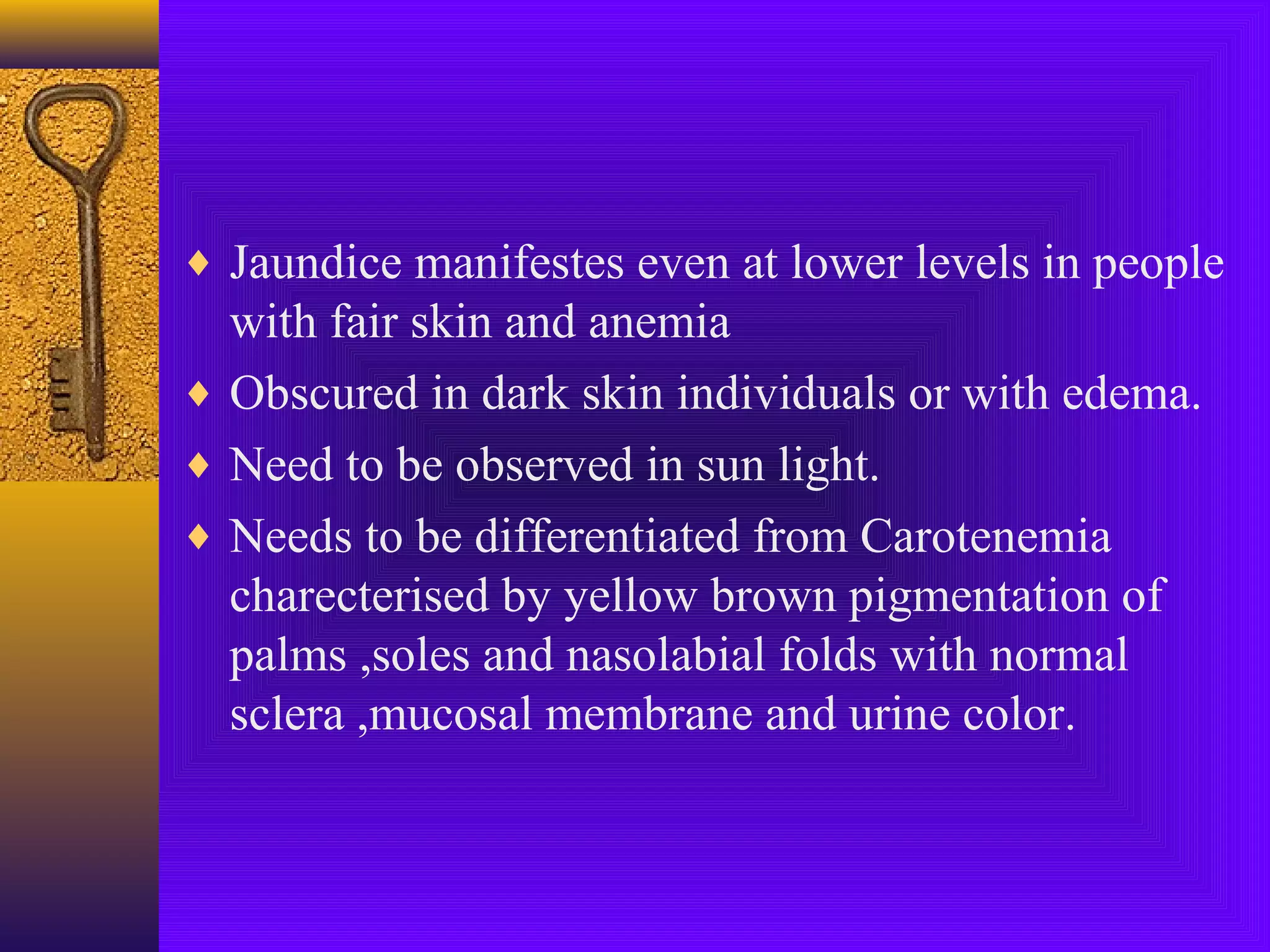
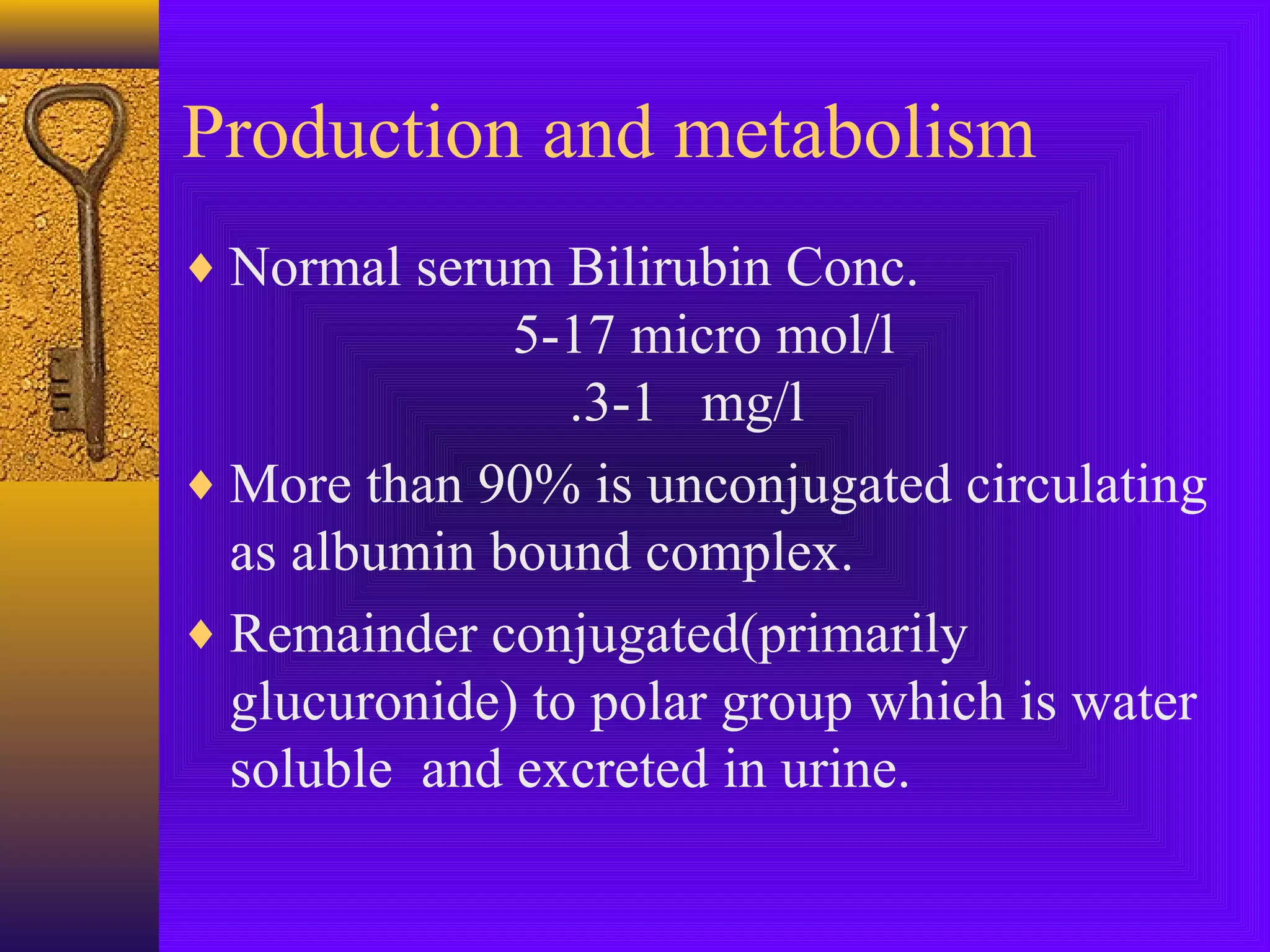
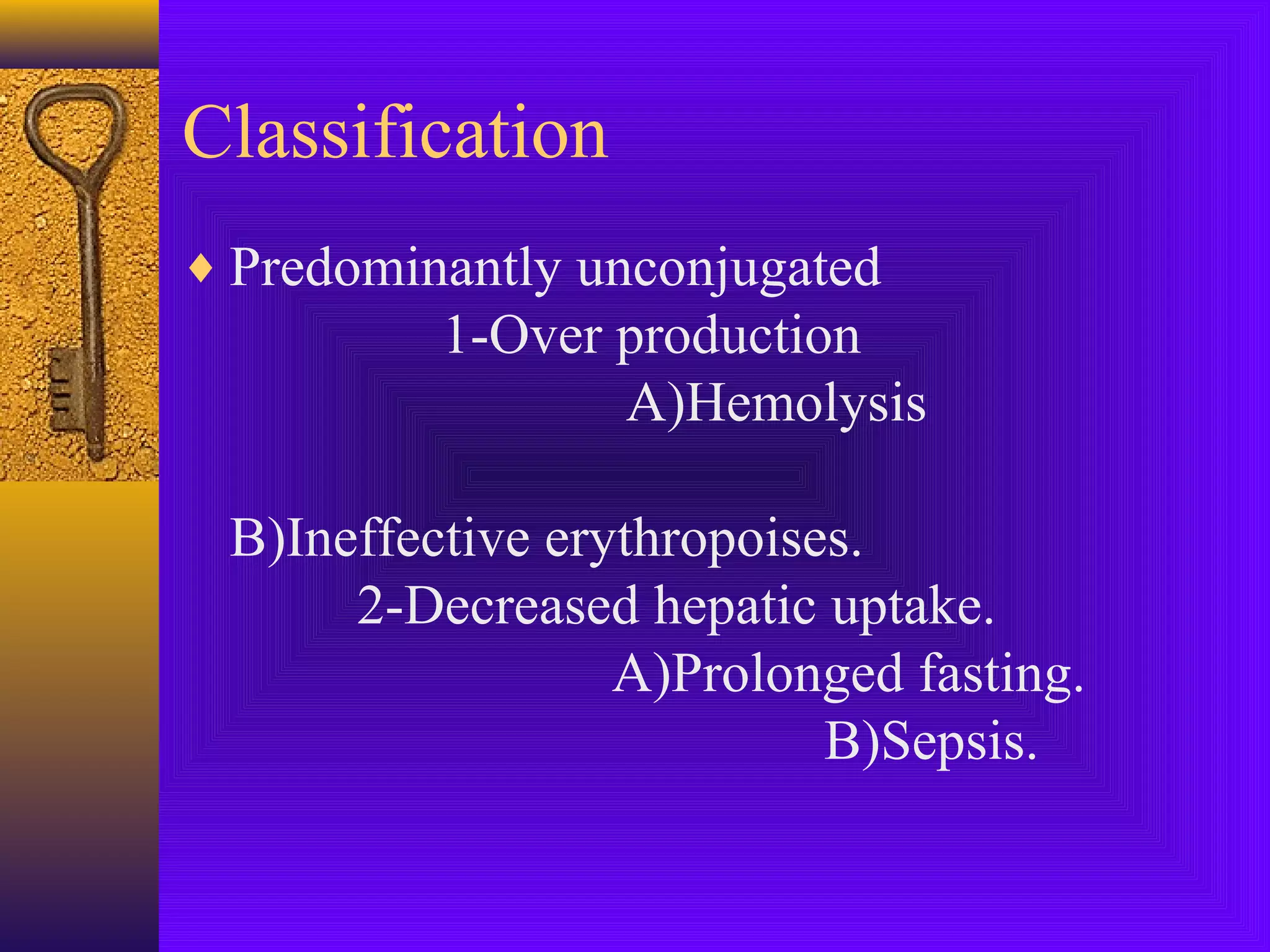
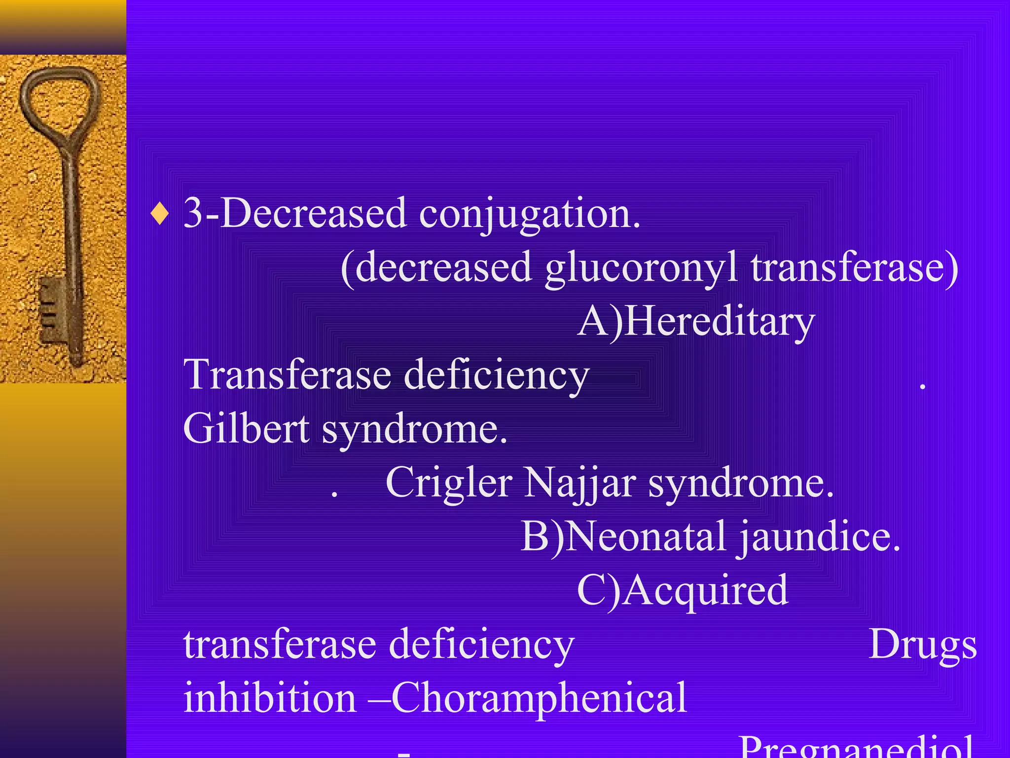
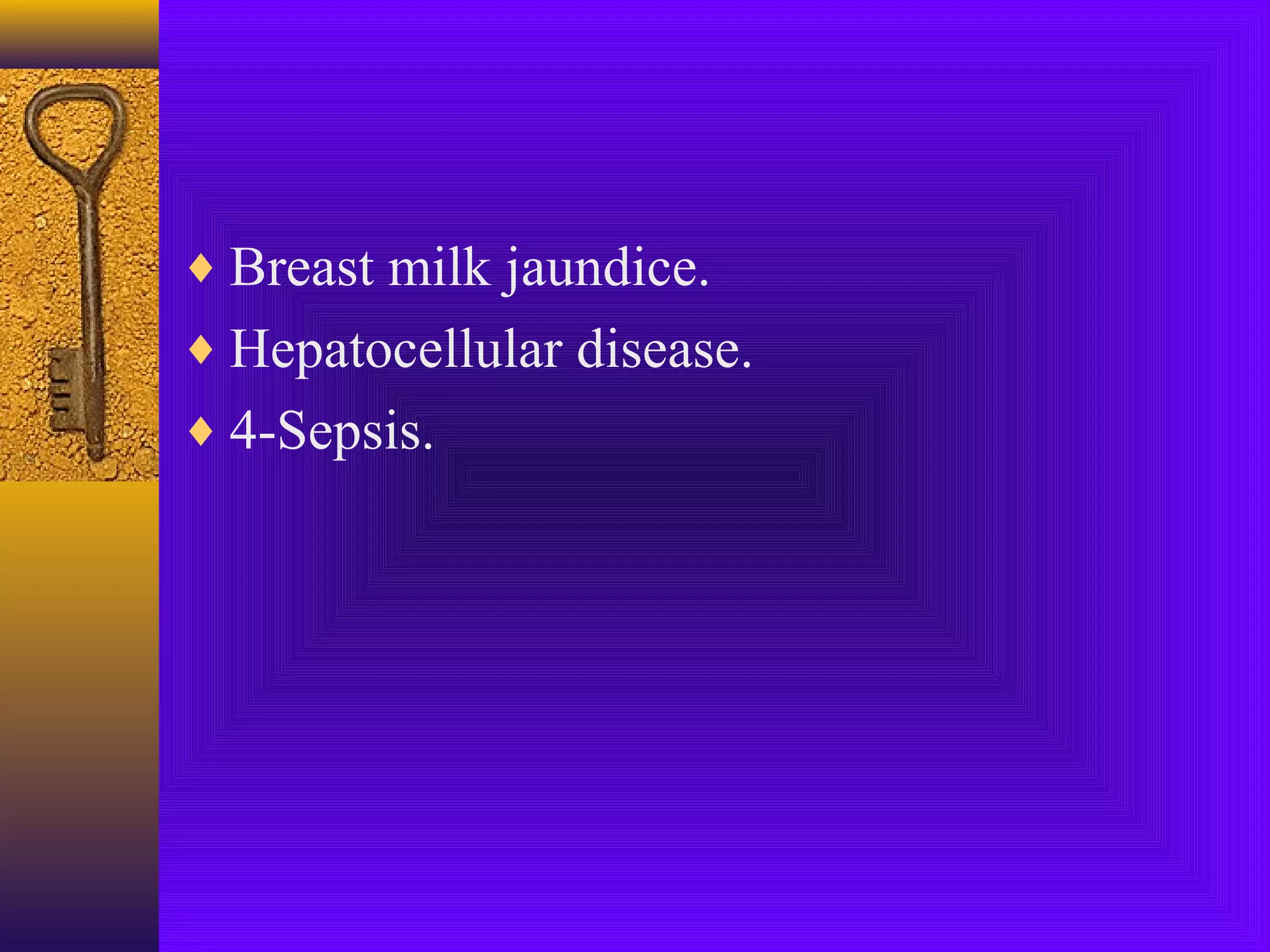
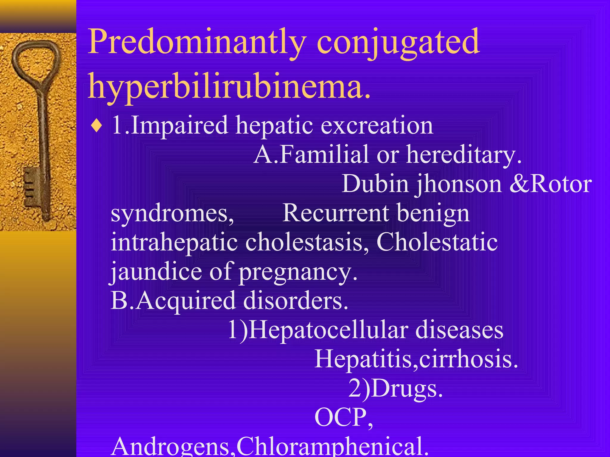
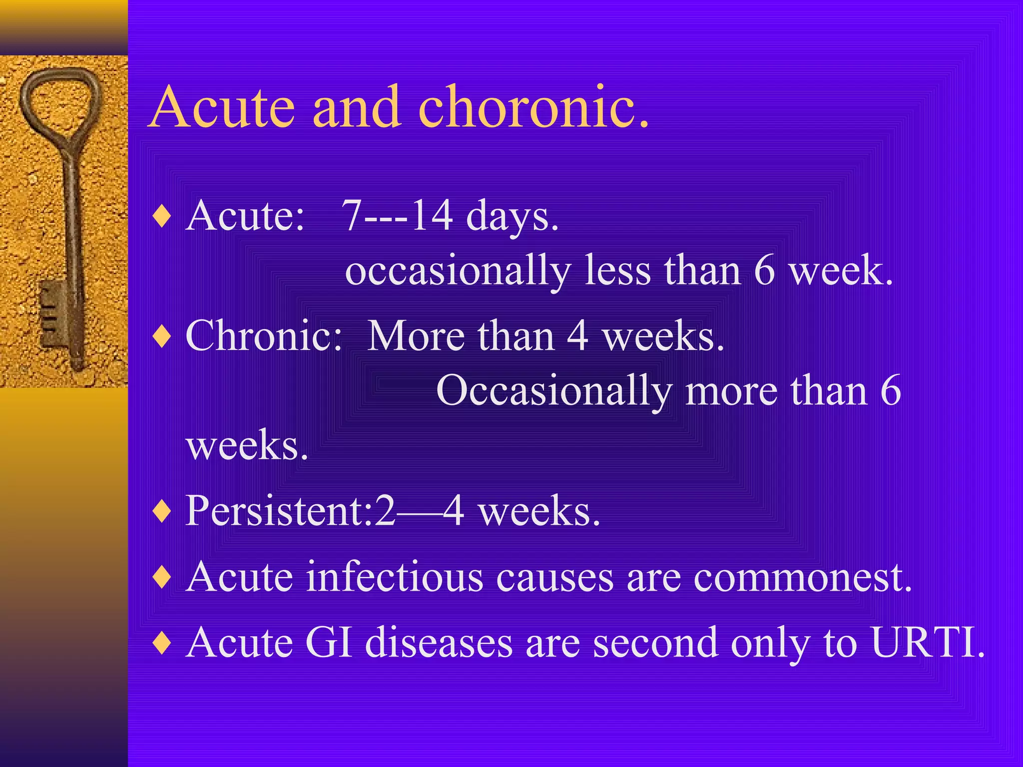
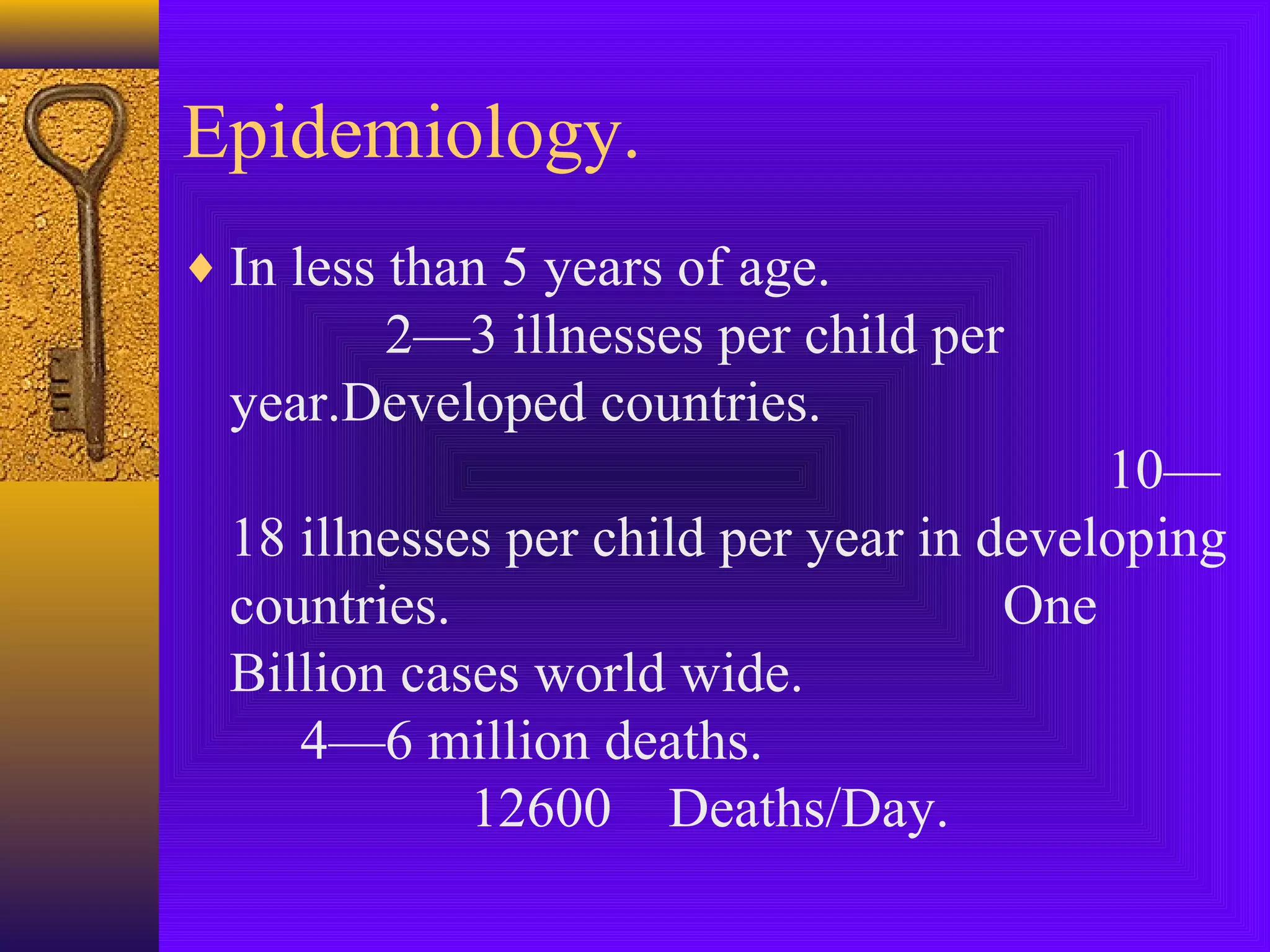
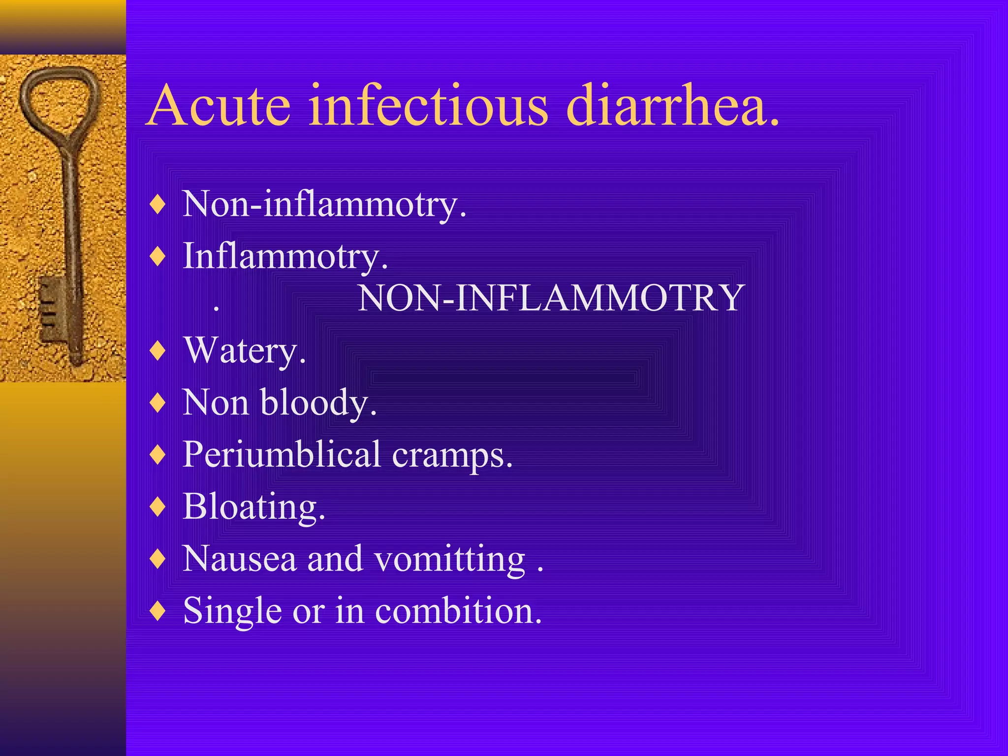
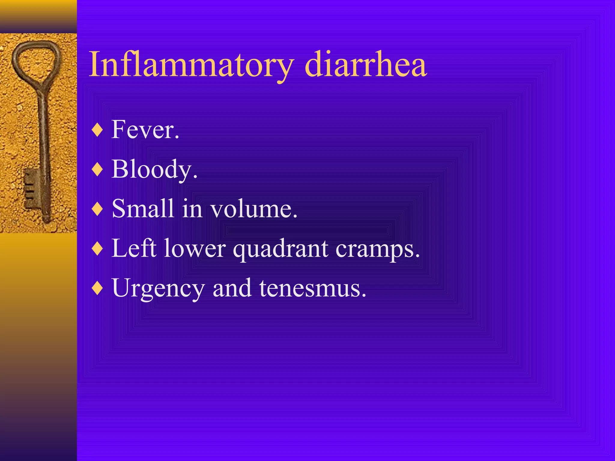
SYMPTOMATOLOGY IN
GIT PART-11
DR.MUZAMIL JAMIL
ASSOCIATE PROFESSOR
MEDICINE
JAUNDICE
♦ Accumulation of bilirubin.
♦ Yellowish pigmentation of plasma.
♦ Discolouration of heavily perfused tissues

likeskin,sclera and mucous membranes.
♦ Clinically hyperbilrubinemia manifests as
icterus or jaundice.
♦ Serum bilrubin > 34-43 micro mol/l
♦

2.6-2.5 mg/dl
♦ Jaundice manifestes even at lower levels in people

with fair skin and anemia
♦ Obscured in dark skin individuals or with edema.
♦ Need to be observed in sun light.
♦ Needs to be differentiated from Carotenemia
charecterised by yellow brown pigmentation of
palms ,soles and nasolabial folds with normal
sclera ,mucosal membrane and urine color.
Production and metabolism
♦ Normal serum Bilirubin Conc.

5-17 micro mol/l
.3-1 mg/l
♦ More than 90% is unconjugated circulating
as albumin bound complex.
♦ Remainder conjugated(primarily
glucuronide) to polar group which is water
soluble and excreted in urine.
♦ 80% of Bilirubin –RBCs break down.
♦ 15-20%-Ineffective erythropoises and

metabolism of other heme containing
protiens
Metabolism
♦ Hepatic uptake.
♦ Conjugation.
♦ Excretion into bile.
Derangement of bilirubin
metabolism
♦ Over production .
♦ Decreased hepatic uptake.
♦ Decreased hepatic conjugation.
♦ Decreased excretion.
Pathological terms.
♦ Hemolytic.
♦ Hepatocellular.
♦ Obtructive.
Classification
♦ Predominantly unconjugated

1-Over production
A)Hemolysis
B)Ineffective erythropoises.
2-Decreased hepatic uptake.
A)Prolonged fasting.
B)Sepsis.
♦ 3-Decreased conjugation.

(decreased glucoronyl transferase)
A)Hereditary
Transferase deficiency
.
Gilbert syndrome.
. Crigler Najjar syndrome.
B)Neonatal jaundice.
C)Acquired
transferase deficiency
Drugs
inhibition –Choramphenical
♦ Breast milk jaundice.
♦ Hepatocellular disease.
♦ 4-Sepsis.
Predominantly conjugated
hyperbilirubinema.
♦ 1.Impaired hepatic excreation

A.Familial or hereditary.
Dubin jhonson &Rotor
syndromes,
Recurrent benign
intrahepatic cholestasis, Cholestatic
jaundice of pregnancy.
B.Acquired disorders.
1)Hepatocellular diseases
Hepatitis,cirrhosis.
2)Drugs.
OCP,
Androgens,Chloramphenical.
♦ 3)Alcohol

4)Sepsis
5)Post operative
6)Biliary
cirrhosis.
♦ Extrahepatic Biliary obstruction.
Intraductal
Compression of biliary
duct.
–
Evalution of jaundice.
Hyperbilirubinemia.
♦ Hemolysis—In direct Bilirubin.
♦ Hepatobiliary –Direct Bilirubin.
Unconjugated Hyperbilirubinia.
♦ Hemolysis.
♦ Resorbtion of large hematoma.
♦ Bil. Rarely above 5mg%.
♦ Gilbert syndrone is an exception.
♦ Reticulocyte count is high.
♦ Hb is low.
♦ LDH is high.
Conjugated Hyperbilirubinia
Hepatocellular.
Intrahepatic obstruction.
Extra hepatic obstruction.
Approach to patient with
jaundice.
♦ Age.

Young------- Hepatitis.
Old -------Malignancy.
♦ Duration of symptoms.
♦ Abdominal pain.
♦ Fever and other symptoms of active
inflammation.
♦ Appitite change,weight loss or altered
bowels—Malignancy.
♦ Transfusion.(hepatitis B&C).
♦ Use of intravenous drugs.
♦ Sexual contact.
♦ Ethanol.
♦ Travel and immunization.
♦ Drugs.

Cholestsis.Anabolic steroids and chlorpromazine.
Heepatocellular
necrosis. Acetoaminophen,ATT.
♦ Sore throat and rash—

Infectious mononucleosis.
♦ Pruritis—Chronic cholestasis.
Hepatic: Primary Biliary cirrhosis.
.
Sclerosing cholangitis.
Extra hepatic obstruction.
♦ Acholic stools.
♦ Pregnancy.
♦ Past history of jaundice, hepatitis,arthralgias

Prodromal symptoms.
Viral hepatitis.
♦ Previous surgery:Biliary procedures.
.
Stones,strictures.
♦ Pre existing IBD.
♦ Right heart failure.
♦ Skin tatooing.
♦ History of GI bleeding.
♦ Family history.Congenital spherocytosis.
Physical examination.
♦ Excoriation.
♦ Fever and epigastric/RUQ tenderness.
♦ Painless jaundice.
♦ Enlarged tender liver.
♦ Rapidly enlarging liver.
♦ Palpable gall bladder.
♦ Spleenomegaly.
♦ Peripheral stigmas of liver diasease.
♦ Wasting and lymphoadenopathy.
♦ History pointing to malignancy.

Primary tomours in abdomen ,breast
and thyroid should be looked for.
Diarrhea.
♦ Increase in daily stool weight of more than

250gm/24 hours.
♦ Normal bowel frequency ranges between
3times/day to3times/week.
Factors influencing stool
weight ,consistency and
frequency. in diet.
♦ 1.fiber content
♦ 2.Gender.
♦ 3.Ingested medicines.
♦ 4.Exercise.
♦ 5.Stress.
♦ Pseudodiarrhea:

Increased frequency with normal
weight.
IBS ,Proctitis and
Hyperthyroidism.
♦ Incontinence:
Involuntary release of rectal contents.
Acute and choronic.
♦ Acute: 7---14 days.

occasionally less than 6 week.
♦ Chronic: More than 4 weeks.
Occasionally more than 6
weeks.
♦ Persistent:2—4 weeks.
♦ Acute infectious causes are commonest.
♦ Acute GI diseases are second only to URTI.
Epidemiology.
♦ In less than 5 years of age.

2—3 illnesses per child per
year.Developed countries.
10—
18 illnesses per child per year in developing
countries.
One
Billion cases world wide.
4—6 million deaths.
12600 Deaths/Day.
Acute infectious diarrhea.
♦ Non-inflammotry.
♦ Inflammotry.

.
NON-INFLAMMOTRY
♦ Watery.
♦ Non bloody.
♦ Periumblical cramps.
♦ Bloating.
♦ Nausea and vomitting .
♦ Single or in combition.
Inflammatory diarrhea
♦ Fever.
♦ Bloody.
♦ Small in volume.
♦ Left lower quadrant cramps.
♦ Urgency and tenesmus.
Etiology(non-inflammatory)
♦ Viral:Norwalk,Nor walk like and Rota virus
♦ Protozoal: Giardia,cryptosporidium.
♦ Bactrial:

1.Preformed toxins:Styphylococcus
aures, bacellius cereus and clostridium
perfringens 2.Enterotoxin production:Ecoli,vibrio cholera.
Food poisoning
♦ Staphylococcus aureus.
♦ Shortest incubation period.1—6 hours.

Lasts

for less than 12 houres.
♦ Infected human carriers are the source.
♦ If food is left to cool slowly and remains at room
temperature organisms have opportunity to form
toxins.
♦ Out breaks after picnics.
♦ Potatos,salads,mayonnise,cream pastries.
Bacillus cereus.
♦ Short incubation period.

1—6 hours emetic form.
Long
incubation period.
upto 18
hours diarrheal form.
♦ If cooked rice is not refrigerated,heat
resistant spores which have escaped boiling
germinate and produce toxin.Frying before
serving may not destroy these preformed
heat stable toxins.
Clostridium perferingens
♦ Incubation period 8—14 hours.
♦ Heat resistant spores.
♦ Inadequately cooked meat, poultry or

legumes.
♦ self limiting upto 24 hours.
Etiology for infllammatory
diarrhea.
♦ Viral:CMV.
♦ Protozoal:Entamoeba histolytica.
♦ Bacterial:Shigella,salmonella,compylopacte

r jejuni,entero invasive E-coli and vibrio
parahemolytic.
Approach to patient.
♦ HISTORY:

1.Duration.
2.Fever.Infections out side
the gut like malaria.
3.Frequency.May correlate
with dehydration.
4.Abdominal pain.
-Inflammatory
nature.
-RIF
Pain with yersina.
-Bloating with Giardiasis.
♦ 5.Vomiting.

-Acute illness
-Toxin.
-Systemic disease.
-Obstruction.
♦ 6.Tenesmes:shigellosis.
♦ 7.Appearance of stools.
-Blood—Shigellosis.
♦ -Rice watery—Vibrio cholera.

-Bulky white—small intestine.
♦ 8.Common source.
♦ 9.Antibiotic use.
♦ 10.Travel.
Physical examination.
♦ Signs of dehydration—Severity of illness.
♦ MILD.

-Thirst.
-Dry mouth.
-Decreased axillary sweat.
-Decreased urine out put.
-Slight weight loss.
Moderate dehyderation.
♦ Orthostatic hypotension
♦ Skin tenting.
♦ Sunken eye balls.
Severe dehydration.
♦ Hypotension.
♦ Tachycardia.
♦ Confusion.
♦ Frank coma.
Prompt medical evaluation.
♦ Inflammatory diarrhea.
♦ High fever.
♦ Bloody diarrhea.
♦ Abdominal pain.
♦ 6 or more unformed stools/24 hours.
♦ Profuse watery diarrhea.
♦ Severe dehyderatuon.
♦ Elderly or immunocompromised patients.
Chronic diarrhea.
Diarrhea which persists for more than 4
weeks
Needs evaluation to exclude serious
pathology
Most of the causes are noninfectious.
Classification.
♦ 1-Osmotic.
♦ 2-Secreatary.
♦ 3-Inflammatory.
♦ 4.Motility disorders.
♦ 5.Fectitious.
♦ 6.Malabsorptive conditions.
♦ 7.chronic infections.
Osmotic diarrhea.
♦ Results from lack of absorption of orally

ingested solutes (food).Osmotic effect.
♦ Relieved with fasting.
♦ Clinical symptoms are usually becauses of
malabsorption of fat or carbohyderates.
♦ Osmotic causes include lactase deficiency,
drugs like laxatives etc.
Steatorrheal causes.
♦ Intraluminal maldigestion.

.Chronic pancreatitis.
.Decreased bile salts.
.Bacterial over
growth.
♦ Mucosal malabsorption.
.Celiiac
disease.
.Tropical sprue.
Secreatary diarrhea.
♦ Excreation of large ammount more than

1 litre/day.
♦ No effect with fasting.
♦ Abnormal fluid and electrolyte transport.
♦ Harmones mediated.
♦ Causes may include Carcinoid, Zollinger
ellison syndrome, Medullary carcinoma of
thyroid and extensive gut recsection.
Inflammatory diarrhea.
♦ Fever.
♦ Abdominal pain and tenderness.
♦ Hematochezia.
♦ Patients may have toxic looks.
♦ Extra intestinal manefestation may be

present.
♦ Causes include IBD,malignancy,radiation
enterits.
Motility disorders
♦ Systemic disorders like diabetes and

hyperthyroidism.
♦ Previous gut surgery.
♦ Irritable bowel.
♦ Fecal impaction.
♦ Neurological disorders.
♦ FECTITIOUS DIARRHEA:Laxative abuse
Approach to patients.
♦ History.
♦ Symptoms and signs of inflammation.
♦ Extra intestinal manefestations.
♦ Perepheral edema or ascitis.
♦ Type of stools-intestinal malabsoption.
♦ Flatulence.
♦ Weight loss.
♦ Systemic manifestations like flushing.
♦ Autonomic dysfunctions like postural drop

and disordered sweating in diabetes.
♦ Diarrhea alternating with constipation-IBS.
♦ Effects of malabsorption like anemia,
bleeding tendency,osteopenia,amenorrhea
and infertility should be looked for.
Common causes
♦ Abdominal tuberculosis.
♦ Coeliac disease.
♦ Inflammatory bowel disease.
♦ Giardiasis.
♦ Tropical sprue.
♦ Colonic malignancy.
Symptomatology-GIT-1

Symptomatology-GIT-1

  • 1.
    SYMPTOMATOLOGY IN GIT PART-11 DR.MUZAMILJAMIL ASSOCIATE PROFESSOR MEDICINE
  • 2.
    JAUNDICE ♦ Accumulation ofbilirubin. ♦ Yellowish pigmentation of plasma. ♦ Discolouration of heavily perfused tissues likeskin,sclera and mucous membranes. ♦ Clinically hyperbilrubinemia manifests as icterus or jaundice. ♦ Serum bilrubin > 34-43 micro mol/l ♦ 2.6-2.5 mg/dl
  • 3.
    ♦ Jaundice manifesteseven at lower levels in people with fair skin and anemia ♦ Obscured in dark skin individuals or with edema. ♦ Need to be observed in sun light. ♦ Needs to be differentiated from Carotenemia charecterised by yellow brown pigmentation of palms ,soles and nasolabial folds with normal sclera ,mucosal membrane and urine color.
  • 4.
    Production and metabolism ♦Normal serum Bilirubin Conc. 5-17 micro mol/l .3-1 mg/l ♦ More than 90% is unconjugated circulating as albumin bound complex. ♦ Remainder conjugated(primarily glucuronide) to polar group which is water soluble and excreted in urine.
  • 5.
    ♦ 80% ofBilirubin –RBCs break down. ♦ 15-20%-Ineffective erythropoises and metabolism of other heme containing protiens
  • 6.
    Metabolism ♦ Hepatic uptake. ♦Conjugation. ♦ Excretion into bile.
  • 7.
    Derangement of bilirubin metabolism ♦Over production . ♦ Decreased hepatic uptake. ♦ Decreased hepatic conjugation. ♦ Decreased excretion.
  • 8.
    Pathological terms. ♦ Hemolytic. ♦Hepatocellular. ♦ Obtructive.
  • 9.
    Classification ♦ Predominantly unconjugated 1-Overproduction A)Hemolysis B)Ineffective erythropoises. 2-Decreased hepatic uptake. A)Prolonged fasting. B)Sepsis.
  • 10.
    ♦ 3-Decreased conjugation. (decreasedglucoronyl transferase) A)Hereditary Transferase deficiency . Gilbert syndrome. . Crigler Najjar syndrome. B)Neonatal jaundice. C)Acquired transferase deficiency Drugs inhibition –Choramphenical
  • 11.
    ♦ Breast milkjaundice. ♦ Hepatocellular disease. ♦ 4-Sepsis.
  • 12.
    Predominantly conjugated hyperbilirubinema. ♦ 1.Impairedhepatic excreation A.Familial or hereditary. Dubin jhonson &Rotor syndromes, Recurrent benign intrahepatic cholestasis, Cholestatic jaundice of pregnancy. B.Acquired disorders. 1)Hepatocellular diseases Hepatitis,cirrhosis. 2)Drugs. OCP, Androgens,Chloramphenical.
  • 13.
    ♦ 3)Alcohol 4)Sepsis 5)Post operative 6)Biliary cirrhosis. ♦Extrahepatic Biliary obstruction. Intraductal Compression of biliary duct. –
  • 14.
    Evalution of jaundice. Hyperbilirubinemia. ♦Hemolysis—In direct Bilirubin. ♦ Hepatobiliary –Direct Bilirubin.
  • 15.
    Unconjugated Hyperbilirubinia. ♦ Hemolysis. ♦Resorbtion of large hematoma. ♦ Bil. Rarely above 5mg%. ♦ Gilbert syndrone is an exception. ♦ Reticulocyte count is high. ♦ Hb is low. ♦ LDH is high.
  • 16.
  • 17.
    Approach to patientwith jaundice. ♦ Age. Young------- Hepatitis. Old -------Malignancy. ♦ Duration of symptoms. ♦ Abdominal pain. ♦ Fever and other symptoms of active inflammation. ♦ Appitite change,weight loss or altered bowels—Malignancy.
  • 18.
    ♦ Transfusion.(hepatitis B&C). ♦Use of intravenous drugs. ♦ Sexual contact. ♦ Ethanol. ♦ Travel and immunization. ♦ Drugs. Cholestsis.Anabolic steroids and chlorpromazine. Heepatocellular necrosis. Acetoaminophen,ATT.
  • 19.
    ♦ Sore throatand rash— Infectious mononucleosis. ♦ Pruritis—Chronic cholestasis. Hepatic: Primary Biliary cirrhosis. . Sclerosing cholangitis. Extra hepatic obstruction. ♦ Acholic stools. ♦ Pregnancy.
  • 20.
    ♦ Past historyof jaundice, hepatitis,arthralgias Prodromal symptoms. Viral hepatitis. ♦ Previous surgery:Biliary procedures. . Stones,strictures. ♦ Pre existing IBD. ♦ Right heart failure. ♦ Skin tatooing. ♦ History of GI bleeding. ♦ Family history.Congenital spherocytosis.
  • 21.
    Physical examination. ♦ Excoriation. ♦Fever and epigastric/RUQ tenderness. ♦ Painless jaundice. ♦ Enlarged tender liver. ♦ Rapidly enlarging liver. ♦ Palpable gall bladder. ♦ Spleenomegaly.
  • 22.
    ♦ Peripheral stigmasof liver diasease. ♦ Wasting and lymphoadenopathy. ♦ History pointing to malignancy. Primary tomours in abdomen ,breast and thyroid should be looked for.
  • 23.
    Diarrhea. ♦ Increase indaily stool weight of more than 250gm/24 hours. ♦ Normal bowel frequency ranges between 3times/day to3times/week.
  • 24.
    Factors influencing stool weight,consistency and frequency. in diet. ♦ 1.fiber content ♦ 2.Gender. ♦ 3.Ingested medicines. ♦ 4.Exercise. ♦ 5.Stress.
  • 25.
    ♦ Pseudodiarrhea: Increased frequencywith normal weight. IBS ,Proctitis and Hyperthyroidism. ♦ Incontinence: Involuntary release of rectal contents.
  • 26.
    Acute and choronic. ♦Acute: 7---14 days. occasionally less than 6 week. ♦ Chronic: More than 4 weeks. Occasionally more than 6 weeks. ♦ Persistent:2—4 weeks. ♦ Acute infectious causes are commonest. ♦ Acute GI diseases are second only to URTI.
  • 27.
    Epidemiology. ♦ In lessthan 5 years of age. 2—3 illnesses per child per year.Developed countries. 10— 18 illnesses per child per year in developing countries. One Billion cases world wide. 4—6 million deaths. 12600 Deaths/Day.
  • 28.
    Acute infectious diarrhea. ♦Non-inflammotry. ♦ Inflammotry. . NON-INFLAMMOTRY ♦ Watery. ♦ Non bloody. ♦ Periumblical cramps. ♦ Bloating. ♦ Nausea and vomitting . ♦ Single or in combition.
  • 29.
    Inflammatory diarrhea ♦ Fever. ♦Bloody. ♦ Small in volume. ♦ Left lower quadrant cramps. ♦ Urgency and tenesmus.
  • 30.
    Etiology(non-inflammatory) ♦ Viral:Norwalk,Nor walklike and Rota virus ♦ Protozoal: Giardia,cryptosporidium. ♦ Bactrial: 1.Preformed toxins:Styphylococcus aures, bacellius cereus and clostridium perfringens 2.Enterotoxin production:Ecoli,vibrio cholera.
  • 31.
    Food poisoning ♦ Staphylococcusaureus. ♦ Shortest incubation period.1—6 hours. Lasts for less than 12 houres. ♦ Infected human carriers are the source. ♦ If food is left to cool slowly and remains at room temperature organisms have opportunity to form toxins. ♦ Out breaks after picnics. ♦ Potatos,salads,mayonnise,cream pastries.
  • 32.
    Bacillus cereus. ♦ Shortincubation period. 1—6 hours emetic form. Long incubation period. upto 18 hours diarrheal form. ♦ If cooked rice is not refrigerated,heat resistant spores which have escaped boiling germinate and produce toxin.Frying before serving may not destroy these preformed heat stable toxins.
  • 33.
    Clostridium perferingens ♦ Incubationperiod 8—14 hours. ♦ Heat resistant spores. ♦ Inadequately cooked meat, poultry or legumes. ♦ self limiting upto 24 hours.
  • 34.
    Etiology for infllammatory diarrhea. ♦Viral:CMV. ♦ Protozoal:Entamoeba histolytica. ♦ Bacterial:Shigella,salmonella,compylopacte r jejuni,entero invasive E-coli and vibrio parahemolytic.
  • 35.
    Approach to patient. ♦HISTORY: 1.Duration. 2.Fever.Infections out side the gut like malaria. 3.Frequency.May correlate with dehydration. 4.Abdominal pain. -Inflammatory nature. -RIF Pain with yersina. -Bloating with Giardiasis.
  • 36.
    ♦ 5.Vomiting. -Acute illness -Toxin. -Systemicdisease. -Obstruction. ♦ 6.Tenesmes:shigellosis. ♦ 7.Appearance of stools. -Blood—Shigellosis.
  • 37.
    ♦ -Rice watery—Vibriocholera. -Bulky white—small intestine. ♦ 8.Common source. ♦ 9.Antibiotic use. ♦ 10.Travel.
  • 38.
    Physical examination. ♦ Signsof dehydration—Severity of illness. ♦ MILD. -Thirst. -Dry mouth. -Decreased axillary sweat. -Decreased urine out put. -Slight weight loss.
  • 39.
    Moderate dehyderation. ♦ Orthostatichypotension ♦ Skin tenting. ♦ Sunken eye balls.
  • 40.
    Severe dehydration. ♦ Hypotension. ♦Tachycardia. ♦ Confusion. ♦ Frank coma.
  • 41.
    Prompt medical evaluation. ♦Inflammatory diarrhea. ♦ High fever. ♦ Bloody diarrhea. ♦ Abdominal pain. ♦ 6 or more unformed stools/24 hours. ♦ Profuse watery diarrhea. ♦ Severe dehyderatuon. ♦ Elderly or immunocompromised patients.
  • 42.
    Chronic diarrhea. Diarrhea whichpersists for more than 4 weeks Needs evaluation to exclude serious pathology Most of the causes are noninfectious.
  • 43.
    Classification. ♦ 1-Osmotic. ♦ 2-Secreatary. ♦3-Inflammatory. ♦ 4.Motility disorders. ♦ 5.Fectitious. ♦ 6.Malabsorptive conditions. ♦ 7.chronic infections.
  • 44.
    Osmotic diarrhea. ♦ Resultsfrom lack of absorption of orally ingested solutes (food).Osmotic effect. ♦ Relieved with fasting. ♦ Clinical symptoms are usually becauses of malabsorption of fat or carbohyderates. ♦ Osmotic causes include lactase deficiency, drugs like laxatives etc.
  • 45.
    Steatorrheal causes. ♦ Intraluminalmaldigestion. .Chronic pancreatitis. .Decreased bile salts. .Bacterial over growth. ♦ Mucosal malabsorption. .Celiiac disease. .Tropical sprue.
  • 46.
    Secreatary diarrhea. ♦ Excreationof large ammount more than 1 litre/day. ♦ No effect with fasting. ♦ Abnormal fluid and electrolyte transport. ♦ Harmones mediated. ♦ Causes may include Carcinoid, Zollinger ellison syndrome, Medullary carcinoma of thyroid and extensive gut recsection.
  • 47.
    Inflammatory diarrhea. ♦ Fever. ♦Abdominal pain and tenderness. ♦ Hematochezia. ♦ Patients may have toxic looks. ♦ Extra intestinal manefestation may be present. ♦ Causes include IBD,malignancy,radiation enterits.
  • 48.
    Motility disorders ♦ Systemicdisorders like diabetes and hyperthyroidism. ♦ Previous gut surgery. ♦ Irritable bowel. ♦ Fecal impaction. ♦ Neurological disorders. ♦ FECTITIOUS DIARRHEA:Laxative abuse
  • 49.
    Approach to patients. ♦History. ♦ Symptoms and signs of inflammation. ♦ Extra intestinal manefestations. ♦ Perepheral edema or ascitis. ♦ Type of stools-intestinal malabsoption. ♦ Flatulence. ♦ Weight loss.
  • 50.
    ♦ Systemic manifestationslike flushing. ♦ Autonomic dysfunctions like postural drop and disordered sweating in diabetes. ♦ Diarrhea alternating with constipation-IBS. ♦ Effects of malabsorption like anemia, bleeding tendency,osteopenia,amenorrhea and infertility should be looked for.
  • 51.
    Common causes ♦ Abdominaltuberculosis. ♦ Coeliac disease. ♦ Inflammatory bowel disease. ♦ Giardiasis. ♦ Tropical sprue. ♦ Colonic malignancy.