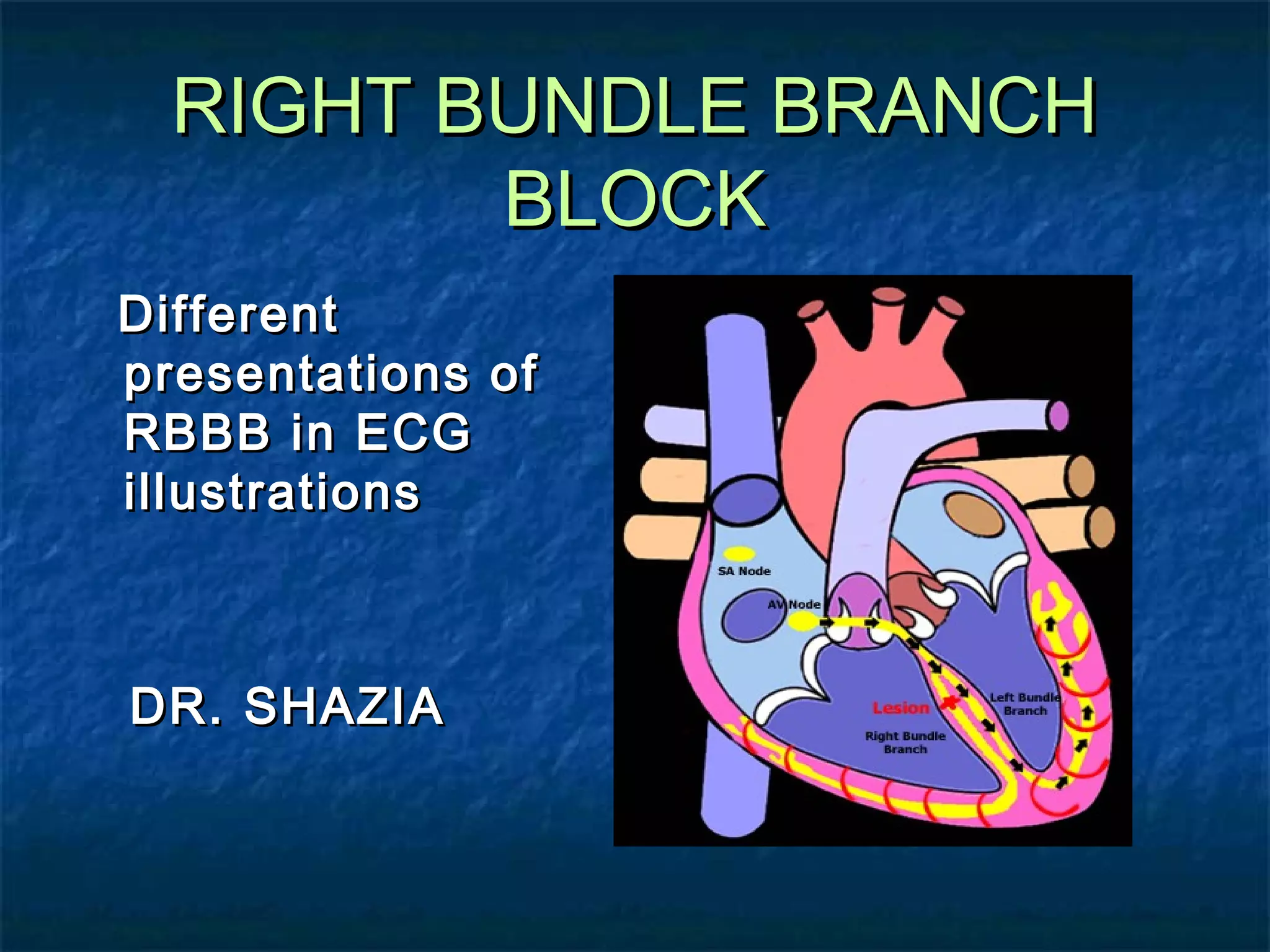
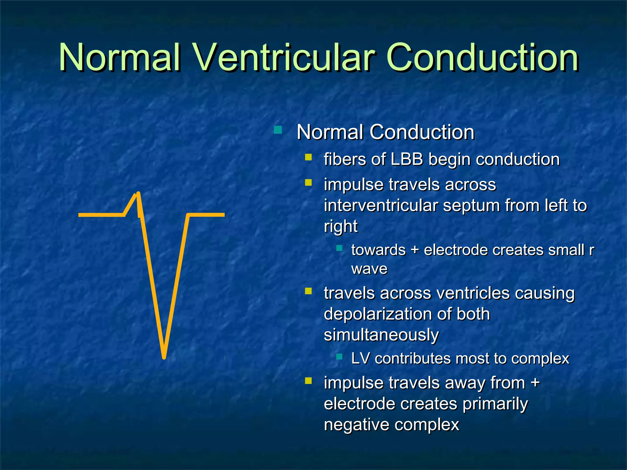
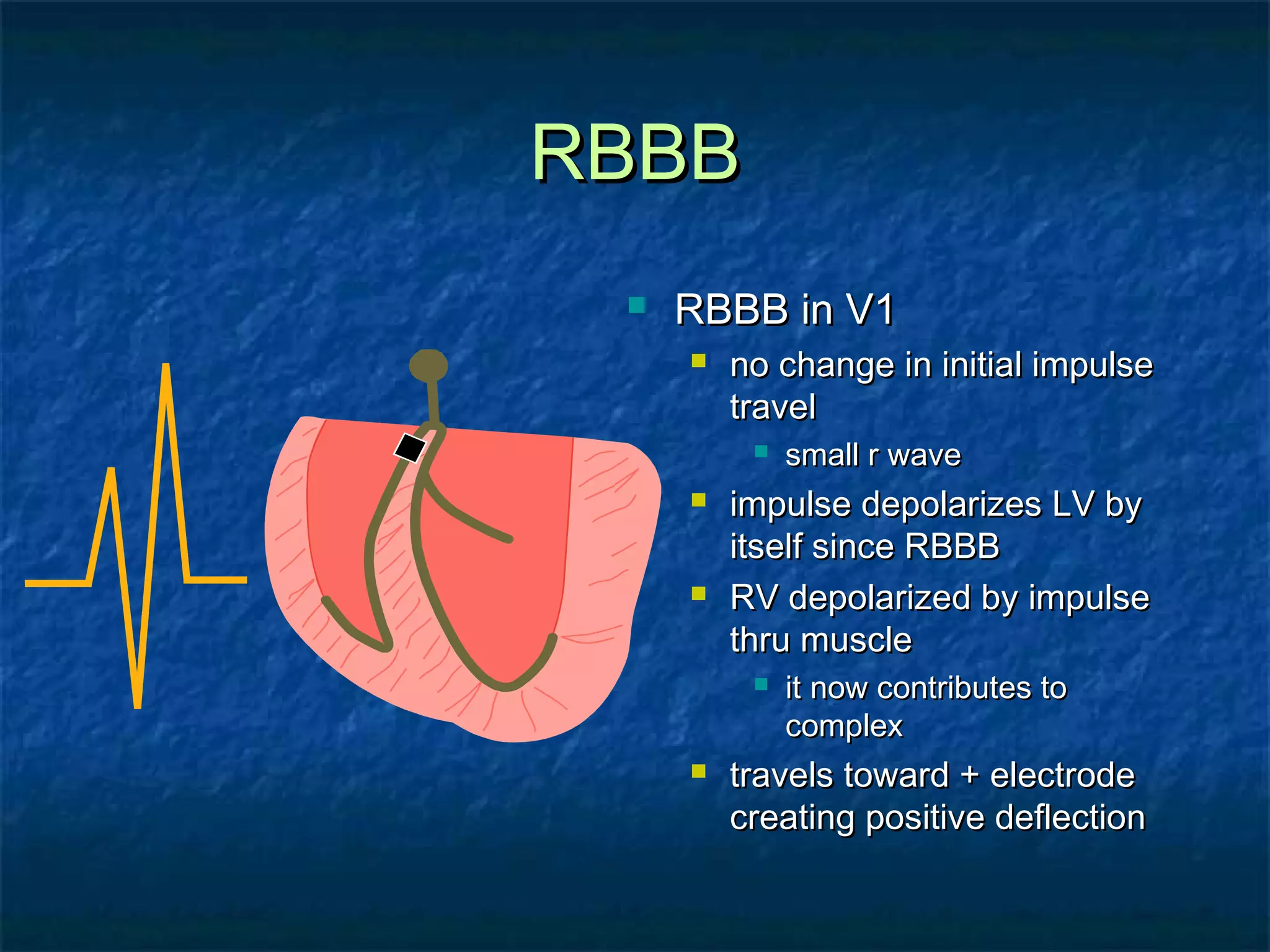
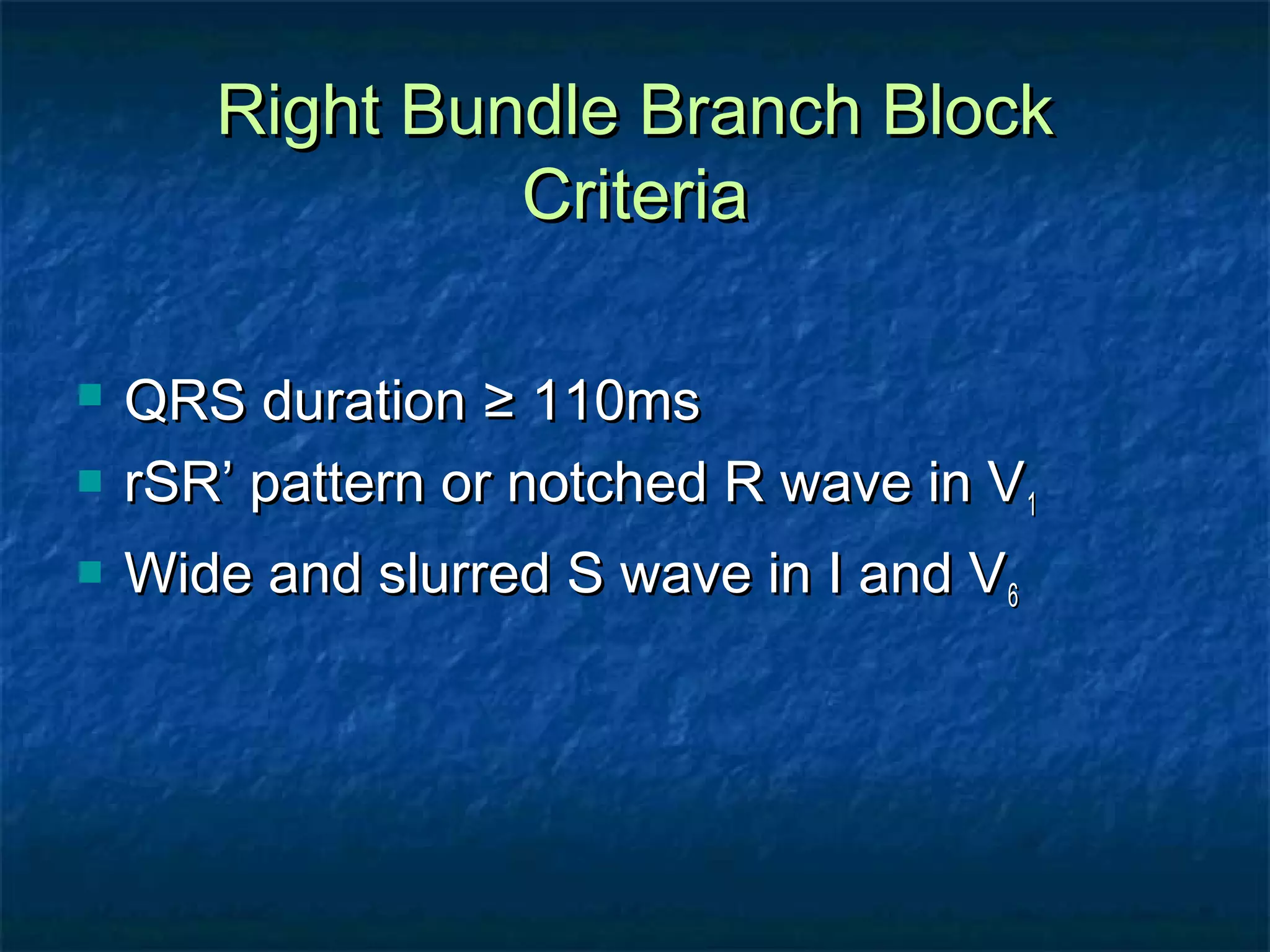
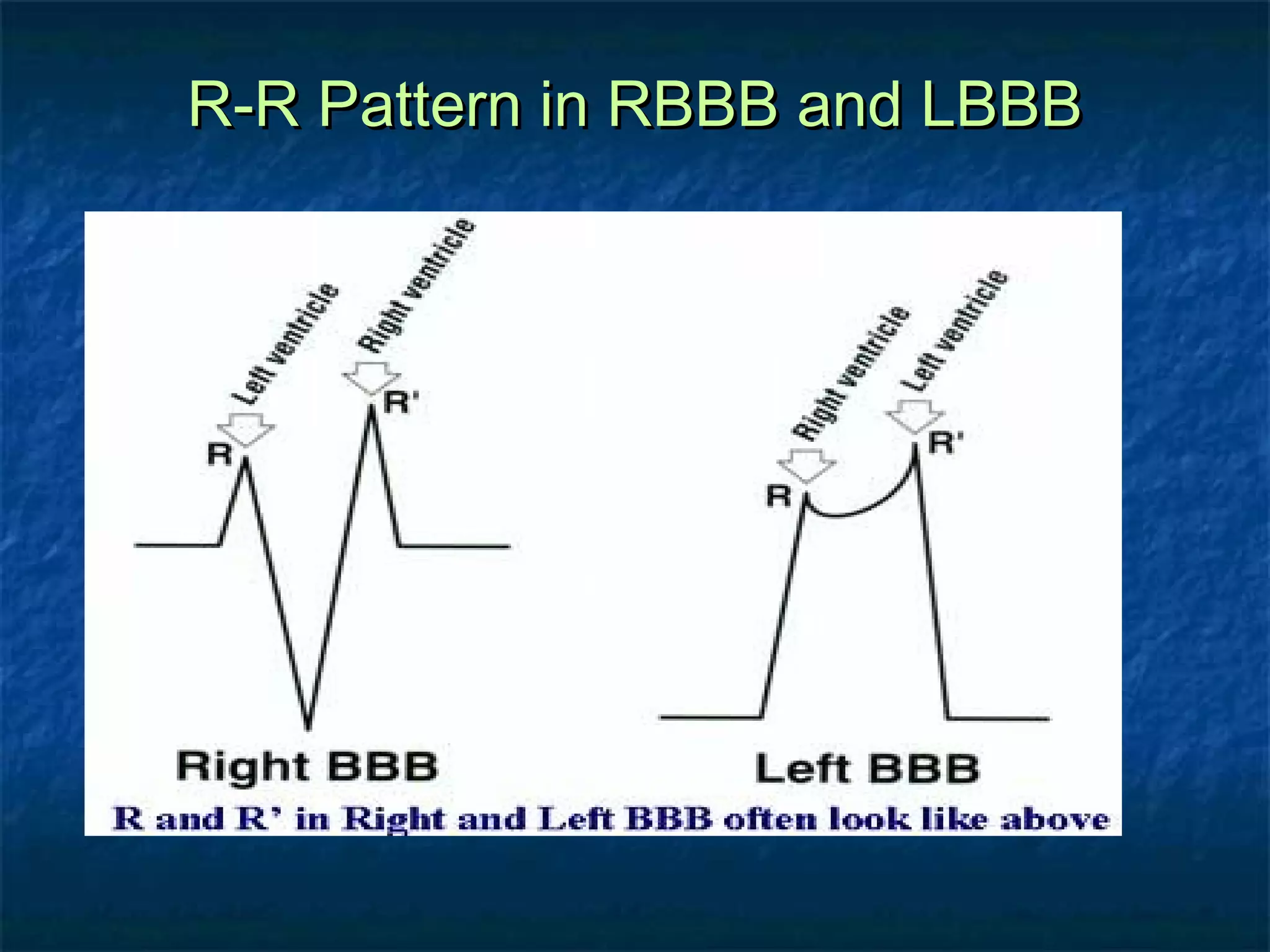
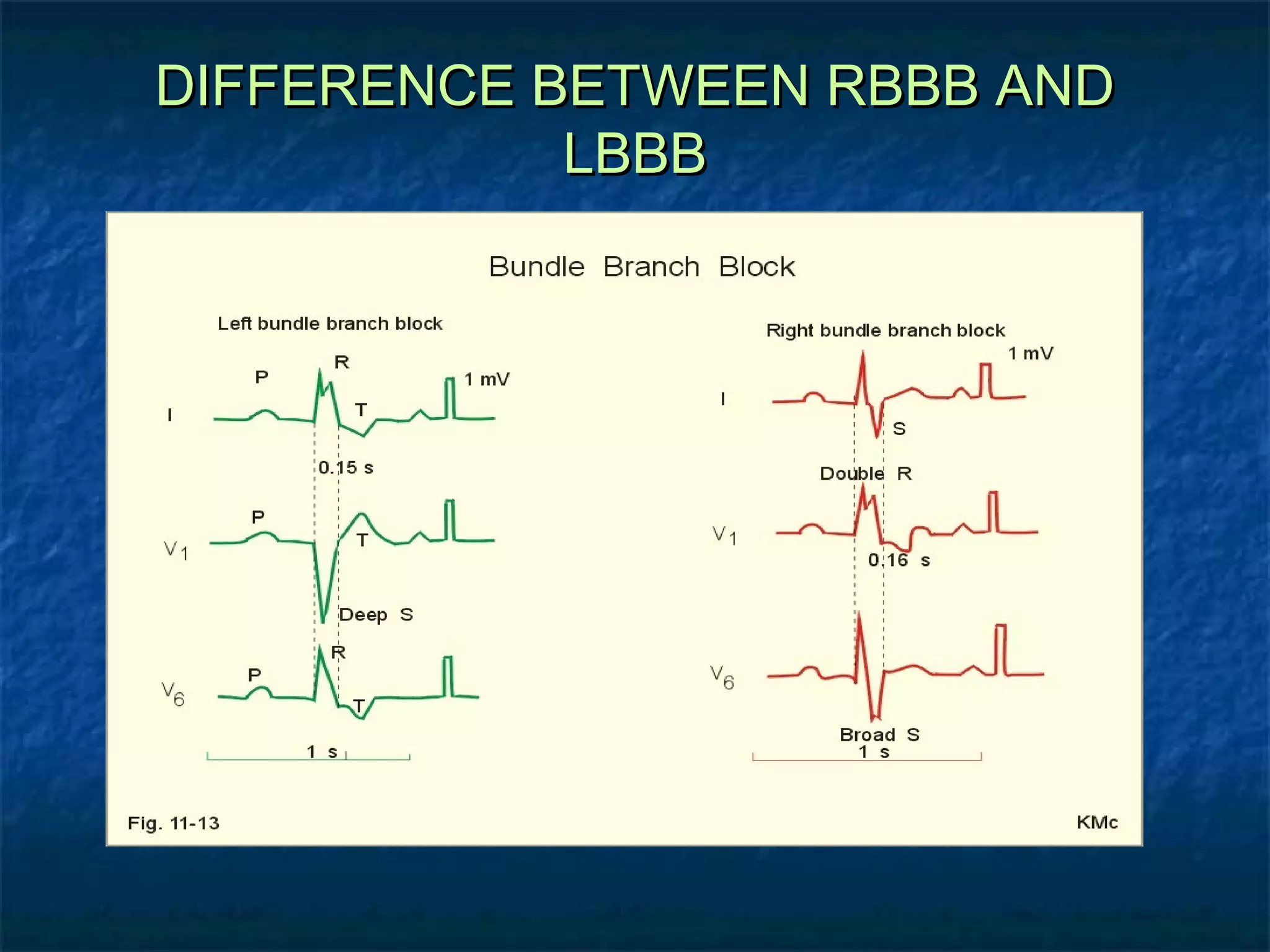
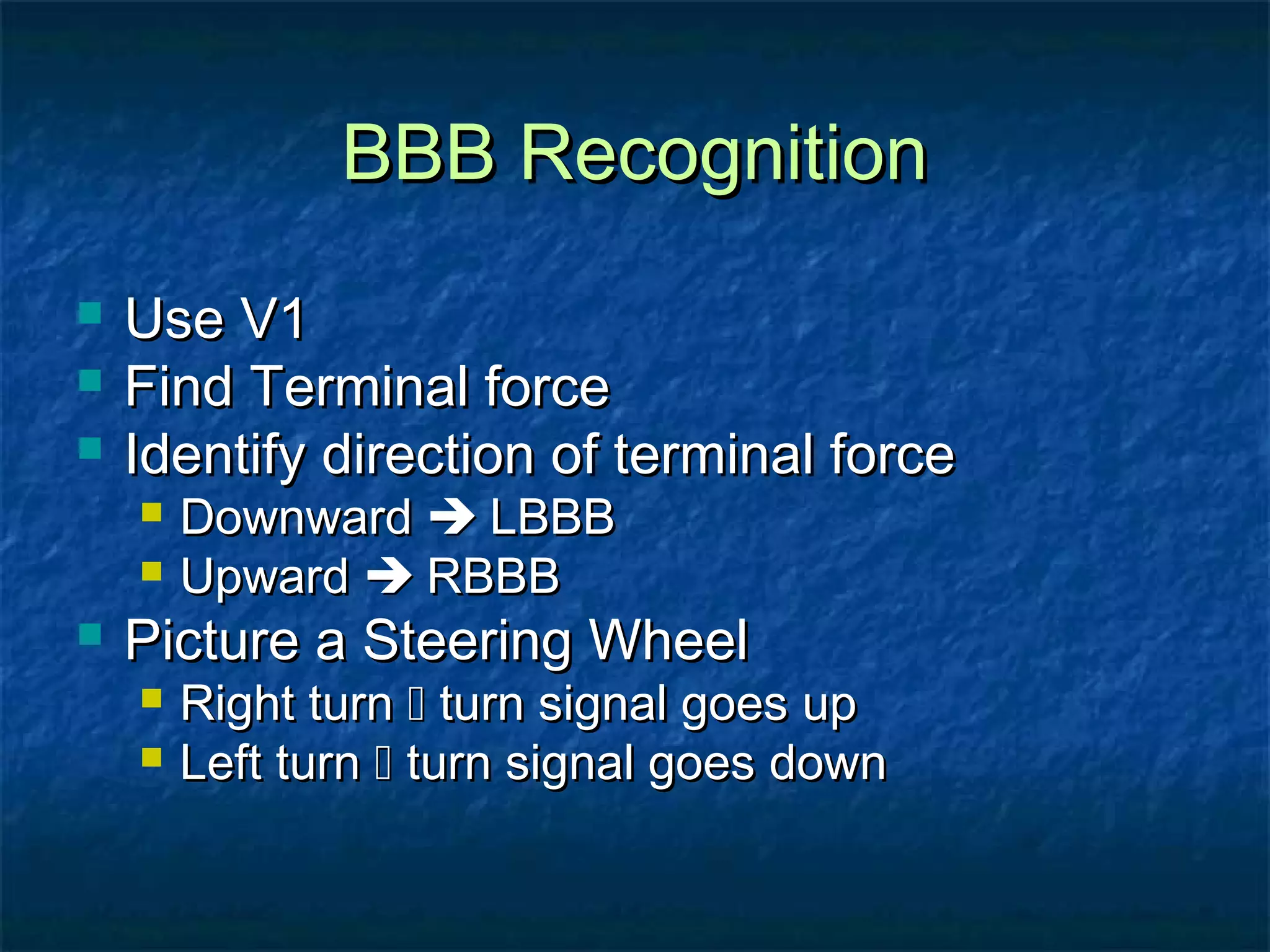
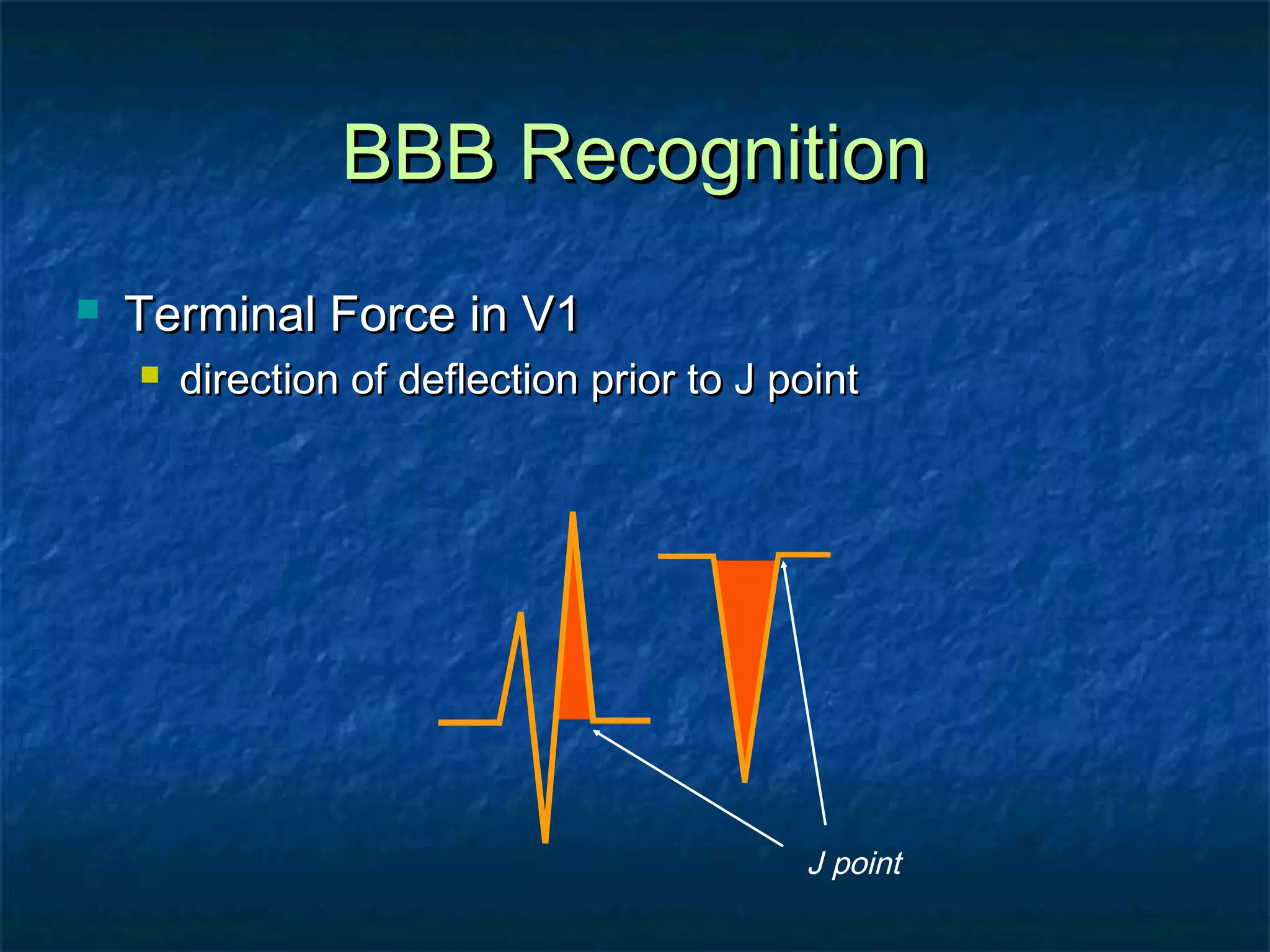
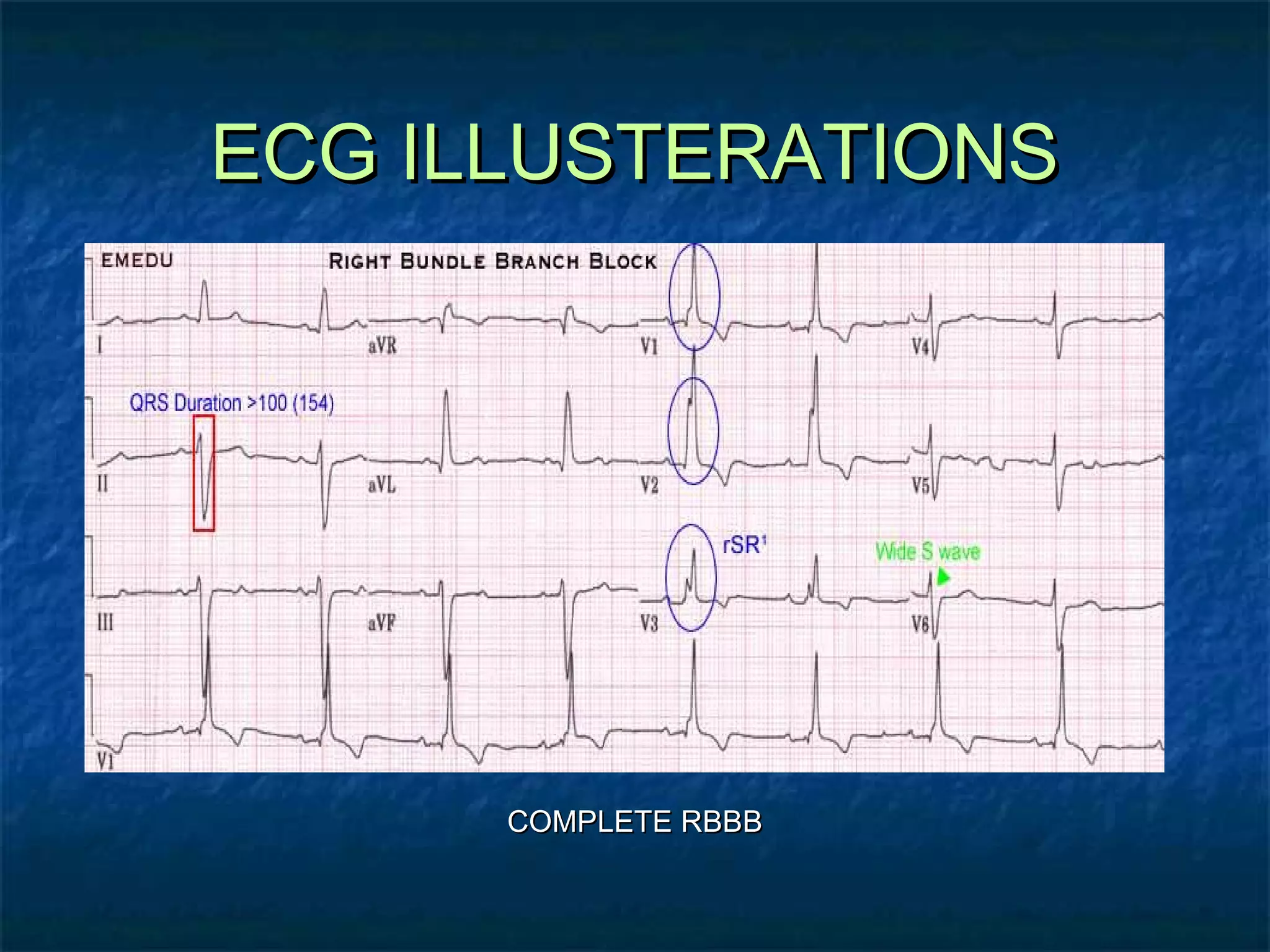
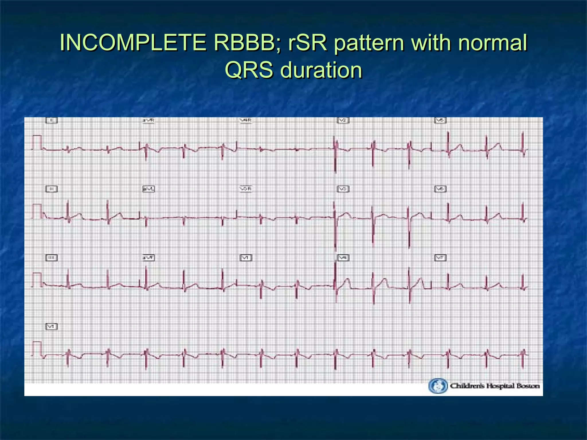
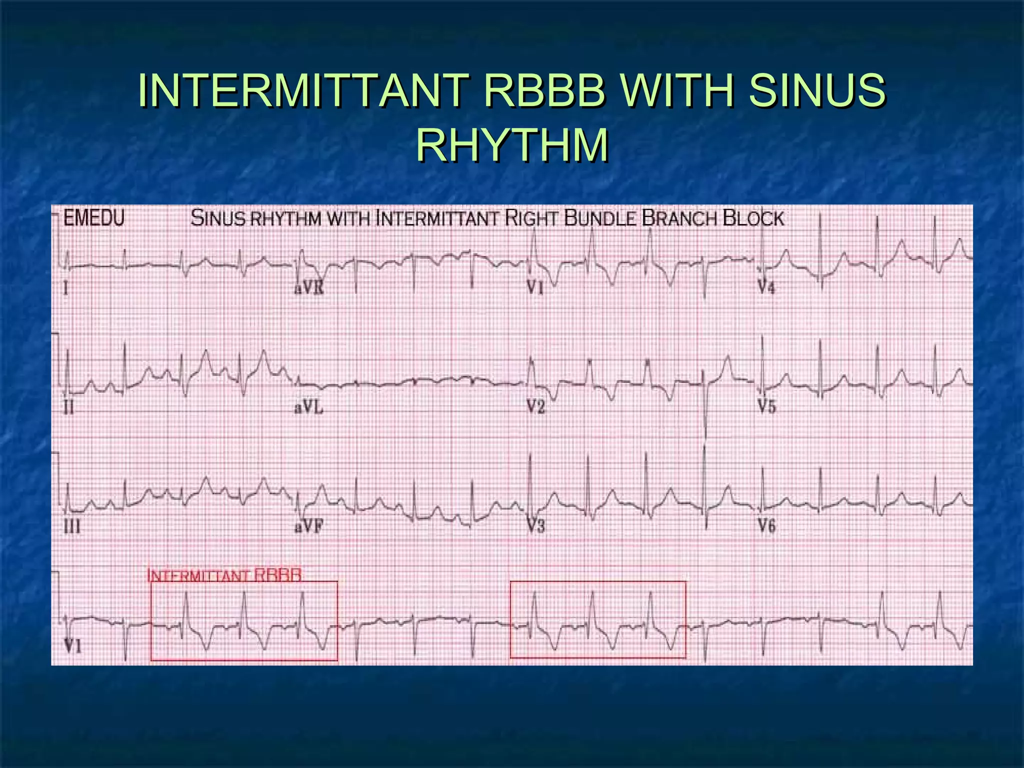
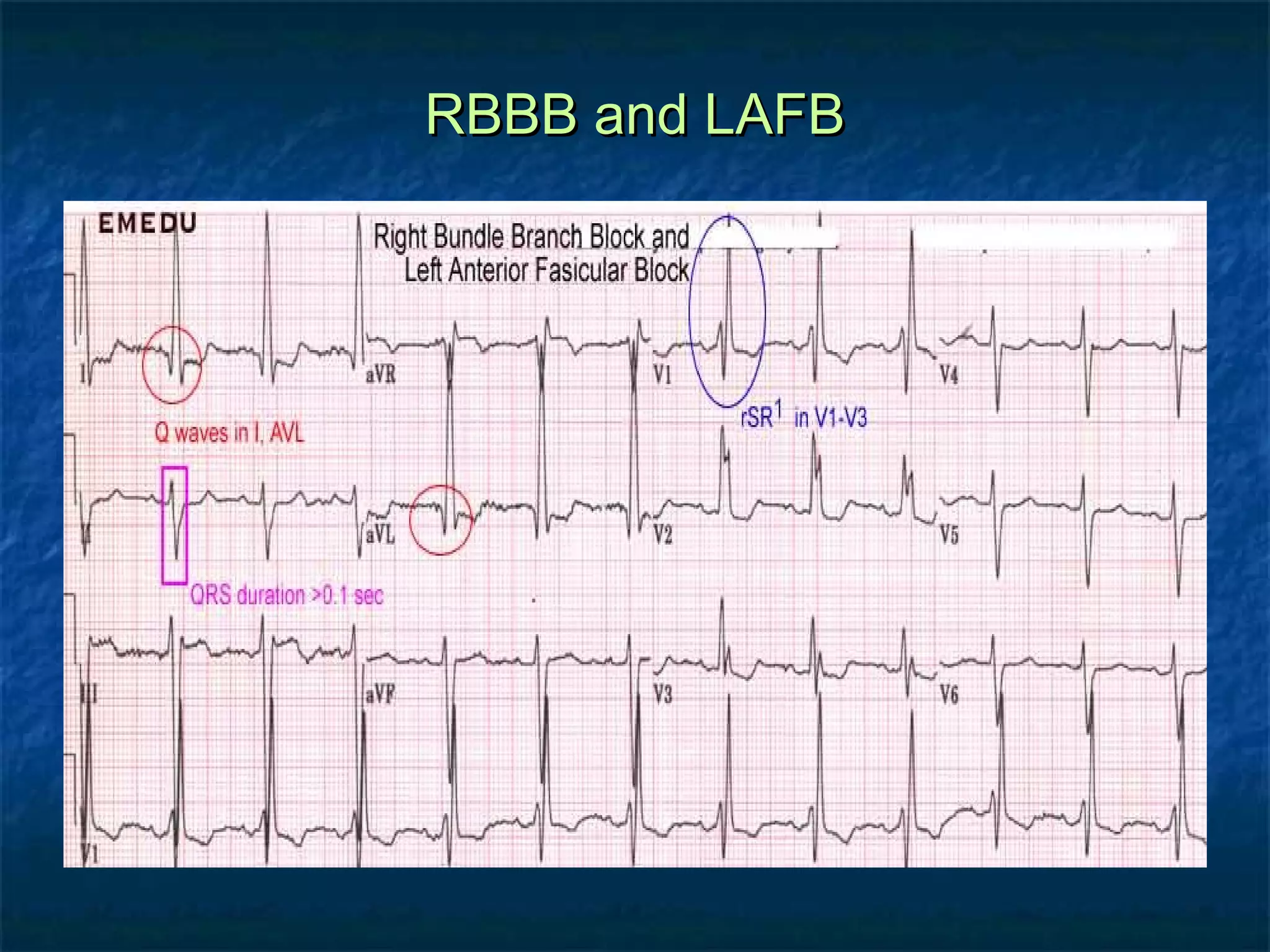
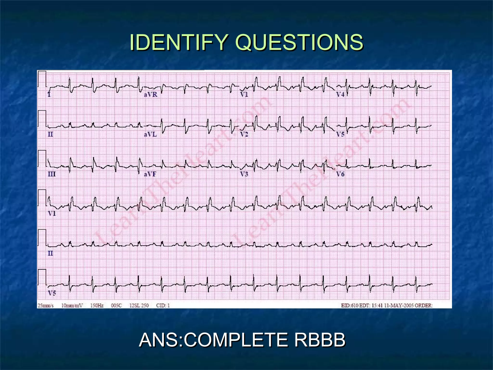
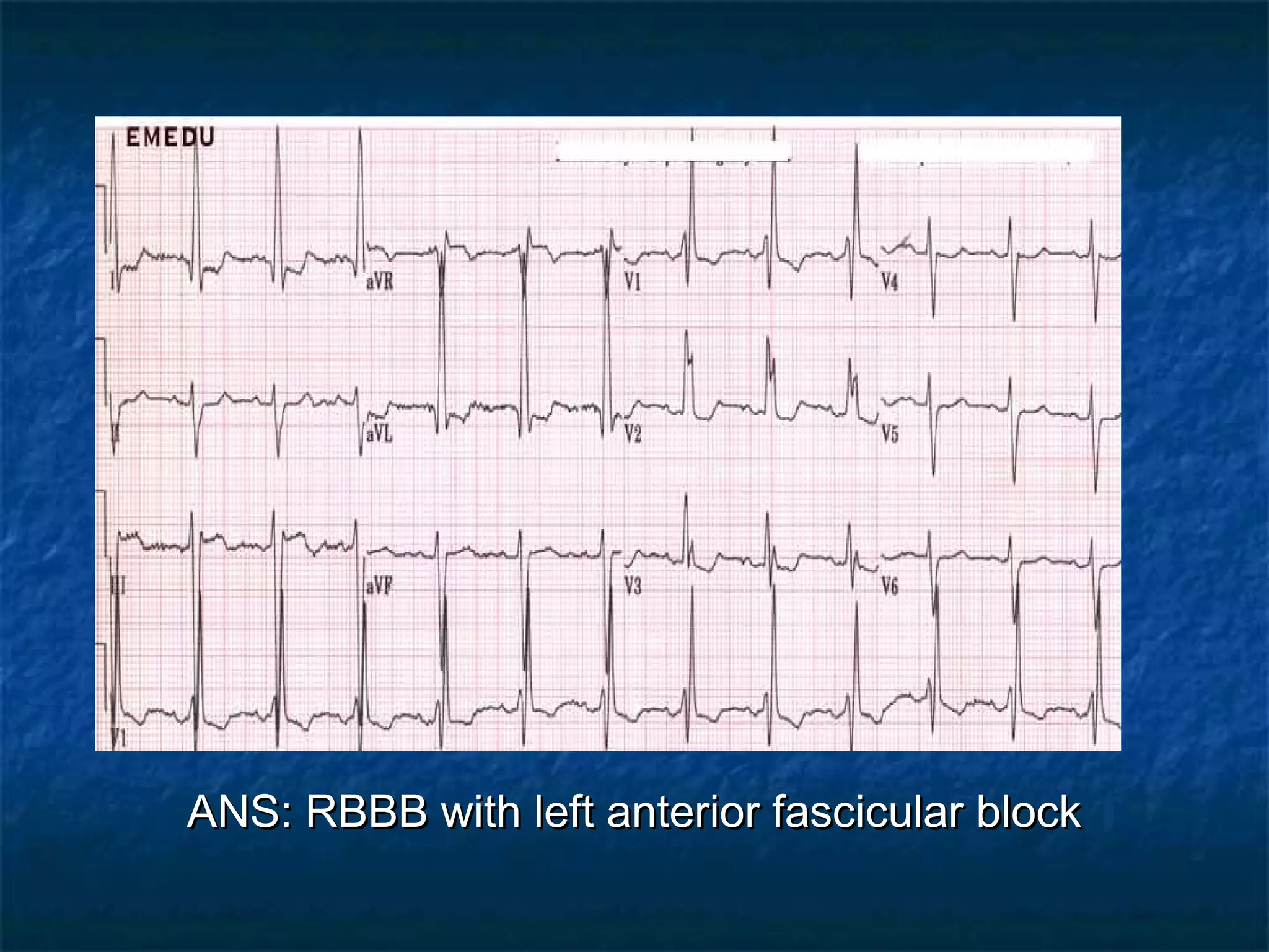
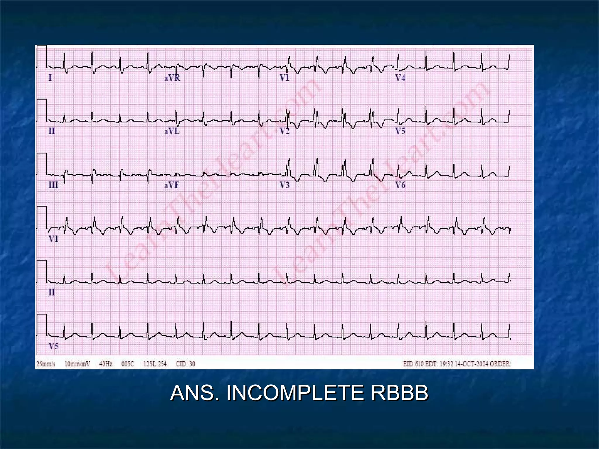
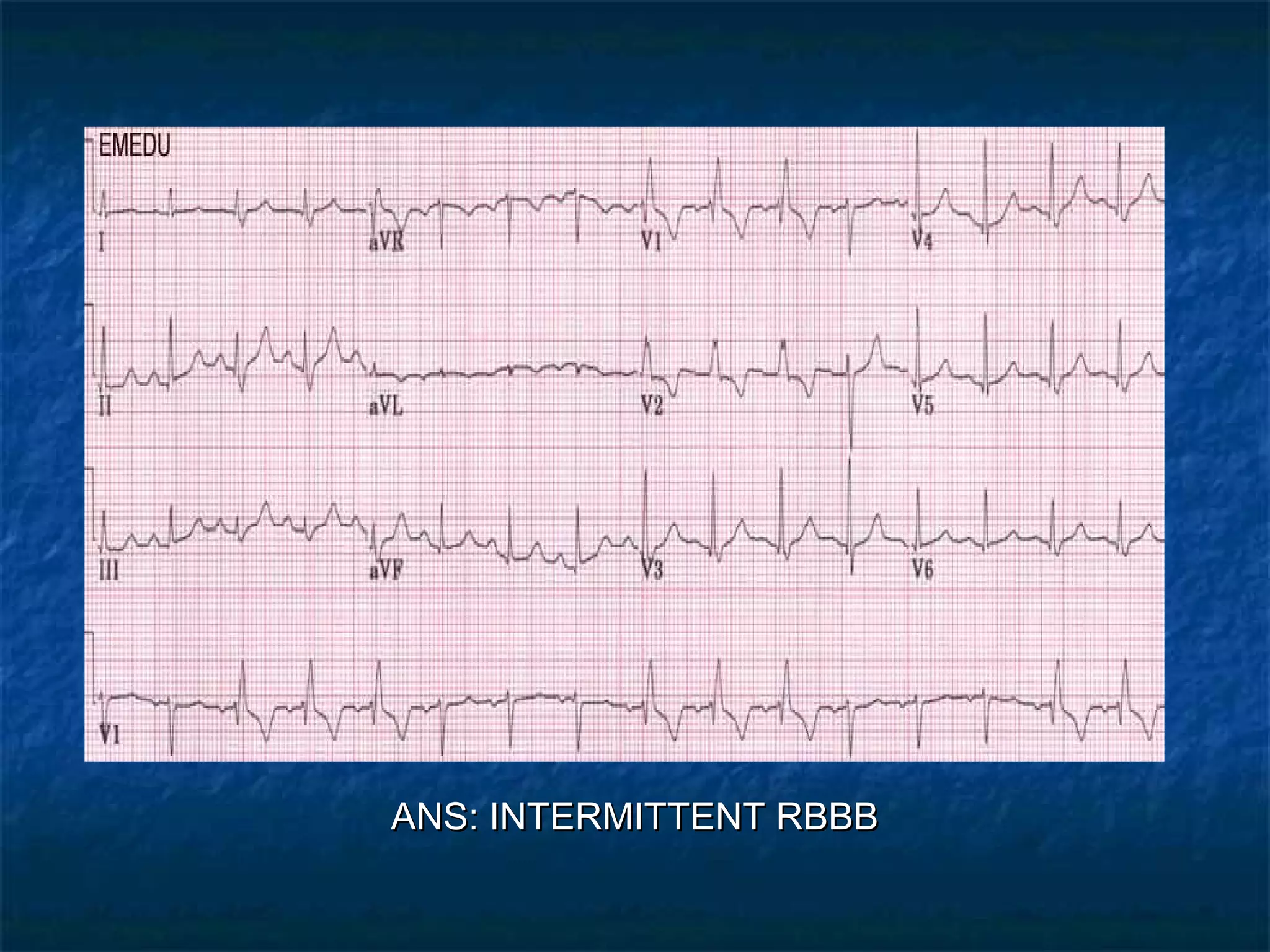
RIGHT BUNDLE BRANCH
BLOCK
Different
presentations of
RBBB in ECG
illustrations

DR. SHAZIA
Normal Ventricular Conduction


Normal Conduction



fibers of LBB begin conduction
impulse travels across
interventricular septum from left to
right




travels across ventricles causing
depolarization of both
simultaneously




towards + electrode creates small r
wave

LV contributes most to complex

impulse travels away from +
electrode creates primarily
negative complex
RBBB


RBBB in V1


no change in initial impulse
travel






impulse depolarizes LV by
itself since RBBB
RV depolarized by impulse
thru muscle




small r wave

it now contributes to
complex

travels toward + electrode
creating positive deflection
Right Bundle Branch Block
Criteria




QRS duration ≥ 110ms
rSR’ pattern or notched R wave in V 1
Wide and slurred S wave in I and V 6
R-R Pattern in RBBB and LBBB
DIFFERENCE BETWEEN RBBB AND
LBBB
BBB Recognition




Use V1
Find Terminal force
Identify direction of terminal force





Downward  LBBB
Upward  RBBB

Picture a Steering Wheel



Right turn  turn signal goes up
Left turn  turn signal goes down
BBB Recognition


Terminal Force in V1


direction of deflection prior to J point

J point
ECG ILLUSTERATIONS

COMPLETE RBBB
INCOMPLETE RBBB; rSR pattern with normal
QRS duration
INTERMITTANT RBBB WITH SINUS
RHYTHM
RBBB and LAFB
IDENTIFY QUESTIONS

ANS:COMPLETE RBBB
ANS: RBBB with left anterior fascicular block
ANS. INCOMPLETE RBBB
ANS: INTERMITTENT RBBB

Right bundle branch block