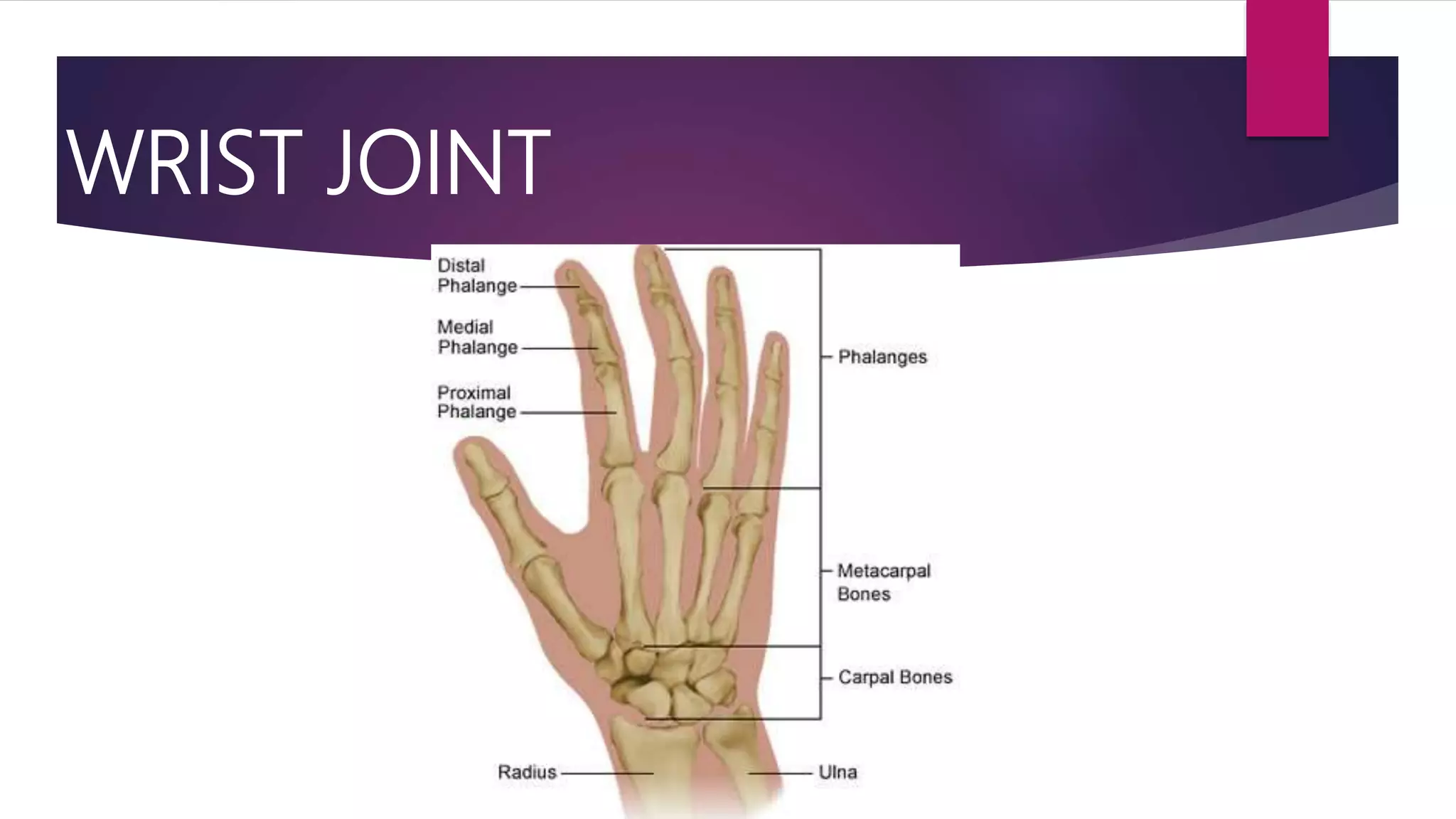
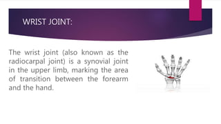
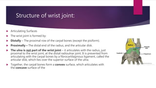
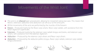
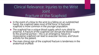
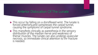
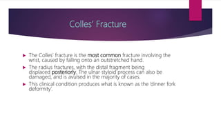
WRIST JOINT
WRIST JOINT:
The wrist joint (also known as the
radiocarpal joint) is a synovial joint
in the upper limb, marking the area
of transition between the forearm
and the hand.
Structure of wrist joint:
 Articulating Surfaces
 The wrist joint is formed by:
 Distally – The proximal row of the carpal bones (except the pisiform).
 Proximally – The distal end of the radius, and the articular disk.
 The ulna is not part of the wrist joint – it articulates with the radius, just
proximal to the wrist joint, at the distal radioulnar joint. It is prevented from
articulating with the carpal bones by a fibrocartilaginous ligament, called the
articular disk, which lies over the superior surface of the ulna.
 Together, the carpal bones form a convex surface, which articulates with
the concave surface of the 6 TeachMeAnatomy.com [CC-BY-NC-
BLOOD SUPPLY:
The wrist joint receives blood from branches of the dorsal and palmar carpal
arches, which are derived from the ulnar and radial arteries (for more
information, see Blood Supply to the Upper Limb)
• Median nerve – Anterior
interosseous branch.
• Radial nerve – Posterior
interosseous branch.
• Ulnar nerve – deep and dorsal
branches.
• Stability of the Wrist Joint
 The joint capsule and ligaments contribute to the stability of the wrist.
 Joint capsule: Like any synovial joint, the capsule is dual layered. The fibrous outer layer
attaches to the radius, ulna and the proximal row of the carpal bones. The internal layer is
comprised of a synovial membrane, secreting synovial fluid which lubricates the joint.
 Ligaments: There are four ligaments of note in the wrist joint, one for each side of the joint
 Palmar radiocarpal – It is found on the palmar (anterior) side of the hand. It passes from
the radius to both rows of carpal bones. Its function, apart from increasing stability, is to
ensure that the hand follows the forearm during supination.
 Dorsal radiocarpal – It is found on the dorsum (posterior) side of the hand. It passes from
the radius to both rows of carpal bones. It contributes to the stability of the wrist, but also
ensures that the hand follows the forearm during pronation.
 Ulnar collateral – Runs from the ulnar styloid process to the triquetrum and pisiform.
Works in union with the other collateral ligament to prevent excessive lateral joint
displacement.
 Radial collateral – Runs from the radial styloid process to the scaphoid and trapezium.
Works in union with the other collateral ligament to prevent excessive lateral joint
displacement.
Movements of the Wrist Joint
 The wrist is an ellipsoid type synovial joint, allowing for movement along two axes. This means that
flexion, extension, adduction and abduction can all occur at the wrist joint.
 All the movements of the wrist are performed by the muscles of the forearm.
 Flexion – Produced mainly by the flexor carpi ulnaris, flexor carpi radialis, with assistance from the
flexor digitorum superficialis.
 Extension – Produced mainly by the extensor carpi radialis longus and brevis, and extensor carpi
ulnaris, with assistance from the extensor digitorum.
 Adduction – Produced by the extensor carpi ulnaris and flexor carpi ulnaris
 Abduction – Produced by the abductor pollicis longus, flexor carpi radialis, extensor carpi radialis
longus and brevis.
Clinical Relevance: Injuries to the Wrist
Joint
Fracture of the Scaphoid
 In the event of a blow to the wrist (e.g falling on an outstretched
hand), the scaphoid takes most of the force. A fractured
scaphoid is more common in the younger population.
 The scaphoid has a unique blood supply, which runs distal to
proximal. A fracture of the scaphoid can disrupt the blood supply
to the proximal portion – this is an emergency. Failure to
revascularise scaphoid can lead to avascular necrosis, and future
arthritis for the patient.
 The main clinical sign of the scaphoid fracture is tenderness in the
anatomical snuffboxt.hecom thyo.ger population.
Anterior Dislocation Of The Lunate
 This occur by falling on a dorsiflexed wrist. The lunate is
forced anteriorly,and cpmpresses the carpal tunnel,
causing the symptoms of carpal tunnel syndrome.
 This manyfests clinically as paresthesia in the sensory
distribution of the median nerve and weakness of
thenar muscles. The lunate can also undergo avascular
necrosis, so immediate clinical attention to thr fracture
is needed.
Colles’ Fracture
 The Colles’ fracture is the most common fracture involving the
wrist, caused by falling onto an outstretched hand.
 The radius fractures, with the distal fragment being
displaced posteriorly. The ulnar styloid process can also be
damaged, and is avulsed in the majority of cases.
 This clinical condition produces what is known as the ‘dinner fork
deformity’.
distal fragment being displaced posteriorly. The ulnar styloid process can also be damaged, and is avulsed
in the majority of cases.
This clinical condition produces what is known as the ‘dinner fork deformity’

Wrist Joint

  • 1.
  • 2.
    WRIST JOINT: The wristjoint (also known as the radiocarpal joint) is a synovial joint in the upper limb, marking the area of transition between the forearm and the hand.
  • 3.
    Structure of wristjoint:  Articulating Surfaces  The wrist joint is formed by:  Distally – The proximal row of the carpal bones (except the pisiform).  Proximally – The distal end of the radius, and the articular disk.  The ulna is not part of the wrist joint – it articulates with the radius, just proximal to the wrist joint, at the distal radioulnar joint. It is prevented from articulating with the carpal bones by a fibrocartilaginous ligament, called the articular disk, which lies over the superior surface of the ulna.  Together, the carpal bones form a convex surface, which articulates with the concave surface of the 6 TeachMeAnatomy.com [CC-BY-NC-
  • 4.
    BLOOD SUPPLY: The wristjoint receives blood from branches of the dorsal and palmar carpal arches, which are derived from the ulnar and radial arteries (for more information, see Blood Supply to the Upper Limb) • Median nerve – Anterior interosseous branch. • Radial nerve – Posterior interosseous branch. • Ulnar nerve – deep and dorsal branches.
  • 5.
    • Stability ofthe Wrist Joint  The joint capsule and ligaments contribute to the stability of the wrist.  Joint capsule: Like any synovial joint, the capsule is dual layered. The fibrous outer layer attaches to the radius, ulna and the proximal row of the carpal bones. The internal layer is comprised of a synovial membrane, secreting synovial fluid which lubricates the joint.  Ligaments: There are four ligaments of note in the wrist joint, one for each side of the joint  Palmar radiocarpal – It is found on the palmar (anterior) side of the hand. It passes from the radius to both rows of carpal bones. Its function, apart from increasing stability, is to ensure that the hand follows the forearm during supination.  Dorsal radiocarpal – It is found on the dorsum (posterior) side of the hand. It passes from the radius to both rows of carpal bones. It contributes to the stability of the wrist, but also ensures that the hand follows the forearm during pronation.  Ulnar collateral – Runs from the ulnar styloid process to the triquetrum and pisiform. Works in union with the other collateral ligament to prevent excessive lateral joint displacement.  Radial collateral – Runs from the radial styloid process to the scaphoid and trapezium. Works in union with the other collateral ligament to prevent excessive lateral joint displacement.
  • 6.
    Movements of theWrist Joint  The wrist is an ellipsoid type synovial joint, allowing for movement along two axes. This means that flexion, extension, adduction and abduction can all occur at the wrist joint.  All the movements of the wrist are performed by the muscles of the forearm.  Flexion – Produced mainly by the flexor carpi ulnaris, flexor carpi radialis, with assistance from the flexor digitorum superficialis.  Extension – Produced mainly by the extensor carpi radialis longus and brevis, and extensor carpi ulnaris, with assistance from the extensor digitorum.  Adduction – Produced by the extensor carpi ulnaris and flexor carpi ulnaris  Abduction – Produced by the abductor pollicis longus, flexor carpi radialis, extensor carpi radialis longus and brevis.
  • 7.
    Clinical Relevance: Injuriesto the Wrist Joint Fracture of the Scaphoid  In the event of a blow to the wrist (e.g falling on an outstretched hand), the scaphoid takes most of the force. A fractured scaphoid is more common in the younger population.  The scaphoid has a unique blood supply, which runs distal to proximal. A fracture of the scaphoid can disrupt the blood supply to the proximal portion – this is an emergency. Failure to revascularise scaphoid can lead to avascular necrosis, and future arthritis for the patient.  The main clinical sign of the scaphoid fracture is tenderness in the anatomical snuffboxt.hecom thyo.ger population.
  • 8.
    Anterior Dislocation OfThe Lunate  This occur by falling on a dorsiflexed wrist. The lunate is forced anteriorly,and cpmpresses the carpal tunnel, causing the symptoms of carpal tunnel syndrome.  This manyfests clinically as paresthesia in the sensory distribution of the median nerve and weakness of thenar muscles. The lunate can also undergo avascular necrosis, so immediate clinical attention to thr fracture is needed.
  • 9.
    Colles’ Fracture  TheColles’ fracture is the most common fracture involving the wrist, caused by falling onto an outstretched hand.  The radius fractures, with the distal fragment being displaced posteriorly. The ulnar styloid process can also be damaged, and is avulsed in the majority of cases.  This clinical condition produces what is known as the ‘dinner fork deformity’. distal fragment being displaced posteriorly. The ulnar styloid process can also be damaged, and is avulsed in the majority of cases. This clinical condition produces what is known as the ‘dinner fork deformity’