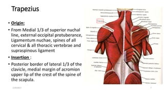
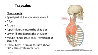
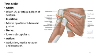
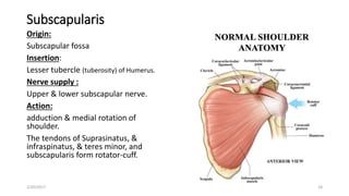
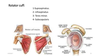
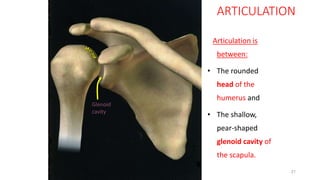
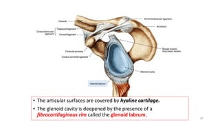
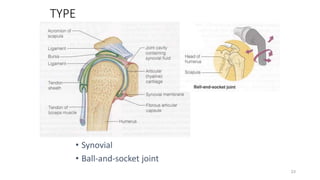
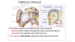
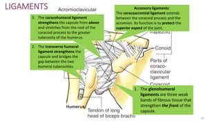
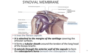
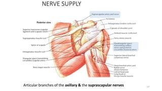
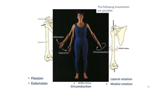
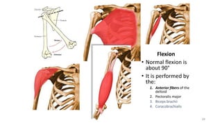
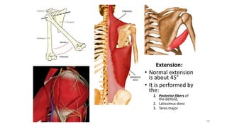
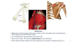
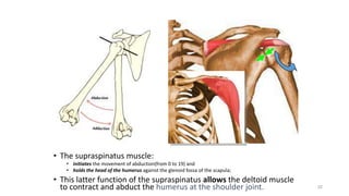
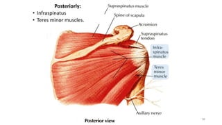
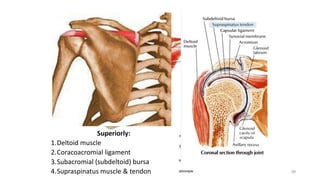
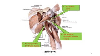
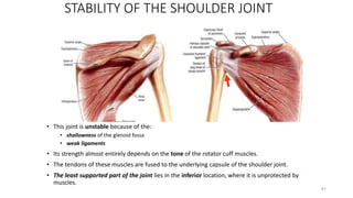
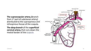
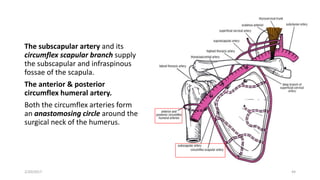
SHOULDER REGION
EssamEldin AbdelHady Salama
2/20/2017 1
Objectives
• Identification of the muscles attached to the scapula.
• Shoulder joint
2/20/2017 2
Scapular muscles
The Muscles Connecting the Upper Extremity to the Vertebral Column
The Muscles Connecting the Upper Extremity to the Anterior and Lateral
Thoracic Walls
The Muscles and Fasciæ of the Shoulder
2/20/2017 3
The Muscles Connecting the Upper Extremity to the Vertebral
Column
• Trapezius.
• Rhomboideus major.
• Latissimus dorsi.
• Rhomboideus minor.
• Levator scapulæ.
2/20/2017 4
The Muscles Connecting the Upper Extremity to
the Anterior and Lateral Thoracic Walls
• The muscles of the anterior and lateral thoracic regions are:
• Pectoralis major.
• Subclavius.
• Pectoralis minor.
• Serratus anterior.
2/20/2017 5
The Muscles and Fasciæ of the Shoulder
• Deltoid.
• Infraspinatus.
• Subscapularis.
• Teres minor.
• Supraspinatus.
• Teres major.
2/20/2017 6
Trapezius
• Origin:
• From Medial 1/3 of superior nuchal
line, external occipital protuberance,
Ligamentum nuchae, spines of all
cervical & all thoracic vertebrae and
supraspinous ligament
• Insertion :
• Posterior border of lateral 1/3 of the
clavicle, medial margin of acromion
upper lip of the crest of the spine of
the scapula.
2/20/2017 7
• Nerve supply:
• Spinal part of the accessory nerve &
• C 3,4
• Actions:
• Upper fibers: elevate the shoulder
• Lower fibers: depress the shoulder.
• Middle fibers: brace back (retraction) of
shoulder
• It also, helps in raising the arm above
90° with (serratus anterior).
Trapezius
2/20/2017 8
Levator scapulae
• Origin:
• Transverse processes of
upper 4 cervical vertebrae.
• Insertion:
• Medial border of scapula.
• Nerve supply:
• C3,4 & 5
• Action:
• Elevate the medial border of
the scapula.
2/20/2017 9
Rhomboid minor
• Origin:
• Ligamentum nuchae & spines
of C 7& T1
• Insertion:
• Medial border of the scapula.
• Nerve supply:
• C4& 5
• Action:
• Raises the medial border of
scapula upward & medially.
2/20/2017 10
Rhomboid Major
• Origin:
• Spines of T2,T3,T4 & T5.
• Insertion:
• Medial border of scapula.
• Nerve supply:
• C4 & 5.
• Action:
• Raises the medial border of
the scapula upward and
medially.
2/20/2017 11
Deltoid
Origin:
Anterior border of the lateral third of the clavicle
Lateral margin of acromion
Lower lip of the crest of the spine of the scapula
Insertion:
deltoid tuberosity of the humerus
Nerve supply:
Axillary nerve
Action:
Anterior fibers, help in flexion and medial rotation of the
arm
Abduction of the arm by the acromial fibers
Posterior fibers help extension of the arm
2/20/2017 12
Supraspinatus
Origin:
supraspinous fossa.
Insertion:
top of greater tubercle.
Nerve supply:
Suprascapular N.
Action:
Starts abduction from
0 to 15.
Steadies head of humerus.2/20/2017 13
Infraspinatus
Origin:
infraspinous fossa
Insertion:
middle impression of greater
tubercle.
Nerve supply:
Suprascapular nerve.
Action:
adduction & Steadies head of
humerus
2/20/2017 14
2/20/2017 15
Teres minor
• Origin:
Upper 2/3 of lateral border of
scapula
• Insertion:
lower impression of the
greater tubercle.
• Nerve supply
Axillary nerve
• Action:
adduction & Lateral rotation.
2/20/2017 16
Teres Major
• Origin:
• lower 1/3 of lateral border of
scapula.
• Insertion:
• Medial lip of intertubercular
groove
• Nerve:
• lower subscapular n.
• Action:
• Adduction, medial rotation
and extension.
2/20/2017 17
Subscapularis
Origin:
Subscapular fossa
Insertion:
Lesser tubercle (tuberosity) of Humerus.
Nerve supply :
Upper & lower subscapular nerve.
Action:
adduction & medial rotation of
shoulder.
The tendons of Suprasinatus, &
infraspinatus, & teres minor, and
subscapularis form rotator-cuff.
2/20/2017 18
Rotator cuff:
1-Supraspinatus.
2- infraspinatus
3- Teres minor.
4- Subscapularis
2/20/2017 19
SHOULDER JOINT
2/20/2017 20
21
ARTICULATION
Articulation is
between:
• The rounded
head of the
humerus and
• The shallow,
pear-shaped
glenoid cavity of
the scapula.
Glenoid
cavity
22
• The articular surfaces are covered by hyaline cartilage.
• The glenoid cavity is deepened by the presence of a
fibrocartilaginous rim called the glenoid labrum.
23
TYPE
• Synovial
• Ball-and-socket joint
24
FIBROUS CAPSULE
• The fibrous capsule surrounds the joint and is attached:
Medially to the margin of the glenoid cavity outside the labrum;
Laterally to the anatomic neck of the humerus.
• The capsule is thin and lax, allowing a wide range of movement.
25
LIGAMENTS
1. The glenohumeral
ligaments are three weak
bands of fibrous tissue that
strengthen the front of the
capsule.
2. The transverse humeral
ligament strengthens the
capsule and bridges the
gap between the two
humeral tuberosities.
3. The coracohumeral ligament
strengthens the capsule from above
and stretches from the root of the
coracoid process to the greater
tuberosity of the humerus.
Accessory ligaments:
The coracoacromial ligament extends
between the coracoid process and the
acromion. Its function is to protect the
superior aspect of the joint.
26
SYNOVIAL MEMBRANE
• It lines the fibrous capsule.
• It is attached to the margins of the cartilage covering the
articular surfaces.
• It forms a tubular sheath around the tendon of the long head
of the biceps brachii.
• It extends through the anterior wall of the capsule to form
the subscapularis bursa beneath the subscapularis muscle.
27
NERVE SUPPLY
Articular branches of the axillary & the suprascapular nerves
28
• Flexion
• Extension
• Abduction
• Adduction
• Lateral rotation
• Medial rotation
Circumduction
The following movements
are possible:
29
Flexion
• Normal flexion is
about 90°
• It is performed by
the:
1. Anterior fibers of the
deltoid
2. Pectoralis major
3. Biceps brachii
4. Coracobrachialis
30
Extension:
• Normal extension
is about 45°
• It is performed by
the:
1. Posterior fibers of
the deltoid,
2. Latissimus dorsi
3. Teres major
31
Abduction:
• Abduction of the upper limb occurs both at the shoulder joint and between
the scapula and the thoracic wall.
• It is initiated by supraspinatus from 0 to 18
• Then from 19 to 90 by the middle fibers of the deltoid.
• Then above 90 by rotation of the scapula by 2 muscles ( Trapezius & S.A..)
32
• The supraspinatus muscle:
• initiates the movement of abduction(from 0 to 19) and
• holds the head of the humerus against the glenoid fossa of the scapula;
• This latter function of the supraspinatus allows the deltoid muscle
to contract and abduct the humerus at the shoulder joint.
33
Adduction:
• Normally the upper
limb can be swung
45° across the front
of the chest.
• This is performed by:
1. pectoralis major
2. latissimus dorsi
3. teres major
4. teres minor
34
Lateral rotation:
• Normal lateral
rotation is about 40
to 45°.
• This is performed by
the:
1. infraspinatus
2. teres minor
3. the posterior fibers of the
deltoid muscle
35
Medial rotation:
• Normal medial rotation
is about 55°.
• This is performed by
the:
1. subscapularis
2. latissimus dorsi
3. teres major
4. anterior fibers of the deltoid.
36
Circumduction:
This is a movement in
which the distal end of
the humerus moves in
circular motion while
the proximal end
remains stable
• It is formed by
flexion,
abduction,
extension and
adduction.
Successively
Relation
2/20/2017 37
38
Posteriorly:
• Infraspinatus
• Teres minor muscles.
39
Superiorly:
1.Deltoid muscle
2.Coracoacromial ligament
3.Subacromial (subdeltoid) bursa
4.Supraspinatus muscle & tendon
40
Inferiorly:
1. the long head of
the triceps muscle
2. the axillary
nerve
3. the posterior
circumflex humeral
vessels
41
STABILITY OF THE SHOULDER JOINT
• This joint is unstable because of the:
• shallowness of the glenoid fossa
• weak ligaments
• Its strength almost entirely depends on the tone of the rotator cuff muscles.
• The tendons of these muscles are fused to the underlying capsule of the shoulder joint.
• The least supported part of the joint lies in the inferior location, where it is unprotected by
muscles.
ANASTOMOSES
AROUND THE
SCAPULAR REGIONS
2/20/2017 42
The suprascapular artery, (branch
from 1st part of subclavian artery)
distributed to the supraspinous and
infraspinous fossae of the scapula.
The deep branch of the superficial
cervical artery, that runs down the
medial border of the scapula.
2/20/2017 43
The subscapular artery and its
circumflex scapular branch supply
the subscapular and infraspinous
fossae of the scapula.
The anterior & posterior
circumflex humeral artery.
Both the circumflex arteries form
an anastomosing circle around the
surgical neck of the humerus.
2/20/2017 44

Shoulder region

  • 1.
  • 2.
    Objectives • Identification ofthe muscles attached to the scapula. • Shoulder joint 2/20/2017 2
  • 3.
    Scapular muscles The MusclesConnecting the Upper Extremity to the Vertebral Column The Muscles Connecting the Upper Extremity to the Anterior and Lateral Thoracic Walls The Muscles and Fasciæ of the Shoulder 2/20/2017 3
  • 4.
    The Muscles Connectingthe Upper Extremity to the Vertebral Column • Trapezius. • Rhomboideus major. • Latissimus dorsi. • Rhomboideus minor. • Levator scapulæ. 2/20/2017 4
  • 5.
    The Muscles Connectingthe Upper Extremity to the Anterior and Lateral Thoracic Walls • The muscles of the anterior and lateral thoracic regions are: • Pectoralis major. • Subclavius. • Pectoralis minor. • Serratus anterior. 2/20/2017 5
  • 6.
    The Muscles andFasciæ of the Shoulder • Deltoid. • Infraspinatus. • Subscapularis. • Teres minor. • Supraspinatus. • Teres major. 2/20/2017 6
  • 7.
    Trapezius • Origin: • FromMedial 1/3 of superior nuchal line, external occipital protuberance, Ligamentum nuchae, spines of all cervical & all thoracic vertebrae and supraspinous ligament • Insertion : • Posterior border of lateral 1/3 of the clavicle, medial margin of acromion upper lip of the crest of the spine of the scapula. 2/20/2017 7
  • 8.
    • Nerve supply: •Spinal part of the accessory nerve & • C 3,4 • Actions: • Upper fibers: elevate the shoulder • Lower fibers: depress the shoulder. • Middle fibers: brace back (retraction) of shoulder • It also, helps in raising the arm above 90° with (serratus anterior). Trapezius 2/20/2017 8
  • 9.
    Levator scapulae • Origin: •Transverse processes of upper 4 cervical vertebrae. • Insertion: • Medial border of scapula. • Nerve supply: • C3,4 & 5 • Action: • Elevate the medial border of the scapula. 2/20/2017 9
  • 10.
    Rhomboid minor • Origin: •Ligamentum nuchae & spines of C 7& T1 • Insertion: • Medial border of the scapula. • Nerve supply: • C4& 5 • Action: • Raises the medial border of scapula upward & medially. 2/20/2017 10
  • 11.
    Rhomboid Major • Origin: •Spines of T2,T3,T4 & T5. • Insertion: • Medial border of scapula. • Nerve supply: • C4 & 5. • Action: • Raises the medial border of the scapula upward and medially. 2/20/2017 11
  • 12.
    Deltoid Origin: Anterior border ofthe lateral third of the clavicle Lateral margin of acromion Lower lip of the crest of the spine of the scapula Insertion: deltoid tuberosity of the humerus Nerve supply: Axillary nerve Action: Anterior fibers, help in flexion and medial rotation of the arm Abduction of the arm by the acromial fibers Posterior fibers help extension of the arm 2/20/2017 12
  • 13.
    Supraspinatus Origin: supraspinous fossa. Insertion: top ofgreater tubercle. Nerve supply: Suprascapular N. Action: Starts abduction from 0 to 15. Steadies head of humerus.2/20/2017 13
  • 14.
    Infraspinatus Origin: infraspinous fossa Insertion: middle impressionof greater tubercle. Nerve supply: Suprascapular nerve. Action: adduction & Steadies head of humerus 2/20/2017 14
  • 15.
  • 16.
    Teres minor • Origin: Upper2/3 of lateral border of scapula • Insertion: lower impression of the greater tubercle. • Nerve supply Axillary nerve • Action: adduction & Lateral rotation. 2/20/2017 16
  • 17.
    Teres Major • Origin: •lower 1/3 of lateral border of scapula. • Insertion: • Medial lip of intertubercular groove • Nerve: • lower subscapular n. • Action: • Adduction, medial rotation and extension. 2/20/2017 17
  • 18.
    Subscapularis Origin: Subscapular fossa Insertion: Lesser tubercle(tuberosity) of Humerus. Nerve supply : Upper & lower subscapular nerve. Action: adduction & medial rotation of shoulder. The tendons of Suprasinatus, & infraspinatus, & teres minor, and subscapularis form rotator-cuff. 2/20/2017 18
  • 19.
    Rotator cuff: 1-Supraspinatus. 2- infraspinatus 3-Teres minor. 4- Subscapularis 2/20/2017 19
  • 20.
  • 21.
    21 ARTICULATION Articulation is between: • Therounded head of the humerus and • The shallow, pear-shaped glenoid cavity of the scapula. Glenoid cavity
  • 22.
    22 • The articularsurfaces are covered by hyaline cartilage. • The glenoid cavity is deepened by the presence of a fibrocartilaginous rim called the glenoid labrum.
  • 23.
  • 24.
    24 FIBROUS CAPSULE • Thefibrous capsule surrounds the joint and is attached: Medially to the margin of the glenoid cavity outside the labrum; Laterally to the anatomic neck of the humerus. • The capsule is thin and lax, allowing a wide range of movement.
  • 25.
    25 LIGAMENTS 1. The glenohumeral ligamentsare three weak bands of fibrous tissue that strengthen the front of the capsule. 2. The transverse humeral ligament strengthens the capsule and bridges the gap between the two humeral tuberosities. 3. The coracohumeral ligament strengthens the capsule from above and stretches from the root of the coracoid process to the greater tuberosity of the humerus. Accessory ligaments: The coracoacromial ligament extends between the coracoid process and the acromion. Its function is to protect the superior aspect of the joint.
  • 26.
    26 SYNOVIAL MEMBRANE • Itlines the fibrous capsule. • It is attached to the margins of the cartilage covering the articular surfaces. • It forms a tubular sheath around the tendon of the long head of the biceps brachii. • It extends through the anterior wall of the capsule to form the subscapularis bursa beneath the subscapularis muscle.
  • 27.
    27 NERVE SUPPLY Articular branchesof the axillary & the suprascapular nerves
  • 28.
    28 • Flexion • Extension •Abduction • Adduction • Lateral rotation • Medial rotation Circumduction The following movements are possible:
  • 29.
    29 Flexion • Normal flexionis about 90° • It is performed by the: 1. Anterior fibers of the deltoid 2. Pectoralis major 3. Biceps brachii 4. Coracobrachialis
  • 30.
    30 Extension: • Normal extension isabout 45° • It is performed by the: 1. Posterior fibers of the deltoid, 2. Latissimus dorsi 3. Teres major
  • 31.
    31 Abduction: • Abduction ofthe upper limb occurs both at the shoulder joint and between the scapula and the thoracic wall. • It is initiated by supraspinatus from 0 to 18 • Then from 19 to 90 by the middle fibers of the deltoid. • Then above 90 by rotation of the scapula by 2 muscles ( Trapezius & S.A..)
  • 32.
    32 • The supraspinatusmuscle: • initiates the movement of abduction(from 0 to 19) and • holds the head of the humerus against the glenoid fossa of the scapula; • This latter function of the supraspinatus allows the deltoid muscle to contract and abduct the humerus at the shoulder joint.
  • 33.
    33 Adduction: • Normally theupper limb can be swung 45° across the front of the chest. • This is performed by: 1. pectoralis major 2. latissimus dorsi 3. teres major 4. teres minor
  • 34.
    34 Lateral rotation: • Normallateral rotation is about 40 to 45°. • This is performed by the: 1. infraspinatus 2. teres minor 3. the posterior fibers of the deltoid muscle
  • 35.
    35 Medial rotation: • Normalmedial rotation is about 55°. • This is performed by the: 1. subscapularis 2. latissimus dorsi 3. teres major 4. anterior fibers of the deltoid.
  • 36.
    36 Circumduction: This is amovement in which the distal end of the humerus moves in circular motion while the proximal end remains stable • It is formed by flexion, abduction, extension and adduction. Successively
  • 37.
  • 38.
  • 39.
    39 Superiorly: 1.Deltoid muscle 2.Coracoacromial ligament 3.Subacromial(subdeltoid) bursa 4.Supraspinatus muscle & tendon
  • 40.
    40 Inferiorly: 1. the longhead of the triceps muscle 2. the axillary nerve 3. the posterior circumflex humeral vessels
  • 41.
    41 STABILITY OF THESHOULDER JOINT • This joint is unstable because of the: • shallowness of the glenoid fossa • weak ligaments • Its strength almost entirely depends on the tone of the rotator cuff muscles. • The tendons of these muscles are fused to the underlying capsule of the shoulder joint. • The least supported part of the joint lies in the inferior location, where it is unprotected by muscles.
  • 42.
  • 43.
    The suprascapular artery,(branch from 1st part of subclavian artery) distributed to the supraspinous and infraspinous fossae of the scapula. The deep branch of the superficial cervical artery, that runs down the medial border of the scapula. 2/20/2017 43
  • 44.
    The subscapular arteryand its circumflex scapular branch supply the subscapular and infraspinous fossae of the scapula. The anterior & posterior circumflex humeral artery. Both the circumflex arteries form an anastomosing circle around the surgical neck of the humerus. 2/20/2017 44