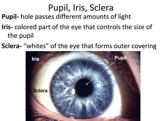
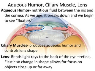
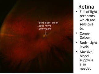
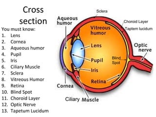
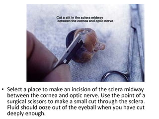
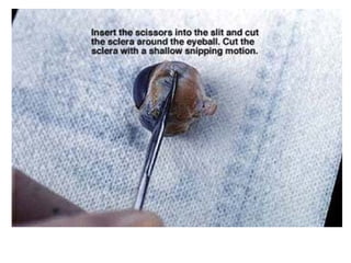
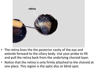
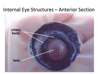
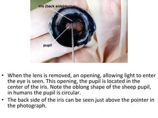
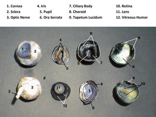
Eye Eye Eye !
• By the end of the dissection students will be
able to identify various parts of the eye.
EYE
The eye is an
extension of
the brain
It helps us take
in light
information
from our
surroundings.
This is how!
Eye brain
proximity
• Can you see :
• the optic
nerve bundle?
• Spinal cord?
What are the parts of the eye?
• Let’s use a diagram to help us get familiar with
the parts and pronounce them correctly!
Cornea
Cornea- transparent membrane that covers iris and
pupil. Focuses light on the retina.
Pupil, Iris, Sclera
Pupil- hole passes different amounts of light
Iris- colored part of the eye that controls the size of
the pupil
Sclera- “whites” of the eye that forms outer covering
Aqueous Humor, Ciliary Muscle, Lens
Aqueous Humor- nutritious fluid between the iris and
the cornea. As we age, it breaks down and we begin
to see “floaters”
Ciliary Muscles- produces aqueous humor and
controls lens shape
Lens- Bends light rays to the back of the eye –retina.
Elastic so change in shape allows for focus on
objects close up or far away
Retina
• Full of light
receptors
which are
sensitive
to:
• Cones-
Colour
• Rods- Light
levels
• Massive
blood
supply is
also
needed
Blind Spot- site of
optic nerve
connection
Vitreous Humor, Blind Spot, Optic Nerve,
Vitreous Humor- transparent jelly-like fluid that fills
the eye and refracts light
Blind Spot- area where optic
nerve attaches. No retina is
located there so information
cannot be picked up; “Blind”
Optic Nerve- sends messages
picked up by retina to the brain
Choroid Layer, Tapetum lucidum
Choroid Layer- lies between the
sclera and the retina it provides
the blood supply to the eye.
Tapetum lucidum- iridescent film
under the retina that provides
animals with “night vision”
Eye Dissection
• Before we go over the dissection, let’s review
the parts of the eye and their function
Cross
section
You must know:
1. Lens
2. Cornea
3. Aqueous humor
4. Pupil
5. Iris
6. Ciliary Muscle
7. Sclera
8. Vitreous Humor
9. Retina
10. Blind Spot
11. Choroid Layer
12. Optic Nerve
13. Tapetum Lucidum
Sclera
Blind
Spot
Ciliary
Choroid Layer
Taptem lucidum
Cut away fat using scissors
Anterior External Eye Structure
Posterior External Eye Structures
• Select a place to make an incision of the sclera midway
between the cornea and optic nerve. Use the point of a
surgical scissors to make a small cut through the sclera.
Fluid should ooze out of the eyeball when you have cut
deeply enough.
• Arrange the two hemispheres of the eye as you see in the
photograph.
• Observe the semi-fluid vitreous humor that fills the central
cavity of the eye. It is transparent in the living eye but might
be cloudy in the preserved specimen
• The retina lines the the posterior cavity of the eye and
extends forward to the ciliary body. Use your probe to lift
and pull the retina back from the underlying choroid layer.
• Notice that the retina is only firmly attached to the choroid at
one place. This region is the optic disc or blind spot.
Internal Eye Structure – Posterior Section
Internal Eye Structures – Anterior Section
Remove the lens and place against
newspaper to see that it is a
magnifier!
• When the lens is removed, an opening, allowing light to enter
the eye is seen. This opening, the pupil is located in the
center of the iris. Note the oblong shape of the sheep pupil,
in humans the pupil is circular.
• The back side of the iris can be seen just above the pointer in
the photograph.
Can you identify the parts? You will
need to to get credit during the lab
1. Cornea 4. Iris 7. Ciliary Body 10. Retina
2. Sclera 5. Pupil 8. Choroid 11. Lens
3. Optic Nerve 6. Ora Serrata 9. Tapetum Lucidum 12. Vitreous Humor
1. Cornea
2. Sclera
3. Optic Nerve
4. Iris
5. Pupil
6. Ora Serrata (you do not
have to know this
structure!)
7. Ciliary Body
8. Choroid
9. Tapetum Lucidum
10. Retina
11. Lens
12. Vitreous Humor
Clean up!
• Follow clean up procedures:
– Place all remains in a zip lock bag and place it in your
box.
– All dissecting instruments, tray and gel must be
thoroughly washed in water.
– Remove the paper towel from the tray and dispose of
it in the trash bag.
– All used gloves are to be disposed of in the trash bag.
– All goggles returned to the designated place.
– Lab stations are clean and free of water/waste.
– Each person washes their hands with soap and water.

Sheep eye dissection

  • 1.
    Eye Eye Eye! • By the end of the dissection students will be able to identify various parts of the eye.
  • 2.
    EYE The eye isan extension of the brain It helps us take in light information from our surroundings. This is how!
  • 3.
    Eye brain proximity • Canyou see : • the optic nerve bundle? • Spinal cord?
  • 4.
    What are theparts of the eye? • Let’s use a diagram to help us get familiar with the parts and pronounce them correctly!
  • 5.
    Cornea Cornea- transparent membranethat covers iris and pupil. Focuses light on the retina.
  • 6.
    Pupil, Iris, Sclera Pupil-hole passes different amounts of light Iris- colored part of the eye that controls the size of the pupil Sclera- “whites” of the eye that forms outer covering
  • 7.
    Aqueous Humor, CiliaryMuscle, Lens Aqueous Humor- nutritious fluid between the iris and the cornea. As we age, it breaks down and we begin to see “floaters” Ciliary Muscles- produces aqueous humor and controls lens shape Lens- Bends light rays to the back of the eye –retina. Elastic so change in shape allows for focus on objects close up or far away
  • 8.
    Retina • Full oflight receptors which are sensitive to: • Cones- Colour • Rods- Light levels • Massive blood supply is also needed Blind Spot- site of optic nerve connection
  • 9.
    Vitreous Humor, BlindSpot, Optic Nerve, Vitreous Humor- transparent jelly-like fluid that fills the eye and refracts light Blind Spot- area where optic nerve attaches. No retina is located there so information cannot be picked up; “Blind” Optic Nerve- sends messages picked up by retina to the brain
  • 10.
    Choroid Layer, Tapetumlucidum Choroid Layer- lies between the sclera and the retina it provides the blood supply to the eye. Tapetum lucidum- iridescent film under the retina that provides animals with “night vision”
  • 11.
    Eye Dissection • Beforewe go over the dissection, let’s review the parts of the eye and their function
  • 12.
    Cross section You must know: 1.Lens 2. Cornea 3. Aqueous humor 4. Pupil 5. Iris 6. Ciliary Muscle 7. Sclera 8. Vitreous Humor 9. Retina 10. Blind Spot 11. Choroid Layer 12. Optic Nerve 13. Tapetum Lucidum Sclera Blind Spot Ciliary Choroid Layer Taptem lucidum
  • 13.
    Cut away fatusing scissors
  • 14.
  • 15.
  • 17.
    • Select aplace to make an incision of the sclera midway between the cornea and optic nerve. Use the point of a surgical scissors to make a small cut through the sclera. Fluid should ooze out of the eyeball when you have cut deeply enough.
  • 19.
    • Arrange thetwo hemispheres of the eye as you see in the photograph. • Observe the semi-fluid vitreous humor that fills the central cavity of the eye. It is transparent in the living eye but might be cloudy in the preserved specimen
  • 20.
    • The retinalines the the posterior cavity of the eye and extends forward to the ciliary body. Use your probe to lift and pull the retina back from the underlying choroid layer. • Notice that the retina is only firmly attached to the choroid at one place. This region is the optic disc or blind spot.
  • 21.
    Internal Eye Structure– Posterior Section
  • 23.
    Internal Eye Structures– Anterior Section
  • 24.
    Remove the lensand place against newspaper to see that it is a magnifier!
  • 25.
    • When thelens is removed, an opening, allowing light to enter the eye is seen. This opening, the pupil is located in the center of the iris. Note the oblong shape of the sheep pupil, in humans the pupil is circular. • The back side of the iris can be seen just above the pointer in the photograph.
  • 27.
    Can you identifythe parts? You will need to to get credit during the lab
  • 28.
    1. Cornea 4.Iris 7. Ciliary Body 10. Retina 2. Sclera 5. Pupil 8. Choroid 11. Lens 3. Optic Nerve 6. Ora Serrata 9. Tapetum Lucidum 12. Vitreous Humor
  • 29.
    1. Cornea 2. Sclera 3.Optic Nerve 4. Iris 5. Pupil 6. Ora Serrata (you do not have to know this structure!) 7. Ciliary Body 8. Choroid 9. Tapetum Lucidum 10. Retina 11. Lens 12. Vitreous Humor
  • 30.
    Clean up! • Followclean up procedures: – Place all remains in a zip lock bag and place it in your box. – All dissecting instruments, tray and gel must be thoroughly washed in water. – Remove the paper towel from the tray and dispose of it in the trash bag. – All used gloves are to be disposed of in the trash bag. – All goggles returned to the designated place. – Lab stations are clean and free of water/waste. – Each person washes their hands with soap and water.