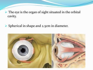
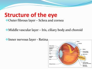
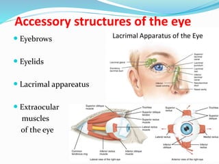
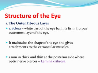
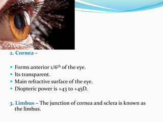
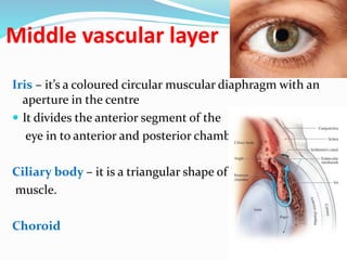
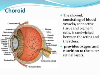
By Dr Satish S Hadimani
Dept of shalakya tantra
SGR Ayurved Mahavidyalaya,
Solapur
 The eye is the organ of sight situated in the orbital
cavity.
 Spherical in shape and 2.5cm in diameter.
Structure of the eye
Outer fibrous layer – Sclera and cornea
Middle vascular layer – Iris, ciliary body and choroid
Inner nervous layer - Retina
Layers of eye
Interior of the Eyeball
Aqueous
humour
Lens
Vitreous
Accessory structures of the eye
 Eyebrows
 Eyelids
 Lacrimal appareatus
 Extraocular
muscles
of the eye
Structure of eye
Structure of the Eye
1. The Outer Fibrous Layer
 1. Sclera – white part of the eye ball. Its firm, fibrous
outermost layer of the eye.
 It maintains the shape of the eye and gives
attachments to the extraocular muscles.
 1 mm in thick and thin at the posterior side where
optic nerve pierces – Lamina cribrosa
2. Cornea –
 Forms anterior 1/6th of the eye.
 Its transparent.
 Main refractive surface of the eye.
 Diopteric power is +43 to +45D.
3. Limbus – The junction of cornea and sclera is known as
the limbus.
Middle vascular layer
Iris – it’s a coloured circular muscular diaphragm with an
aperture in the centre
 It divides the anterior segment of the
eye in to anterior and posterior chambers
Ciliary body – it is a triangular shape of
muscle.
Choroid
Choroid
 The choroid,
consisting of blood
vessels, connective
tissue and pigment
cells, is sandwiched
between the retina and
the sclera.
 provides oxygen and
nutrition to the outer
retinal layers.
Lens
Retina
 At the back of the retina
 Contains 2 types of photoreceptors - rods and cones
 Converts light signal into an electrical signal that is
transmitted to brain through optic nerve.
Fundus image
Interior of the Eyeball
 Aqueous humour
 Lens
 Vitreous
Aqueous humour
 fills the anterior and
posterior chambers.
 The anterior
chamber is the
space between the
cornea and the iris.
 Behind the iris and
in front of the lens
is the posterior
chamber.
 They are
connected by the
pupil.
Lens
Lens
 The discus-like lens comprises a mass of
long cells known as fibres. At the centre
these fibres are compacted into a hard
nucleus surrounded by less dense fibres, the
cortex.
 The lens is relatively dehydrated and its
fibres contain special proteins. This is why it
is transparent.
Vitreous body
Vitreous body
The vitreous body is 99% water but,
vitally, also contains collagen fibrils and
hyaluronan, which impart cohesion and
a gel-like consistency.
The vitreous is adherent to the retina at
certain points, particularly at the optic
disc and at the ora serrata.
Accessory structures of the eye
 Eyebrows
 Eyelids
 Lacrimal appareatus
 Extraocular
muscles
of the eye
Accessory structures of the eye
Eye brows :
 These are two arched ridges of the
supraorbital margin of the frontal bone.
 These protects the eye from sweat, dust and
other foreign bodies.
Eye lids and eye lashes
 The eyelids are two movable folds of tissue situated
above and below the front of each eye.
 There are short curved hair, called eyelashes situated
on their free edges.
 A thin covering of skin
 Three muscles – orbicularis oculi, levator palpebrae
superioris and muller’s muscles
 A sheet of dense connective tissue and tarsal plate
 A lining of the conjunctiva
Lacrimal apparatus
 Lacrimal gland and its ducts
 Accessory lacrimal glands
 Lacrimal canaliculi
 Lacrimal sac
 Nasolacrimal duct
Extraocular muscles
 Medial rectus muscle
 Lateral rectus muscle
 Superior rectus muscle
 Inferior rectus muscle
 Superior oblique muscle
 Inferior oblique muscle
Blood supply
Carotid artery
Ophthalmic artery
Ciliary artery and central retinal artery
Eye
Artery supply
Blood supply
Nerve supply
 Motor nerves
 3rd cranial nerve – Levator palpebrae
Superior rectus
Medial rectus
Inferior rectus
Inferior oblique
Sphincter pupillae
Ciliary muscle
 4th cranial nerve - superior oblique muscle
 6th cranial nerve – lateral rectus muscle
 7th cranial nerve – orbicularis oculi muscle
 Sensory nerve – ophthalmic division supplies the whole eye
Autonomic nerves
Sympathetic nerve supply
 Iris - dilator pupillae
 Ciliary body
 Muller’s muscle
 Lacrimal gland
Parasympathetic nerve supply
 Iris – sphincter pupillae
 Ciliary body
 Lacrimal gland
Eye general anatomy

Eye general anatomy

  • 1.
    By Dr SatishS Hadimani Dept of shalakya tantra SGR Ayurved Mahavidyalaya, Solapur
  • 4.
     The eyeis the organ of sight situated in the orbital cavity.  Spherical in shape and 2.5cm in diameter.
  • 6.
    Structure of theeye Outer fibrous layer – Sclera and cornea Middle vascular layer – Iris, ciliary body and choroid Inner nervous layer - Retina
  • 7.
  • 8.
    Interior of theEyeball Aqueous humour Lens Vitreous
  • 10.
    Accessory structures ofthe eye  Eyebrows  Eyelids  Lacrimal appareatus  Extraocular muscles of the eye
  • 11.
  • 12.
    Structure of theEye 1. The Outer Fibrous Layer  1. Sclera – white part of the eye ball. Its firm, fibrous outermost layer of the eye.  It maintains the shape of the eye and gives attachments to the extraocular muscles.  1 mm in thick and thin at the posterior side where optic nerve pierces – Lamina cribrosa
  • 13.
    2. Cornea – Forms anterior 1/6th of the eye.  Its transparent.  Main refractive surface of the eye.  Diopteric power is +43 to +45D. 3. Limbus – The junction of cornea and sclera is known as the limbus.
  • 14.
    Middle vascular layer Iris– it’s a coloured circular muscular diaphragm with an aperture in the centre  It divides the anterior segment of the eye in to anterior and posterior chambers Ciliary body – it is a triangular shape of muscle. Choroid
  • 15.
    Choroid  The choroid, consistingof blood vessels, connective tissue and pigment cells, is sandwiched between the retina and the sclera.  provides oxygen and nutrition to the outer retinal layers.
  • 16.
  • 17.
    Retina  At theback of the retina  Contains 2 types of photoreceptors - rods and cones  Converts light signal into an electrical signal that is transmitted to brain through optic nerve.
  • 19.
  • 20.
    Interior of theEyeball  Aqueous humour  Lens  Vitreous
  • 21.
    Aqueous humour  fillsthe anterior and posterior chambers.  The anterior chamber is the space between the cornea and the iris.  Behind the iris and in front of the lens is the posterior chamber.  They are connected by the pupil.
  • 22.
  • 23.
    Lens  The discus-likelens comprises a mass of long cells known as fibres. At the centre these fibres are compacted into a hard nucleus surrounded by less dense fibres, the cortex.  The lens is relatively dehydrated and its fibres contain special proteins. This is why it is transparent.
  • 24.
  • 25.
    Vitreous body The vitreousbody is 99% water but, vitally, also contains collagen fibrils and hyaluronan, which impart cohesion and a gel-like consistency. The vitreous is adherent to the retina at certain points, particularly at the optic disc and at the ora serrata.
  • 26.
    Accessory structures ofthe eye  Eyebrows  Eyelids  Lacrimal appareatus  Extraocular muscles of the eye
  • 28.
    Accessory structures ofthe eye Eye brows :  These are two arched ridges of the supraorbital margin of the frontal bone.  These protects the eye from sweat, dust and other foreign bodies.
  • 29.
    Eye lids andeye lashes  The eyelids are two movable folds of tissue situated above and below the front of each eye.  There are short curved hair, called eyelashes situated on their free edges.  A thin covering of skin  Three muscles – orbicularis oculi, levator palpebrae superioris and muller’s muscles  A sheet of dense connective tissue and tarsal plate  A lining of the conjunctiva
  • 31.
    Lacrimal apparatus  Lacrimalgland and its ducts  Accessory lacrimal glands  Lacrimal canaliculi  Lacrimal sac  Nasolacrimal duct
  • 32.
    Extraocular muscles  Medialrectus muscle  Lateral rectus muscle  Superior rectus muscle  Inferior rectus muscle  Superior oblique muscle  Inferior oblique muscle
  • 33.
    Blood supply Carotid artery Ophthalmicartery Ciliary artery and central retinal artery Eye Artery supply
  • 34.
  • 37.
    Nerve supply  Motornerves  3rd cranial nerve – Levator palpebrae Superior rectus Medial rectus Inferior rectus Inferior oblique Sphincter pupillae Ciliary muscle  4th cranial nerve - superior oblique muscle  6th cranial nerve – lateral rectus muscle  7th cranial nerve – orbicularis oculi muscle  Sensory nerve – ophthalmic division supplies the whole eye
  • 40.
    Autonomic nerves Sympathetic nervesupply  Iris - dilator pupillae  Ciliary body  Muller’s muscle  Lacrimal gland Parasympathetic nerve supply  Iris – sphincter pupillae  Ciliary body  Lacrimal gland