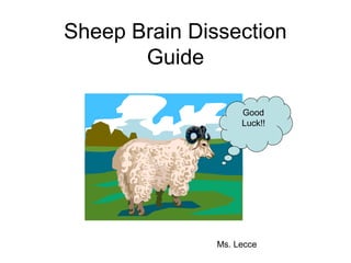
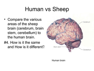
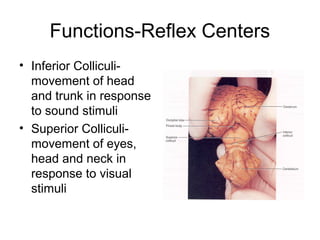
2. To begin
• Get gloves and a rubber apron or lab coat.
Goggles are available if you would like
them.
• Pick up dissection tray, dull probe, pins
and labels
• Obtain a sheep brain and share this with a
partner (or 2)
• Rinse the sheep brain to remove
preservative
3. Meninges of the Brain
Brain is protected by the skull and 3 layers of membranes called
meninges
4. Observe Meninges
• Examine the brain to see if there is a tough outer coat…
This is the DURA MATER (literally means “tough
mother”)
• Now look for the stringy, web-like ARACHNOID MATER
(literally means spider mother) beneath and stuck to the
dura mater. The space under the arachnoid, the
subarachnoid space, is filled with cerebrospinal
fluid and contains blood vessels.
• Look deeper and see an thin transparent membrane that
follows the contour of the ridges (gyri) and valleys (sulci)
of the brain. This is the PIA MATER ( means tender
mother)
5. Questions??
• What is the function of the meninges?
• Describe the following meninges:
Dura mater, Arachnoid mater, and Pia
mater
• What is meningitis?
6. Main Brain Regions
• Identify the cerebrum,
brain stem) and
cerebellum on your
brain
• Draw picture of sheep
brain and label
structures
Cerebellum
Cerebrum
Brain stem
8. Human vs Sheep
• Compare the various
areas of the sheep
brain (cerebrum, brain
stem, cerebellum) to
the human brain.
#4. How is it the same
and How is it different?
Human brain
9. Identify Dorsal Structures
• With the dorsal
side up, identify
the cerebral
hemispheres, gyri,
sulci,and
longitudinal fissure
on your sheep
brain
10. Questions??
• What does the
longitudinal fissure
divide?
• What is the difference
between gyri and
sulci?
• What is the purpose
of the gyri and sulci?
11. Lobes of the Cerebrum
• Find the 4
lobes of the
brain-frontal,
parietal,
occipital,
and temporal
• Using pins
label each lobe
• Teacher check
13. Corpus Callosum
• At the longitudinal fissure, gently separate
the two hemispheres and look down
between them for the thick band of white
fibers. This is the corpus callosum
• What is its function?
14. Identify Ventral Structures
• Place the sheep brain
ventral side up.
• Identify the olfactory
bulb, optic nerve,
optic chiasm
• Using pins label each
structure.
• Draw and label the
structures in lab
writeup
15. Questions??
• How does the human brain’s olfactory
bulb differ from the sheeps?
• Which is able to smell more odors?
• Look at the optic chiasma. What happens
to the optic nerve (vision) at the optic
chiasma?
• Which side of the occipital lobe (right or
left) “sees” for the right eye?
16. Brain Stem
• Look at the three
parts of the brain
stem-the midbrain
(cerebral peduncle
and mammilary
body), pons and
medulla oblongata
• Pin and label parts
• Teacher check
18. Questions??
• Label your diagram with parts of midbrain
and brain stem
• What is the function of the medulla
oblongata and pons?
• Could you survive without them?
• Why or Why not?
19. Identify dorsal structures
• Carefully pull the
cerebellum away from
the cerebrum. Don’t
break any structures
off.
• Identify the pineal
body, superior
colliculi, and inferior
colliculi.
20. Functions-Reflex Centers
• Inferior Colliculi-
movement of head
and trunk in response
to sound stimuli
• Superior Colliculi-
movement of eyes,
head and neck in
response to visual
stimuli
23. Midsagittal Section
• Obtain a brain the has been cut in half
• Locate the brain stem parts: the medulla
oblongata, the pons, cerebrum, and cerebellum
24. Cerebellum
• Look at the cross section of the
cerebellum. The inner structure is called
the arbor vitae (living tree).
Why is this a good name for it?
26. Questions
– What is the function of the ventricles?
– The cerebral aquaduct allows for the
circulation of the cerebral spinal fluid through
the ventricles. What do you think would
happen if the duct became blocked?
– How would that affect brain tissue?