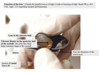
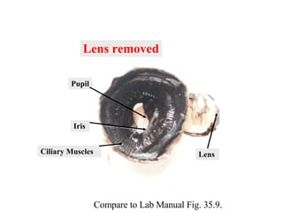
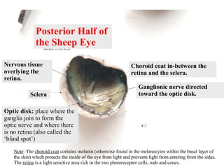
College Level  Sheep Eye Dissection Guide Passaic County Community College Design - Dr. E. Hager Edited by Dr. S.C. Wache
Note the  eyelid  on the anterior half of the eyeball. Note the  fat  around the eyeball. It protects by cushioning the eyeball within the bones that surround the eye Take the fine tipped scissors to cut away the eyelid and extra fat.
Scalpel Cleaned Eyeball Excess Fat and Eyelid
Cornea  – extension of sclera over the lens -opaque in preserved specimen Anterior View of the Sheep Eye
Cornea  Pupil Sclera Anterior View of the Sheep Eye
Optic Nerve – cranial nerve II Posterior View of  the Sheep Eye
Prepare a coronal cut:  Start by making a primary incision into the sclera using a scalpel; then, using the bone cutting scissors, complete the coronal sectioning of the eyeball. Optic Nerve Incision into the tough connective  tissue called the sclera
Sensory  Cranial Nerve II Sclera Lens in the anterior half Vitreous Humor in the posterior half of the eyeball:  this jelly-like fluid   helps maintain shape of the eyeball Function of the lens:  it bends the parallel waves of light; it aids in focusing of light. Read TB, p. 457, Clin. Appl. 12.5 regarding myopia and hyperopia. Note the thickness of the sclera layer.
Anterior Half of  the Sheep Eye Lens Ciliary Body
The lens is easily pushed out. Often, some of the vitreous humor sticks to it. Removal of the Lens Lens Suspensory Ligaments / Ciliary  Muscles  sticking to the lens Vitreous Humor
Lens removed Lens Ciliary Muscles Iris Pupil Compare to Lab Manual Fig. 35.9.
Lens Vitreous Humor Suspensory ligaments attach the  ciliary  muscles to the lens.
Posterior Half of  the Sheep Eye Nervous tissue overlying the retina. Ganglionic nerve directed toward the optic disk. Choroid coat in-between the retina and the sclera. Sclera Optic disk:  place where the ganglia join to form the optic nerve and where there is no retina (also called the ‘blind spot’) Note : The  choroid coat  contains melanin (otherwise found in the melanocytes within the basal layer of  the skin) which protects the inside of the eye from light and prevents light from entering from the sides. The  retina  is a light sensitive area rich in the two photoreceptor cells, rods and cones.
Sclera Tapetum lucidum-  shiny part of the choroid coat. Melanin containing part of the  choroid coat. Retina and nervous tissue  pulled to the side.  Note : The  fovea centralis  (a small rodless area of the retina that affords acute vision) and the  macula lutea  constitute the region of maximum visual acuity and are made up almost completely of cones which provide color vision. Rods are active during night vision. Note : The  choroid coat   also contains the  tapetum lucidum  which reflects light and enhances night vision. It is well developed in cats.  Rhodopsin , a protein within  rods and cones , is excited by photons, the energy-rich particles that make up a light wave. Once excited above threshold, an impulse is sent along the sensory nerve  to the CNS where it is  interpreted in interneurons.

Lab 15 Sheepeyediss

  • 1.
    College Level Sheep Eye Dissection Guide Passaic County Community College Design - Dr. E. Hager Edited by Dr. S.C. Wache
  • 2.
    Note the eyelid on the anterior half of the eyeball. Note the fat around the eyeball. It protects by cushioning the eyeball within the bones that surround the eye Take the fine tipped scissors to cut away the eyelid and extra fat.
  • 3.
    Scalpel Cleaned EyeballExcess Fat and Eyelid
  • 4.
    Cornea –extension of sclera over the lens -opaque in preserved specimen Anterior View of the Sheep Eye
  • 5.
    Cornea PupilSclera Anterior View of the Sheep Eye
  • 6.
    Optic Nerve –cranial nerve II Posterior View of the Sheep Eye
  • 7.
    Prepare a coronalcut: Start by making a primary incision into the sclera using a scalpel; then, using the bone cutting scissors, complete the coronal sectioning of the eyeball. Optic Nerve Incision into the tough connective tissue called the sclera
  • 8.
    Sensory CranialNerve II Sclera Lens in the anterior half Vitreous Humor in the posterior half of the eyeball: this jelly-like fluid helps maintain shape of the eyeball Function of the lens: it bends the parallel waves of light; it aids in focusing of light. Read TB, p. 457, Clin. Appl. 12.5 regarding myopia and hyperopia. Note the thickness of the sclera layer.
  • 9.
    Anterior Half of the Sheep Eye Lens Ciliary Body
  • 10.
    The lens iseasily pushed out. Often, some of the vitreous humor sticks to it. Removal of the Lens Lens Suspensory Ligaments / Ciliary Muscles sticking to the lens Vitreous Humor
  • 11.
    Lens removed LensCiliary Muscles Iris Pupil Compare to Lab Manual Fig. 35.9.
  • 12.
    Lens Vitreous HumorSuspensory ligaments attach the ciliary muscles to the lens.
  • 13.
    Posterior Half of the Sheep Eye Nervous tissue overlying the retina. Ganglionic nerve directed toward the optic disk. Choroid coat in-between the retina and the sclera. Sclera Optic disk: place where the ganglia join to form the optic nerve and where there is no retina (also called the ‘blind spot’) Note : The choroid coat contains melanin (otherwise found in the melanocytes within the basal layer of the skin) which protects the inside of the eye from light and prevents light from entering from the sides. The retina is a light sensitive area rich in the two photoreceptor cells, rods and cones.
  • 14.
    Sclera Tapetum lucidum- shiny part of the choroid coat. Melanin containing part of the choroid coat. Retina and nervous tissue pulled to the side. Note : The fovea centralis (a small rodless area of the retina that affords acute vision) and the macula lutea constitute the region of maximum visual acuity and are made up almost completely of cones which provide color vision. Rods are active during night vision. Note : The choroid coat also contains the tapetum lucidum which reflects light and enhances night vision. It is well developed in cats. Rhodopsin , a protein within rods and cones , is excited by photons, the energy-rich particles that make up a light wave. Once excited above threshold, an impulse is sent along the sensory nerve to the CNS where it is interpreted in interneurons.