Downloaded 486 times
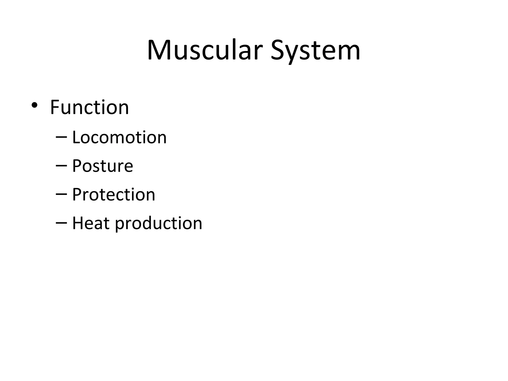
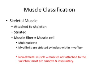


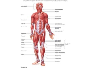
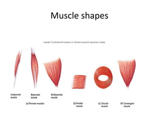
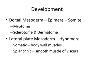
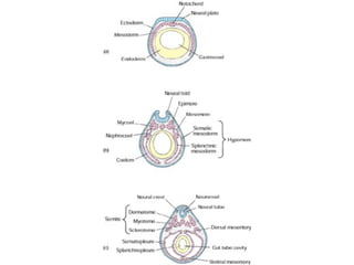
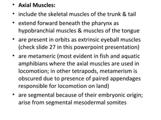
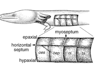

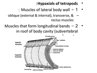
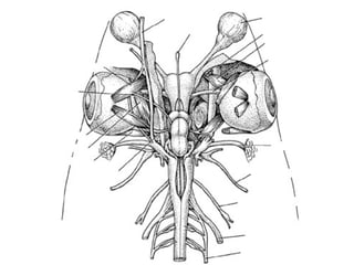
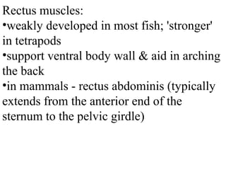
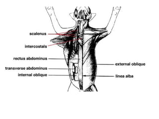
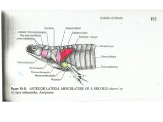
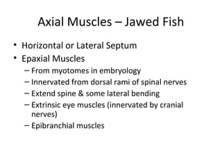
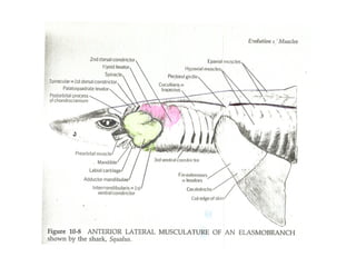
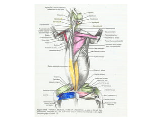
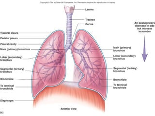

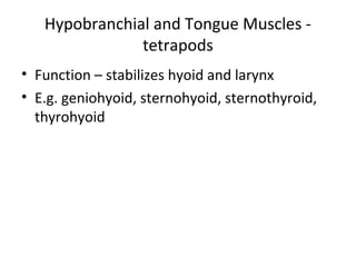
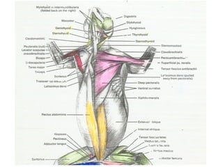
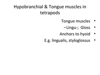


The document discusses the muscular system, describing four main types of vertebrate muscles and their functions. It focuses on axial muscles, including those in the trunk, tail, hypobranchial region, and tongue. In fish, axial muscles are segmented into myomeres, while tetrapods retain some evidence of segmentation. Epaxial muscles extend along the spine dorsally while hypaxial muscles include lateral body wall muscles and subvertebral muscles. Hypobranchial muscles originate in the pharyngeal region and migrate forward in tetrapods.


