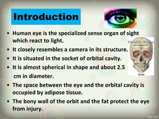
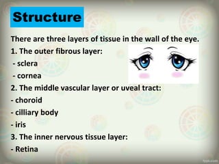
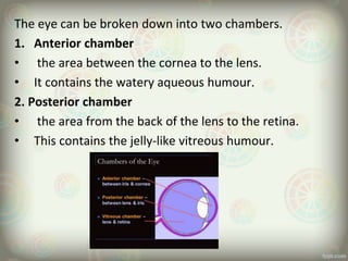
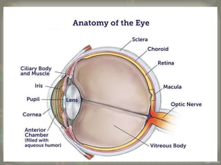
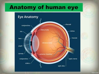
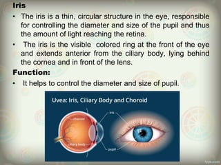
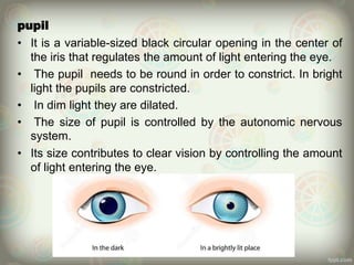
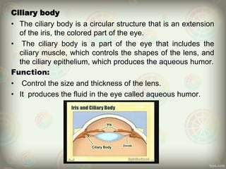
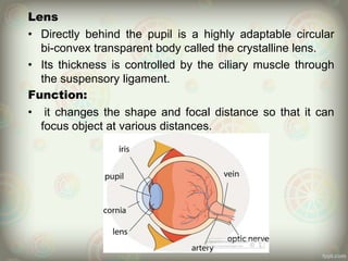
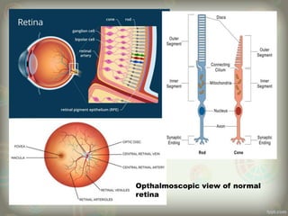
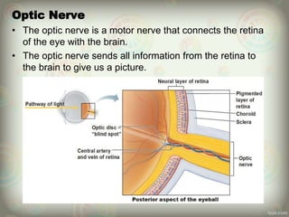
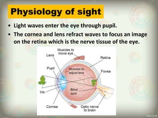
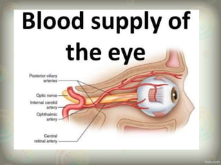
SlideShare a Scribd company logo
HUMAN
EYE
Prepared by Dhirendra
Thakur
General medicine
Table of contents
S.N. Name of topics.
1. Introduction
2. Structure
3. Anatomy of eye
4. Parts of eye
5. Physiology of sight
6. The accessory organs of eye
7. Muscles of eye
8. Lacrimal apparatus
9. Supplies to eye
Introduction
• Human eye is the specialized sense organ of sight
which react to light.
• It closely resembles a camera in its structure.
• It is situated in the socket of orbital cavity.
• It is almost spherical in shape and about 2.5
cm in diameter.
• The space between the eye and the orbital cavity is
occupied by adipose tissue.
• The bony wall of the orbit and the fat protect the eye
from injury.
Structure
There are three layers of tissue in the wall of the eye.
1. The outer fibrous layer:
- sclera
- cornea
2. The middle vascular layer or uveal tract:
- choroid
- cilliary body
- iris
3. The inner nervous tissue layer:
- Retina
The eye can be broken down into two chambers.
1. Anterior chamber
• the area between the cornea to the lens.
• It contains the watery aqueous humour.
2. Posterior chamber
• the area from the back of the lens to the retina.
• This contains the jelly-like vitreous humour.
Anatomy of human eye
Parts of eye
Sclera
• It is the white part of eye.
• It is the opaque, fibrous, tough, protective layer of the
eye.
• At the front of the eye it is continues with the cornea.
• It consists of a firm fibrous membrane that maintains
the shape of the eye and give attachment to the
extrinsic muscle of the eye.
Function:
• It protect the eye ball from dust and other harmful
matter.
Cornea
• The cornea acts as the eye's outermost lens.
• The cornea contributes between 65-75 % of the eye’s total
focusing power.
• The light is pass through the cornea to reach the retina.
• When the light strikes in the cornea, it bends or refracts the
incoming into the lens.
Function
• It helps to shield the rest of the eyes from germs, dust.
• It acts as a window that controls and focus the entry of light
into the eye.
Iris
• The iris is a thin, circular structure in the eye, responsible
for controlling the diameter and size of the pupil and thus
the amount of light reaching the retina.
• The iris is the visible colored ring at the front of the eye
and extends anterior from the ciliary body, lying behind
the cornea and in front of the lens.
Function:
• It helps to control the diameter and size of pupil.
pupil
• It is a variable-sized black circular opening in the center of
the iris that regulates the amount of light entering the eye.
• The pupil needs to be round in order to constrict. In bright
light the pupils are constricted.
• In dim light they are dilated.
• The size of pupil is controlled by the autonomic nervous
system.
• Its size contributes to clear vision by controlling the amount
of light entering the eye.
Choroid
• The choroid lines the posterior five-sixths of the inner
surface of the sclera.
• It is very rich in blood vessels and is deep chocolate
brown in color.
• Light enters the eye through the pupil, stimulates the
sensory receptors in the retina and is then absorbed by
the choroid.
• It is the vascular layer of eye, containing connective
tissue, and lying between retina and sclera.
Function:
• It provide nourishment to the outer layer of the retina
through blood vessels.
Ciliary body
• The ciliary body is a circular structure that is an extension
of the iris, the colored part of the eye.
• The ciliary body is a part of the eye that includes the
ciliary muscle, which controls the shapes of the lens, and
the ciliary epithelium, which produces the aqueous humor.
Function:
• Control the size and thickness of the lens.
• It produces the fluid in the eye called aqueous humor.
Lens
• Directly behind the pupil is a highly adaptable circular
bi-convex transparent body called the crystalline lens.
• Its thickness is controlled by the ciliary muscle through
the suspensory ligament.
Function:
• it changes the shape and focal distance so that it can
focus object at various distances.
Retina
• The retina is a thin layer of tissue that lines the
back of the eyes on the inside.
• It is located near the optic nerve.
• The purpose of the retina is to receive light that
the lens has focused.
Function:
• It convert image or light into electrical impulses.
• It send this impulses to the brain for visual
recognition.
Opthalmoscopic view of normal
retina
Optic Nerve
• The optic nerve is a motor nerve that connects the retina
of the eye with the brain.
• The optic nerve sends all information from the retina to
the brain to give us a picture.
Physiology of sight
• Light waves enter the eye through pupil.
• The cornea and lens refract waves to focus an image
on the retina which is the nerve tissue of the eye.
• Light- sensitive pigments in visual receptor neurons known as
rods and cones in the retina decompose and trigger a
complex series of reactions that initiate nerve impulses.
• Impulses travel through the optic nerves to the optic chiasma.
• Optic disc carries nerve impulses to the brain.
• In the brain, the visual area of the cerebral cortex in the
occipital lobe of the cerebrum interprets vision.
The optic
nerves and
their
pathway
The accessory organs of eye
• The eye brows
• The eye lids
• The eye lashes
• The conjunctiva
• The muscles of the eyes
• The lacrimal apparatus
The eye brows
• These are two arched ridges of the supraorbital
margins of the frontal bone.
• Numerous hairs (eyebrows) project obliquely
from the surface of the skin.
• They protect the eyeball from sweat, dust and
other foreign bodies.
Eye lids and eyelashes
• The eye lids are two moveable folds of tissues
situated above and below the front of each eye.
• On their free edges are short curved hairs, the
eye lashes.
• The eye lids are made up of:
 skin
 arealor tissue
 the circular muscles
 Tarsal plate
 Conunctival lining
Functions
• The eye lids and eye lashes protect the eye from
injury.
• Closure of the eyelids occurs when an object
come close to the eye and when bright light
shines into the eyes this is called the corneal
reflux.
• Blinking at about 3-7 seconds interval spreads
tears and oily secretions over cornea, preventing
drying.
Conjunctiva
• This is a fine transparent membrane that lines
the eyelids and the front of the eyeball.
• It consists of highly vascular columnar
epithelium.
• Corneal conjunctiva consists of avascular
epithelium i.e. without blood vessels.
• There are two canthus they are
• Medial canthus
• Lateral canthus
Extrinsic muscles of the eye:
Name Action Cranial nerve supply
Medial rectus Rotates eyeball inward Oculomotor nerve
(3rd cranial nerve)
Lateral rectus Rotates eyeball outwards Abducent nerve
( 6th cranial nerve)
Superior rectus Rotates eyeball upwards Oculomotor nerve
(3rd cranial nerve)
Inferior rectus Rotates eyeball
downwards
Oculomotor nerve
(3rd cranial nerve)
Superior oblique Rotates eyeball
Downward and outward
Trochlear nerve
(4th cranial nerve)
Inferior oblique Rotates eyeball
Upward and outward
Oculomotor nerve
(3rd cranial nerve )
Extrinsic muscles of the eye:
The Lacrimal Apparatus
For each eye this consists of structures that secrete
tears and drain them from the front of the eyeball:
• 1 lacrimal gland and its ducts
• 2 lacrimal puctum
• 1 lacrimal sac
• 1 nasolacrimal duct
Functions
• Provision of oxygen and nutrients to the
avasular corneal conjunctiva and drainage of
waste.
• Washing away irritating material e.g. dust grit
.
• The bactericidal enzymes lysozyme prevents
microbial infections.
• Its oiliness delays evaporation and prevents
friction or drying of the conjunctiva.
Supplies to eye
Nerve supply
• It is supplied by branches of the ophthalmic nerve.
Blood supply
• The eye supplied with arterial blood by the ciliary arteries and
the central retinal artery.
• These are branches of the ophthalmic artery, branch of the
internal carotid artery.
• Venous drainage is by a number of veins, including the central
retinal veins.
Thank you!!

More Related Content

What's hot

The physiology of human eye
The physiology of human eyeThe physiology of human eye
The physiology of human eye
Haider zaman
 
Anatomy of retina
Anatomy of retinaAnatomy of retina
Anatomy of retina
SAMEEKSHA AGRAWAL
 
Eye ball.pptx
Eye ball.pptxEye ball.pptx
Eye anatomy
Eye anatomyEye anatomy
Eye anatomy
Nabeel Doc
 
Orbit and Extra-Ocular Muscles
Orbit and Extra-Ocular MusclesOrbit and Extra-Ocular Muscles
Orbit and Extra-Ocular Muscles
Sushrut Ahale
 
Anatomy Of The Eye
Anatomy Of The EyeAnatomy Of The Eye
Anatomy Of The Eye
Pave Medicine
 
Anatomy of eye ppt
Anatomy of eye pptAnatomy of eye ppt
Anatomy of eye ppt
Bijukumar Vasupillai
 
Anatomy of iris
Anatomy of irisAnatomy of iris
Anatomy of iris
DARSHAN S M
 
Anatomy of the eye
Anatomy of the eyeAnatomy of the eye
Anatomy of the eye
Dr. Mohammad Mahmoud
 
Ocular anatomy DR TEK
Ocular anatomy DR TEKOcular anatomy DR TEK
Ocular anatomy DR TEK
drtek
 
The physiology of vision part 1
The physiology of vision   part 1The physiology of vision   part 1
The physiology of vision part 1
Santosh Deo
 
Embryology of eye
Embryology of eyeEmbryology of eye
Embryology of eye
Samuel Ponraj
 
Embryology of human eye
Embryology of human eyeEmbryology of human eye
Embryology of human eye
Lhacha
 
Male reproductive system
Male reproductive systemMale reproductive system
Male reproductive system
Neha Bhandari
 
Aqueous Humour
Aqueous HumourAqueous Humour
Aqueous Humour
laraib jameel
 
Anatomy of the eye
Anatomy of the eyeAnatomy of the eye
Anatomy of the eye
DrVarun5179
 
Development of eye
Development of eye  Development of eye
Development of eye
Prabhakar Yadav
 
strabismus
strabismusstrabismus
EXTRA OCULAR MUSCLES ANATOMY
EXTRA OCULAR MUSCLES ANATOMYEXTRA OCULAR MUSCLES ANATOMY
EXTRA OCULAR MUSCLES ANATOMY
Rabia Ammer
 

What's hot (20)

The physiology of human eye
The physiology of human eyeThe physiology of human eye
The physiology of human eye
 
Anatomy of retina
Anatomy of retinaAnatomy of retina
Anatomy of retina
 
Eye ball.pptx
Eye ball.pptxEye ball.pptx
Eye ball.pptx
 
Eye anatomy
Eye anatomyEye anatomy
Eye anatomy
 
Orbit and Extra-Ocular Muscles
Orbit and Extra-Ocular MusclesOrbit and Extra-Ocular Muscles
Orbit and Extra-Ocular Muscles
 
Anatomy Of The Eye
Anatomy Of The EyeAnatomy Of The Eye
Anatomy Of The Eye
 
Anatomy of eye ppt
Anatomy of eye pptAnatomy of eye ppt
Anatomy of eye ppt
 
Anatomy of iris
Anatomy of irisAnatomy of iris
Anatomy of iris
 
Anatomy of the eye
Anatomy of the eyeAnatomy of the eye
Anatomy of the eye
 
Ocular anatomy DR TEK
Ocular anatomy DR TEKOcular anatomy DR TEK
Ocular anatomy DR TEK
 
The physiology of vision part 1
The physiology of vision   part 1The physiology of vision   part 1
The physiology of vision part 1
 
Embryology of eye
Embryology of eyeEmbryology of eye
Embryology of eye
 
Embryology of human eye
Embryology of human eyeEmbryology of human eye
Embryology of human eye
 
Male reproductive system
Male reproductive systemMale reproductive system
Male reproductive system
 
Aqueous Humour
Aqueous HumourAqueous Humour
Aqueous Humour
 
Anatomy of the eye
Anatomy of the eyeAnatomy of the eye
Anatomy of the eye
 
Development of eye
Development of eye  Development of eye
Development of eye
 
Tearfilm & blinking
Tearfilm & blinkingTearfilm & blinking
Tearfilm & blinking
 
strabismus
strabismusstrabismus
strabismus
 
EXTRA OCULAR MUSCLES ANATOMY
EXTRA OCULAR MUSCLES ANATOMYEXTRA OCULAR MUSCLES ANATOMY
EXTRA OCULAR MUSCLES ANATOMY
 

Similar to Presentation on eye

The Human Eye
The Human EyeThe Human Eye
The Human Eye
PriyankaJakhar9
 
anatomyofeye-180217183456.pdf
anatomyofeye-180217183456.pdfanatomyofeye-180217183456.pdf
anatomyofeye-180217183456.pdf
EidTahir
 
Anatomy of eye
Anatomy of eyeAnatomy of eye
Anatomy of eye
sneha chavan
 
The eye (sense of sight organ)
The eye (sense of sight organ)The eye (sense of sight organ)
The eye (sense of sight organ)
Jordan Mwelwa
 
EYE ANATOMY AND PHYSIOLOGY ppt.pptx
EYE  ANATOMY AND PHYSIOLOGY ppt.pptxEYE  ANATOMY AND PHYSIOLOGY ppt.pptx
EYE ANATOMY AND PHYSIOLOGY ppt.pptx
Nitin Sharma
 
Anatomy and Physiology of Special senses
Anatomy and Physiology of Special senses Anatomy and Physiology of Special senses
Anatomy and Physiology of Special senses
DR .PALLAVI PATHANIA
 
Anatomy of sight and the eye
Anatomy of sight and the eyeAnatomy of sight and the eye
Anatomy of sight and the eye
Arsi University, Asella, Ethiopia
 
vision.pptx
vision.pptxvision.pptx
vision.pptx
LuzSan3
 
ANATOMY OF EYE n.pdf
ANATOMY OF EYE n.pdfANATOMY OF EYE n.pdf
ANATOMY OF EYE n.pdf
Pooja Rani
 
Ocular Anatomy.ppt
Ocular Anatomy.pptOcular Anatomy.ppt
Ocular Anatomy.ppt
Medical Knowledge
 
Anatomy and physilogy of eye,nose and throat
Anatomy and physilogy of eye,nose and throatAnatomy and physilogy of eye,nose and throat
Anatomy and physilogy of eye,nose and throat
Deeps Gupta
 
Eye and its disorders in anatomy and physiology
Eye and its disorders in anatomy and physiologyEye and its disorders in anatomy and physiology
Eye and its disorders in anatomy and physiology
Sreenivasa Reddy Thalla
 
Anatomy of eye neelesh
Anatomy of eye neeleshAnatomy of eye neelesh
Anatomy of eye neelesh
Neelesh Kumar
 
Mammalian eye
Mammalian eyeMammalian eye
Mammalian eye
Kankana Choudhury
 
The human eye amatomy
The human eye amatomyThe human eye amatomy
The human eye amatomy
Patel Dharmendra
 
Sensory organs
Sensory organsSensory organs
Sensory organs
Pavithra Pavi
 
ocular anatomy of eye for optometry and optalomolgy
ocular anatomy of eye for optometry and optalomolgyocular anatomy of eye for optometry and optalomolgy
ocular anatomy of eye for optometry and optalomolgy
devaramgehlot
 
ocular_anatomy.pptx
ocular_anatomy.pptxocular_anatomy.pptx
ocular_anatomy.pptx
HimanshuHarsh22ZOO00
 
eye ball anat.pptx
eye ball anat.pptxeye ball anat.pptx
eye ball anat.pptx
Dr Mohammad Amaan
 
The human eye. group 1
The human eye. group 1The human eye. group 1
The human eye. group 1
Domlan Mathias
 

Similar to Presentation on eye (20)

The Human Eye
The Human EyeThe Human Eye
The Human Eye
 
anatomyofeye-180217183456.pdf
anatomyofeye-180217183456.pdfanatomyofeye-180217183456.pdf
anatomyofeye-180217183456.pdf
 
Anatomy of eye
Anatomy of eyeAnatomy of eye
Anatomy of eye
 
The eye (sense of sight organ)
The eye (sense of sight organ)The eye (sense of sight organ)
The eye (sense of sight organ)
 
EYE ANATOMY AND PHYSIOLOGY ppt.pptx
EYE  ANATOMY AND PHYSIOLOGY ppt.pptxEYE  ANATOMY AND PHYSIOLOGY ppt.pptx
EYE ANATOMY AND PHYSIOLOGY ppt.pptx
 
Anatomy and Physiology of Special senses
Anatomy and Physiology of Special senses Anatomy and Physiology of Special senses
Anatomy and Physiology of Special senses
 
Anatomy of sight and the eye
Anatomy of sight and the eyeAnatomy of sight and the eye
Anatomy of sight and the eye
 
vision.pptx
vision.pptxvision.pptx
vision.pptx
 
ANATOMY OF EYE n.pdf
ANATOMY OF EYE n.pdfANATOMY OF EYE n.pdf
ANATOMY OF EYE n.pdf
 
Ocular Anatomy.ppt
Ocular Anatomy.pptOcular Anatomy.ppt
Ocular Anatomy.ppt
 
Anatomy and physilogy of eye,nose and throat
Anatomy and physilogy of eye,nose and throatAnatomy and physilogy of eye,nose and throat
Anatomy and physilogy of eye,nose and throat
 
Eye and its disorders in anatomy and physiology
Eye and its disorders in anatomy and physiologyEye and its disorders in anatomy and physiology
Eye and its disorders in anatomy and physiology
 
Anatomy of eye neelesh
Anatomy of eye neeleshAnatomy of eye neelesh
Anatomy of eye neelesh
 
Mammalian eye
Mammalian eyeMammalian eye
Mammalian eye
 
The human eye amatomy
The human eye amatomyThe human eye amatomy
The human eye amatomy
 
Sensory organs
Sensory organsSensory organs
Sensory organs
 
ocular anatomy of eye for optometry and optalomolgy
ocular anatomy of eye for optometry and optalomolgyocular anatomy of eye for optometry and optalomolgy
ocular anatomy of eye for optometry and optalomolgy
 
ocular_anatomy.pptx
ocular_anatomy.pptxocular_anatomy.pptx
ocular_anatomy.pptx
 
eye ball anat.pptx
eye ball anat.pptxeye ball anat.pptx
eye ball anat.pptx
 
The human eye. group 1
The human eye. group 1The human eye. group 1
The human eye. group 1
 

Recently uploaded

BENIGN PROSTATIC HYPERPLASIA.BPH. BPHpdf
BENIGN PROSTATIC HYPERPLASIA.BPH. BPHpdfBENIGN PROSTATIC HYPERPLASIA.BPH. BPHpdf
BENIGN PROSTATIC HYPERPLASIA.BPH. BPHpdf
DR SETH JOTHAM
 
basicmodesofventilation2022-220313203758.pdf
basicmodesofventilation2022-220313203758.pdfbasicmodesofventilation2022-220313203758.pdf
basicmodesofventilation2022-220313203758.pdf
aljamhori teaching hospital
 
Evaluation of antidepressant activity of clitoris ternatea in animals
Evaluation of antidepressant activity of clitoris ternatea in animalsEvaluation of antidepressant activity of clitoris ternatea in animals
Evaluation of antidepressant activity of clitoris ternatea in animals
Shweta
 
Tom Selleck Health: A Comprehensive Look at the Iconic Actor’s Wellness Journey
Tom Selleck Health: A Comprehensive Look at the Iconic Actor’s Wellness JourneyTom Selleck Health: A Comprehensive Look at the Iconic Actor’s Wellness Journey
Tom Selleck Health: A Comprehensive Look at the Iconic Actor’s Wellness Journey
greendigital
 
Prix Galien International 2024 Forum Program
Prix Galien International 2024 Forum ProgramPrix Galien International 2024 Forum Program
Prix Galien International 2024 Forum Program
Levi Shapiro
 
How STIs Influence the Development of Pelvic Inflammatory Disease.pptx
How STIs Influence the Development of Pelvic Inflammatory Disease.pptxHow STIs Influence the Development of Pelvic Inflammatory Disease.pptx
How STIs Influence the Development of Pelvic Inflammatory Disease.pptx
FFragrant
 
Couples presenting to the infertility clinic- Do they really have infertility...
Couples presenting to the infertility clinic- Do they really have infertility...Couples presenting to the infertility clinic- Do they really have infertility...
Couples presenting to the infertility clinic- Do they really have infertility...
Sujoy Dasgupta
 
ACUTE SCROTUM.....pdf. ACUTE SCROTAL CONDITIOND
ACUTE SCROTUM.....pdf. ACUTE SCROTAL CONDITIONDACUTE SCROTUM.....pdf. ACUTE SCROTAL CONDITIOND
ACUTE SCROTUM.....pdf. ACUTE SCROTAL CONDITIOND
DR SETH JOTHAM
 
Ophthalmology Clinical Tests for OSCE exam
Ophthalmology Clinical Tests for OSCE examOphthalmology Clinical Tests for OSCE exam
Ophthalmology Clinical Tests for OSCE exam
KafrELShiekh University
 
HOT NEW PRODUCT! BIG SALES FAST SHIPPING NOW FROM CHINA!! EU KU DB BK substit...
HOT NEW PRODUCT! BIG SALES FAST SHIPPING NOW FROM CHINA!! EU KU DB BK substit...HOT NEW PRODUCT! BIG SALES FAST SHIPPING NOW FROM CHINA!! EU KU DB BK substit...
HOT NEW PRODUCT! BIG SALES FAST SHIPPING NOW FROM CHINA!! EU KU DB BK substit...
GL Anaacs
 
24 Upakrama.pptx class ppt useful in all
24 Upakrama.pptx class ppt useful in all24 Upakrama.pptx class ppt useful in all
24 Upakrama.pptx class ppt useful in all
DrSathishMS1
 
For Better Surat #ℂall #Girl Service ❤85270-49040❤ Surat #ℂall #Girls
For Better Surat #ℂall #Girl Service ❤85270-49040❤ Surat #ℂall #GirlsFor Better Surat #ℂall #Girl Service ❤85270-49040❤ Surat #ℂall #Girls
For Better Surat #ℂall #Girl Service ❤85270-49040❤ Surat #ℂall #Girls
Savita Shen $i11
 
New Drug Discovery and Development .....
New Drug Discovery and Development .....New Drug Discovery and Development .....
New Drug Discovery and Development .....
NEHA GUPTA
 
New Directions in Targeted Therapeutic Approaches for Older Adults With Mantl...
New Directions in Targeted Therapeutic Approaches for Older Adults With Mantl...New Directions in Targeted Therapeutic Approaches for Older Adults With Mantl...
New Directions in Targeted Therapeutic Approaches for Older Adults With Mantl...
i3 Health
 
NVBDCP.pptx Nation vector borne disease control program
NVBDCP.pptx Nation vector borne disease control programNVBDCP.pptx Nation vector borne disease control program
NVBDCP.pptx Nation vector borne disease control program
Sapna Thakur
 
POST OPERATIVE OLIGURIA and its management
POST OPERATIVE OLIGURIA and its managementPOST OPERATIVE OLIGURIA and its management
POST OPERATIVE OLIGURIA and its management
touseefaziz1
 
Flu Vaccine Alert in Bangalore Karnataka
Flu Vaccine Alert in Bangalore KarnatakaFlu Vaccine Alert in Bangalore Karnataka
Flu Vaccine Alert in Bangalore Karnataka
addon Scans
 
Physiology of Chemical Sensation of smell.pdf
Physiology of Chemical Sensation of smell.pdfPhysiology of Chemical Sensation of smell.pdf
Physiology of Chemical Sensation of smell.pdf
MedicoseAcademics
 
Charaka Samhita Sutra sthana Chapter 15 Upakalpaniyaadhyaya
Charaka Samhita Sutra sthana Chapter 15 UpakalpaniyaadhyayaCharaka Samhita Sutra sthana Chapter 15 Upakalpaniyaadhyaya
Charaka Samhita Sutra sthana Chapter 15 Upakalpaniyaadhyaya
Dr KHALID B.M
 
Ocular injury ppt Upendra pal optometrist upums saifai etawah
Ocular injury  ppt  Upendra pal  optometrist upums saifai etawahOcular injury  ppt  Upendra pal  optometrist upums saifai etawah
Ocular injury ppt Upendra pal optometrist upums saifai etawah
pal078100
 

Recently uploaded (20)

BENIGN PROSTATIC HYPERPLASIA.BPH. BPHpdf
BENIGN PROSTATIC HYPERPLASIA.BPH. BPHpdfBENIGN PROSTATIC HYPERPLASIA.BPH. BPHpdf
BENIGN PROSTATIC HYPERPLASIA.BPH. BPHpdf
 
basicmodesofventilation2022-220313203758.pdf
basicmodesofventilation2022-220313203758.pdfbasicmodesofventilation2022-220313203758.pdf
basicmodesofventilation2022-220313203758.pdf
 
Evaluation of antidepressant activity of clitoris ternatea in animals
Evaluation of antidepressant activity of clitoris ternatea in animalsEvaluation of antidepressant activity of clitoris ternatea in animals
Evaluation of antidepressant activity of clitoris ternatea in animals
 
Tom Selleck Health: A Comprehensive Look at the Iconic Actor’s Wellness Journey
Tom Selleck Health: A Comprehensive Look at the Iconic Actor’s Wellness JourneyTom Selleck Health: A Comprehensive Look at the Iconic Actor’s Wellness Journey
Tom Selleck Health: A Comprehensive Look at the Iconic Actor’s Wellness Journey
 
Prix Galien International 2024 Forum Program
Prix Galien International 2024 Forum ProgramPrix Galien International 2024 Forum Program
Prix Galien International 2024 Forum Program
 
How STIs Influence the Development of Pelvic Inflammatory Disease.pptx
How STIs Influence the Development of Pelvic Inflammatory Disease.pptxHow STIs Influence the Development of Pelvic Inflammatory Disease.pptx
How STIs Influence the Development of Pelvic Inflammatory Disease.pptx
 
Couples presenting to the infertility clinic- Do they really have infertility...
Couples presenting to the infertility clinic- Do they really have infertility...Couples presenting to the infertility clinic- Do they really have infertility...
Couples presenting to the infertility clinic- Do they really have infertility...
 
ACUTE SCROTUM.....pdf. ACUTE SCROTAL CONDITIOND
ACUTE SCROTUM.....pdf. ACUTE SCROTAL CONDITIONDACUTE SCROTUM.....pdf. ACUTE SCROTAL CONDITIOND
ACUTE SCROTUM.....pdf. ACUTE SCROTAL CONDITIOND
 
Ophthalmology Clinical Tests for OSCE exam
Ophthalmology Clinical Tests for OSCE examOphthalmology Clinical Tests for OSCE exam
Ophthalmology Clinical Tests for OSCE exam
 
HOT NEW PRODUCT! BIG SALES FAST SHIPPING NOW FROM CHINA!! EU KU DB BK substit...
HOT NEW PRODUCT! BIG SALES FAST SHIPPING NOW FROM CHINA!! EU KU DB BK substit...HOT NEW PRODUCT! BIG SALES FAST SHIPPING NOW FROM CHINA!! EU KU DB BK substit...
HOT NEW PRODUCT! BIG SALES FAST SHIPPING NOW FROM CHINA!! EU KU DB BK substit...
 
24 Upakrama.pptx class ppt useful in all
24 Upakrama.pptx class ppt useful in all24 Upakrama.pptx class ppt useful in all
24 Upakrama.pptx class ppt useful in all
 
For Better Surat #ℂall #Girl Service ❤85270-49040❤ Surat #ℂall #Girls
For Better Surat #ℂall #Girl Service ❤85270-49040❤ Surat #ℂall #GirlsFor Better Surat #ℂall #Girl Service ❤85270-49040❤ Surat #ℂall #Girls
For Better Surat #ℂall #Girl Service ❤85270-49040❤ Surat #ℂall #Girls
 
New Drug Discovery and Development .....
New Drug Discovery and Development .....New Drug Discovery and Development .....
New Drug Discovery and Development .....
 
New Directions in Targeted Therapeutic Approaches for Older Adults With Mantl...
New Directions in Targeted Therapeutic Approaches for Older Adults With Mantl...New Directions in Targeted Therapeutic Approaches for Older Adults With Mantl...
New Directions in Targeted Therapeutic Approaches for Older Adults With Mantl...
 
NVBDCP.pptx Nation vector borne disease control program
NVBDCP.pptx Nation vector borne disease control programNVBDCP.pptx Nation vector borne disease control program
NVBDCP.pptx Nation vector borne disease control program
 
POST OPERATIVE OLIGURIA and its management
POST OPERATIVE OLIGURIA and its managementPOST OPERATIVE OLIGURIA and its management
POST OPERATIVE OLIGURIA and its management
 
Flu Vaccine Alert in Bangalore Karnataka
Flu Vaccine Alert in Bangalore KarnatakaFlu Vaccine Alert in Bangalore Karnataka
Flu Vaccine Alert in Bangalore Karnataka
 
Physiology of Chemical Sensation of smell.pdf
Physiology of Chemical Sensation of smell.pdfPhysiology of Chemical Sensation of smell.pdf
Physiology of Chemical Sensation of smell.pdf
 
Charaka Samhita Sutra sthana Chapter 15 Upakalpaniyaadhyaya
Charaka Samhita Sutra sthana Chapter 15 UpakalpaniyaadhyayaCharaka Samhita Sutra sthana Chapter 15 Upakalpaniyaadhyaya
Charaka Samhita Sutra sthana Chapter 15 Upakalpaniyaadhyaya
 
Ocular injury ppt Upendra pal optometrist upums saifai etawah
Ocular injury  ppt  Upendra pal  optometrist upums saifai etawahOcular injury  ppt  Upendra pal  optometrist upums saifai etawah
Ocular injury ppt Upendra pal optometrist upums saifai etawah
 

Presentation on eye

  • 2. Table of contents S.N. Name of topics. 1. Introduction 2. Structure 3. Anatomy of eye 4. Parts of eye 5. Physiology of sight 6. The accessory organs of eye 7. Muscles of eye 8. Lacrimal apparatus 9. Supplies to eye
  • 3. Introduction • Human eye is the specialized sense organ of sight which react to light. • It closely resembles a camera in its structure. • It is situated in the socket of orbital cavity. • It is almost spherical in shape and about 2.5 cm in diameter. • The space between the eye and the orbital cavity is occupied by adipose tissue. • The bony wall of the orbit and the fat protect the eye from injury.
  • 4. Structure There are three layers of tissue in the wall of the eye. 1. The outer fibrous layer: - sclera - cornea 2. The middle vascular layer or uveal tract: - choroid - cilliary body - iris 3. The inner nervous tissue layer: - Retina
  • 5. The eye can be broken down into two chambers. 1. Anterior chamber • the area between the cornea to the lens. • It contains the watery aqueous humour. 2. Posterior chamber • the area from the back of the lens to the retina. • This contains the jelly-like vitreous humour.
  • 6.
  • 8. Parts of eye Sclera • It is the white part of eye. • It is the opaque, fibrous, tough, protective layer of the eye. • At the front of the eye it is continues with the cornea. • It consists of a firm fibrous membrane that maintains the shape of the eye and give attachment to the extrinsic muscle of the eye. Function: • It protect the eye ball from dust and other harmful matter.
  • 9. Cornea • The cornea acts as the eye's outermost lens. • The cornea contributes between 65-75 % of the eye’s total focusing power. • The light is pass through the cornea to reach the retina. • When the light strikes in the cornea, it bends or refracts the incoming into the lens. Function • It helps to shield the rest of the eyes from germs, dust. • It acts as a window that controls and focus the entry of light into the eye.
  • 10. Iris • The iris is a thin, circular structure in the eye, responsible for controlling the diameter and size of the pupil and thus the amount of light reaching the retina. • The iris is the visible colored ring at the front of the eye and extends anterior from the ciliary body, lying behind the cornea and in front of the lens. Function: • It helps to control the diameter and size of pupil.
  • 11. pupil • It is a variable-sized black circular opening in the center of the iris that regulates the amount of light entering the eye. • The pupil needs to be round in order to constrict. In bright light the pupils are constricted. • In dim light they are dilated. • The size of pupil is controlled by the autonomic nervous system. • Its size contributes to clear vision by controlling the amount of light entering the eye.
  • 12. Choroid • The choroid lines the posterior five-sixths of the inner surface of the sclera. • It is very rich in blood vessels and is deep chocolate brown in color. • Light enters the eye through the pupil, stimulates the sensory receptors in the retina and is then absorbed by the choroid. • It is the vascular layer of eye, containing connective tissue, and lying between retina and sclera. Function: • It provide nourishment to the outer layer of the retina through blood vessels.
  • 13. Ciliary body • The ciliary body is a circular structure that is an extension of the iris, the colored part of the eye. • The ciliary body is a part of the eye that includes the ciliary muscle, which controls the shapes of the lens, and the ciliary epithelium, which produces the aqueous humor. Function: • Control the size and thickness of the lens. • It produces the fluid in the eye called aqueous humor.
  • 14. Lens • Directly behind the pupil is a highly adaptable circular bi-convex transparent body called the crystalline lens. • Its thickness is controlled by the ciliary muscle through the suspensory ligament. Function: • it changes the shape and focal distance so that it can focus object at various distances.
  • 15. Retina • The retina is a thin layer of tissue that lines the back of the eyes on the inside. • It is located near the optic nerve. • The purpose of the retina is to receive light that the lens has focused. Function: • It convert image or light into electrical impulses. • It send this impulses to the brain for visual recognition.
  • 16. Opthalmoscopic view of normal retina
  • 17. Optic Nerve • The optic nerve is a motor nerve that connects the retina of the eye with the brain. • The optic nerve sends all information from the retina to the brain to give us a picture.
  • 18. Physiology of sight • Light waves enter the eye through pupil. • The cornea and lens refract waves to focus an image on the retina which is the nerve tissue of the eye.
  • 19. • Light- sensitive pigments in visual receptor neurons known as rods and cones in the retina decompose and trigger a complex series of reactions that initiate nerve impulses. • Impulses travel through the optic nerves to the optic chiasma. • Optic disc carries nerve impulses to the brain. • In the brain, the visual area of the cerebral cortex in the occipital lobe of the cerebrum interprets vision.
  • 21. The accessory organs of eye • The eye brows • The eye lids • The eye lashes • The conjunctiva • The muscles of the eyes • The lacrimal apparatus
  • 22. The eye brows • These are two arched ridges of the supraorbital margins of the frontal bone. • Numerous hairs (eyebrows) project obliquely from the surface of the skin. • They protect the eyeball from sweat, dust and other foreign bodies.
  • 23. Eye lids and eyelashes • The eye lids are two moveable folds of tissues situated above and below the front of each eye. • On their free edges are short curved hairs, the eye lashes. • The eye lids are made up of:  skin  arealor tissue  the circular muscles  Tarsal plate  Conunctival lining
  • 24. Functions • The eye lids and eye lashes protect the eye from injury. • Closure of the eyelids occurs when an object come close to the eye and when bright light shines into the eyes this is called the corneal reflux. • Blinking at about 3-7 seconds interval spreads tears and oily secretions over cornea, preventing drying.
  • 25. Conjunctiva • This is a fine transparent membrane that lines the eyelids and the front of the eyeball. • It consists of highly vascular columnar epithelium. • Corneal conjunctiva consists of avascular epithelium i.e. without blood vessels. • There are two canthus they are • Medial canthus • Lateral canthus
  • 26. Extrinsic muscles of the eye: Name Action Cranial nerve supply Medial rectus Rotates eyeball inward Oculomotor nerve (3rd cranial nerve) Lateral rectus Rotates eyeball outwards Abducent nerve ( 6th cranial nerve) Superior rectus Rotates eyeball upwards Oculomotor nerve (3rd cranial nerve) Inferior rectus Rotates eyeball downwards Oculomotor nerve (3rd cranial nerve) Superior oblique Rotates eyeball Downward and outward Trochlear nerve (4th cranial nerve) Inferior oblique Rotates eyeball Upward and outward Oculomotor nerve (3rd cranial nerve )
  • 28. The Lacrimal Apparatus For each eye this consists of structures that secrete tears and drain them from the front of the eyeball: • 1 lacrimal gland and its ducts • 2 lacrimal puctum • 1 lacrimal sac • 1 nasolacrimal duct
  • 29. Functions • Provision of oxygen and nutrients to the avasular corneal conjunctiva and drainage of waste. • Washing away irritating material e.g. dust grit . • The bactericidal enzymes lysozyme prevents microbial infections. • Its oiliness delays evaporation and prevents friction or drying of the conjunctiva.
  • 30. Supplies to eye Nerve supply • It is supplied by branches of the ophthalmic nerve. Blood supply • The eye supplied with arterial blood by the ciliary arteries and the central retinal artery. • These are branches of the ophthalmic artery, branch of the internal carotid artery. • Venous drainage is by a number of veins, including the central retinal veins.
  • 31.