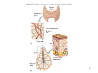
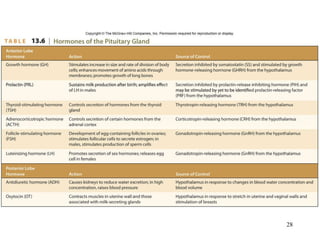
Copyright © The McGraw-Hill Companies, Inc. Permission required for reproduction or display. Chapter 13 Lecture PowerPoint
2401 Anatomy and Physiology I Chapter 13 Susan Gossett [email_address] Department of Biology Paris Junior College
Hole’s Human Anatomy and Physiology Twelfth Edition Shier    Butler    Lewis  Chapter  13 Endocrine System Copyright © The McGraw-Hill Companies, Inc. Permission required for reproduction or display.
13.1: Introduction The endocrine system assists the nervous system with communication and control of the body The cells, tissues, and organs are called endocrine glands  They are ductless They use the bloodstream They secrete hormones There are also similar glands called paracrine and autocrine glands that are quasi-endocrine Other glands that secrete substances are the exocrine glands  They have ducts They deliver their products directly to a specific site
Copyright  ©  The McGraw-Hill Companies, Inc. Permission required for reproduction or display. Hormone secretion (a) (b) Blood flow Skin Duct Thyroid gland Endocrine gland Endocrine cell Exocrine gland (sweat gland) Exocrine cells
13.2: General Characteristics of the Endocrine System The endocrine and nervous systems communicate using chemical signals Neurons release neurotransmitters into a synapse affecting postsynaptic cells Endocrine glands release hormones into the bloodstream to specific target cell receptors
Copyright  ©  The McGraw-Hill Companies, Inc. Permission required for reproduction or display. (a) (b) Nerve impulse Bloodstream Neuron transmits nerve impulse Glandular cells secrete hormone into bloodstream Neurotransmitter released into synapse Post- synaptic cell responds Target cells (cells with hormone receptors) respond to hormone Hormones have no effect on other cells
13.3: Hormone Action Hormones are released into the extracellular spaces surrounding endocrine cells Copyright  ©  The McGraw-Hill Companies, Inc. Permission required for reproduction or display. Hypothalamus Pituitary gland Thyroid gland Thymus Adrenal gland Pancreas Parathyroid gland Pineal gland Kidney Testis (in male) Ovary (in female)
Chemistry of Hormones Chemically, hormones are either: Steroid or steroid-like hormones such as: Sex hormones Adrenal cortex hormones Non-steroid hormones such as: Amines Proteins Peptides Glycoproteins
Copyright  ©  The McGraw-Hill Companies, Inc. Permission required for reproduction or display. O H 3 C (a) Cortisol (d) Oxytocin OH HO C HO (b) Norepinephrine H H OH H C NH 2 C H H C C H H C H H C H H H H H H C C C C C (e) Prostaglandin PGE 2 H OH H H H OH C H H H H C H C C H H C C H H C O OH H H H H C HO H 3 C C O C O C Ala Val Asp His Ser Arg Gly Arg Arg Asp Ser Glu Pro Arg Asp Ala Ser Glu Glu Phe Ile Gly Asp His His Ser Leu Leu Met Ala Pro Pro Ala Ala Glu Glu Leu Asp Ser Gly Glu Glu Glu Arg Gly Cys Cys Asp Glu Ile Pro Leu Gly His Ileu Met Glu Ser Phe Ala Leu Glu Ser Asp Ile Leu Leu Leu Asp Glu Leu Gly Glu S S CH 2 OH Val Lys Lys Lys Lys Val Lys Lys Lys Val Lys Tyr Val Val Lys Lys Lys Try Val Tyr (c) Parathyroid ho rm one (PTH)
Action of Hormones Steroid Hormones Hormone molecule Ribosome Nucleus 1 2 3 5 4 Newly forming protein molecule Cell membrane Hormone-receptor complex Intracellular receptor molecule mRNA mRNA DNA Copyright  ©  The McGraw-Hill Companies, Inc. Permission required for reproduction or display.
Action of Hormones Non-steroid Hormones Cell membrane G protein Cytoplasm Nucleus ATP cAMP 1 2 3 5 4 Membrane-bound receptor molecule Nonsteroid hormone molecule Hormone- receptor complex Adenylate cyclase Protein kinases (inactive) Substrate (inactive) Protein kinases (active) Substrate (active) Cellular changes Copyright  ©  The McGraw-Hill Companies, Inc. Permission required for reproduction or display.
13.1 Clinical Application Using Hormones to Improve  Athletic Performance
Prostaglandins Prostaglandins: Are paracrine substances Are very potent in small amounts Are not stored in cells but synthesized just before release Rapidly inactivate Regulate cellular responses to hormones Can activate or inhibit adenylate cyclase Controls cAMP production Alters a cells response to hormones Has a wide variety of effects
13.4: Control of  Hormonal Secretions Primarily controlled by negative feedback mechanism Hormones can be short-lived or may last for days Hormone secretions are precisely regulated
Control Sources Copyright  ©  The McGraw-Hill Companies, Inc. Permission required for reproduction or display. too high too low Control center Endocrine gland inhibited. Receptors Hormone control mechanism senses change. Effectors Hormone secretion decreased. Stimulus Hormone levels rise or controlled process increases. Response Hormone levels return toward normal. Normal hormone levels Stimulus Hormone levels drop or controlled process decreases. Response Hormone levels return toward normal. Receptors Hormone control mechanism senses change. Effectors Hormone secretion increased. Control center Endocrine gland stimulated.
Control Sources Copyright  ©  The McGraw-Hill Companies, Inc. Permission required for reproduction or display. Nervous system (a) (b) (c) Target cells Action Target cells Action – – – – Anterior pituitary gland Action Hypothalamus Peripheral endocrine gland Target cells Endocrine gland Changing level of substance in plasma Endocrine gland
13.5: Pituitary Gland Lies at the base of the brain in the sella turcica Consists of two distinct portions: Anterior pituitary (adenohypophysis) Posterior pituitary (neurohypophysis)
Optic nerve Sphenoid bone Hypothalamus Optic chiasma Sella turcica Third ventricle Trochlear nerve Basilar artery Anterior cerebral artery Pituitary stalk (Infundibulum) Anterior lobe of pituitary gland Sphenoidal sinus Posterior lobe of pituitary gland Oculomotor nerve Copyright  ©  The McGraw-Hill Companies, Inc. Permission required for reproduction or display.
Anterior Pituitary Hormones Hypothalamic releasing hormones stimulate cells of anterior pituitary to release hormones Nerve impulses from hypothalamus stimulate nerve endings in the posterior pituitary gland to release hormones Hypophyseal veins Optic chiasma Capillary bed Posterior lobe of pituitary gland Third ventricle Hypothalamus Anterior lobe of pituitary gland Capillary bed Hypophyseal portal veins Secretory cells of anterior pituitary gland Neurosecretory cells that secrete releasing hormones Superior hypophyseal artery Neurosecretory cells that secrete posterior pituitary hormones Inferior hypophyseal artery Sella turcica of sphenoid bone Copyright  ©  The McGraw-Hill Companies, Inc. Permission required for reproduction or display.
Anterior pituitary Peripheral endocrine gland Hypothalamus Target cells (Hormone 3) + – – – + + Inhibition Stimulation Releasing hormone (Hormone 1) Anterior pituitary hormone (Hormone 2) Copyright  ©  The McGraw-Hill Companies, Inc. Permission required for reproduction or display. Copyright  ©  The McGraw-Hill Companies, Inc. Permission required for reproduction or display. © Michael Ross/Photo Researchers, Inc.   Secretory cells
Copyright  ©  The McGraw-Hill Companies, Inc. Permission required for reproduction or display. Thyroid Mammary gland Ovary Testis Bone Muscle Hormones from Anterior Pituitary Hormones from Hypothalamus GHRH Growth  hormone- releasing hormone SS Somatostatin PRF Prolactin- releasing factor PIH Prolactin- release inhibiting  hormone TRH Thyrotropin- releasing hormone CRH Corticotropin- releasing hormone GnRH Gonadotropin- releasing hormone GH Growth hormone PRL Prolactin TSH Thyroid- stimulating hormone ACTH Adrenocorticotropic hormone LH Luteinizing hormone FSH Follicle- stimulating hormone Adipose tissue Adrenal cortex
13.2 Clinical Application Growth Hormone Ups and Downs
Posterior Pituitary Hormones Structurally consists of nerve fibers and neuroglia v. glandular epithelial cells of the anterior pituitary gland The nerve fibers originate in the hypothalamus Two hormones are produced: Antidiuretic hormone (vasopressin) Oxytocin
13.6: Thyroid Gland The thyroid gland has two lateral lobes and lies just below the larynx It produces three hormones: T3 (thyroxine) T4 (triiodothyronine) Calcitonin
Structure of the Gland Copyright  ©  The McGraw-Hill Companies, Inc. Permission required for reproduction or display. Follicular cells Colloid Extrafollicular cells © Fred Hossler/Visuals Unlimited   Larynx Colloid Isthmus (a) (b) Thyroid gland Follicular cell Extrafollicular cell Copyright  ©  The McGraw-Hill Companies, Inc. Permission required for reproduction or display.
Thyroid Hormones Copyright  ©  The McGraw-Hill Companies, Inc. Permission required for reproduction or display. OH O I I CH 2 I I OH O I CH 2 I I NH 2 CHCOOH NH 2 CHCOOH Thyroxine (T 4 ) Triiodothyronine (T 3 )
Copyright  ©  The McGraw-Hill Companies, Inc. Permission required for reproduction or display. © Mediscan/Visuals Unlimited   © Mediscan/Visuals Unlimited   © Mediscan/Visuals Unlimited
13.7: Parathyroid Glands The parathyroid glands are on the posterior surface of the thyroid gland There are typically four parathyroid glands It secretes one hormone: PTH (parathyroid hormone or parathormone)
Structure of the Glands Posterior view Esophagus Pharynx Thyroid gland Parathyroid glands Trachea Copyright  ©  The McGraw-Hill Companies, Inc. Permission required for reproduction or display. Secretory cells Capillaries Copyright  ©  The McGraw-Hill Companies, Inc. Permission required for reproduction or display. © R. Calentine/Visuals Unlimited
Parathyroid Hormone Copyright  ©  The McGraw-Hill Companies, Inc. Permission required for reproduction or display. Liver Intestinal enzymes Ultraviolet light in skin Kidney Stimulated by PTH Hydroxycholecalciferol Foods Ca +2 Ca +2 Ca +2 Cholesterol Provitamin D Vitamin D (Cholecalciferol) Also obtained directly from foods Dihydroxycholecalciferol (active form of vitamin D) Controls absorption of calcium in intestine
Copyright  ©  The McGraw-Hill Companies, Inc. Permission required for reproduction or display. PTH Ca +2 + PTH PTH Ca +2 + Ca +2 Bloodstream – Stimulation Inhibition Release into bloodstream Parathyroid glands (on posterior of thyroid gland) Decreased blood calcium stimulates parathyroid hormone secretion Increased blood calcium inhibits PTH secretion Kidneys conserve Ca +2  and  activate Vitamin D Bone releases Ca +2 Intestine absorbs Ca +2 Active Vitamin D
13.8: Adrenal Glands The adrenal glands are closely associated with the kidneys The gland sits like a cap on each kidney Hormones are secreted from two different areas of the gland, the adrenal cortex and the adrenal medulla Numerous hormones are secreted by the adrenal glands
Structure of the Glands Cortex Medulla Capsule Zona glomerulosa Zona fasciculata Zona reticularis Chromaffin cells Copyright  ©  The McGraw-Hill Companies, Inc. Permission required for reproduction or display. © Ed Reschke   Adrenal gland Kidney Adrenal cortex (a) Zona  lomerulosa Connective tissue capsule (b) Zona fasciculata Zona reticularis Adrenal medulla Adrenal cortex Adrenal medulla Surface of adrenal gland Copyright  ©  The McGraw-Hill Companies, Inc. Permission required for reproduction or display.
Hormones of the Adrenal Medulla
Hormones of the Adrenal Cortex
13.3 Clinical Application Disorders of the Adrenal Cortex
13.1 From Science to Technology Treating Diabetes
13.9: Pancreas The pancreas has two major types of secretory tissue This is why it is a dual functioning organ as both an exocrine gland and endocrine gland Three hormones are secreted from the islet cells: Alpha cells secrete glucagon Beta cells insulin Delta cells secrete somatostatin
Structure of the Gland Copyright  ©  The McGraw-Hill Companies, Inc. Permission required for reproduction or display. Pancreatic islet (Islet of Langerhans) From Kent M. Van De Graaff and Stuart Ira Fox, Concepts of Human Anatomy and Physiology, 2nd ed.  ©1989 Wm. C. Brown Publishers, Dubuque, Iowa.  All Rights Reserved.  Reprinted with permission   Copyright  ©  The McGraw-Hill Companies, Inc. Permission required for reproduction or display. Gallbladder Common bile duct Pancreatic duct Pancreas Duct Capillary Small intestine Digestive enzyme- secreting cells Pancreatic islet (Islet of Langerhans) Hormone-secreting islet cells
Hormones of the Pancreatic Islets
Copyright  ©  The McGraw-Hill Companies, Inc. Permission required for reproduction or display. too high too low Control center Beta cells secrete insulin Receptors Beta cells detect a rise in blood glucose Effectors Insulin •  Promotes movement  of glucose into certain cells •  Stimulates formation of glycogen from glucose Stimulus Rise in blood glucose Response Blood glucose drops toward normal (and inhibits insulin secretion) Normal blood glucose concentration Stimulus Drop in blood glucose Response Blood glucose rises toward normal (and inhibits glucagon secretion) Receptors Alpha cells detect a drop in blood glucose Effectors Glucagon •  Stimulates cells to break down glycogen into glucose •  Stimulates cells to convert noncarbohydrates into glucose Control center Alpha cells secrete glucagon
13.4 Clinical Application Diabetes Mellitus
13.10: Other Endocrine Glands Pineal Gland Secretes melatonin Regulates circadian rhythms Thymus Gland Secretes thymosins Promotes development of certain lymphocytes Important in role of immunity Reproductive Organs Ovaries produce estrogens and progesterone Testes produce testosterone Placenta produces estrogens, progesterone, and gonadotropin Other organs : digestive glands, heart, and kidney
13.11: Stress and Its Effects Survival depends on maintaining homeostasis Factors that change the internal environment are potentially life-threatening Sensing such dangers directs nerve impulses to the hypothalamus This can trigger a loss of homeostasis
Types of Stress Two types of stress: Physical stress Psychological stress
Responses to Stress Copyright  ©  The McGraw-Hill Companies, Inc. Permission required for reproduction or display. Sympathetic impulses CRH released ACTH released Cortisol released Long-term adjustment or resistance stage •  Increase in blood concentration of amino acids. •  Increased release of fatty acids. •  Blood glucose increases. •  Blood glycerol and fatty acids increase. •  Heart rate increases. •  Blood pressure rises. •  Breathing rate increases. • Air passages dilate. •  Pupils dilate. Anterior pituitary Hypothalamus Adrenal cortex Adrenal medulla Neural signals Hormonal signals Stress results from changes in the external environment Signals from sensory receptors Epinephrine and norepinephrine released Norepinephrine released Short-term “fight or flight” or alarm stage. •  Blood flow redistributes. •  Increased glucose formed from noncarbohydrates—amino acids (from proteins) and glycerol (from fats).
13.12: Lifespan Changes Endocrine glands decrease in size Muscular strength decreases as GH levels decrease ADH levels increase due to slower break down in liver and kidneys Calcitonin levels decrease; increase risk of osteoporosis PTH level changes contribute to risk of osteoporosis Insulin resistance may develop Changes in melatonin secretion affect the body clock Thymosin production declines increasing risk of infections
Important Points in Chapter 13: Outcomes to be Assessed 13.1: Introduction  Define hormone. Distinguish between endocrine and exocrine glands. 13.2: General Characteristics of the Endocrine System Explain what makes a cell a target cell for a hormone. List some important functions of hormones. 13.3: Hormone Action Describe how hormones can be classified according to their chemical composition. Explain how steroid and non-steroid hormones affect their target cells.
Important Points in Chapter 13: Outcomes to be Assessed 13.4: Control of Hormone Secretion Discuss how negative feedback mechanisms regulate hormone secretion. Explain how the nervous system controls hormone secretion. 13.5-13.10: Pituitary Gland – Other Endocrine Glands Name and describe the locations of the major endocrine glands and list the hormones that they secrete. Describe the actions of the various hormones and their contributions to homeostasis. Explain how the secretion of each hormone is regulated.
Important Points in Chapter 13: Outcomes to be Assessed 13.11: Stress and Its Effects Distinguish between physical and psychological stress. Describe the general stress response. 13.12: Lifespan Changes Describe some of the changes associated with aging of the endocrine system.
Quiz 13 Complete Quiz 13 now! Read Chapter 14.

Chapter 13 Endocrine System

  • 1.
    Copyright © TheMcGraw-Hill Companies, Inc. Permission required for reproduction or display. Chapter 13 Lecture PowerPoint
  • 2.
    2401 Anatomy andPhysiology I Chapter 13 Susan Gossett [email_address] Department of Biology Paris Junior College
  • 3.
    Hole’s Human Anatomyand Physiology Twelfth Edition Shier  Butler  Lewis Chapter 13 Endocrine System Copyright © The McGraw-Hill Companies, Inc. Permission required for reproduction or display.
  • 4.
    13.1: Introduction Theendocrine system assists the nervous system with communication and control of the body The cells, tissues, and organs are called endocrine glands They are ductless They use the bloodstream They secrete hormones There are also similar glands called paracrine and autocrine glands that are quasi-endocrine Other glands that secrete substances are the exocrine glands They have ducts They deliver their products directly to a specific site
  • 5.
    Copyright © The McGraw-Hill Companies, Inc. Permission required for reproduction or display. Hormone secretion (a) (b) Blood flow Skin Duct Thyroid gland Endocrine gland Endocrine cell Exocrine gland (sweat gland) Exocrine cells
  • 6.
    13.2: General Characteristicsof the Endocrine System The endocrine and nervous systems communicate using chemical signals Neurons release neurotransmitters into a synapse affecting postsynaptic cells Endocrine glands release hormones into the bloodstream to specific target cell receptors
  • 7.
    Copyright © The McGraw-Hill Companies, Inc. Permission required for reproduction or display. (a) (b) Nerve impulse Bloodstream Neuron transmits nerve impulse Glandular cells secrete hormone into bloodstream Neurotransmitter released into synapse Post- synaptic cell responds Target cells (cells with hormone receptors) respond to hormone Hormones have no effect on other cells
  • 9.
    13.3: Hormone ActionHormones are released into the extracellular spaces surrounding endocrine cells Copyright © The McGraw-Hill Companies, Inc. Permission required for reproduction or display. Hypothalamus Pituitary gland Thyroid gland Thymus Adrenal gland Pancreas Parathyroid gland Pineal gland Kidney Testis (in male) Ovary (in female)
  • 10.
    Chemistry of HormonesChemically, hormones are either: Steroid or steroid-like hormones such as: Sex hormones Adrenal cortex hormones Non-steroid hormones such as: Amines Proteins Peptides Glycoproteins
  • 11.
    Copyright © The McGraw-Hill Companies, Inc. Permission required for reproduction or display. O H 3 C (a) Cortisol (d) Oxytocin OH HO C HO (b) Norepinephrine H H OH H C NH 2 C H H C C H H C H H C H H H H H H C C C C C (e) Prostaglandin PGE 2 H OH H H H OH C H H H H C H C C H H C C H H C O OH H H H H C HO H 3 C C O C O C Ala Val Asp His Ser Arg Gly Arg Arg Asp Ser Glu Pro Arg Asp Ala Ser Glu Glu Phe Ile Gly Asp His His Ser Leu Leu Met Ala Pro Pro Ala Ala Glu Glu Leu Asp Ser Gly Glu Glu Glu Arg Gly Cys Cys Asp Glu Ile Pro Leu Gly His Ileu Met Glu Ser Phe Ala Leu Glu Ser Asp Ile Leu Leu Leu Asp Glu Leu Gly Glu S S CH 2 OH Val Lys Lys Lys Lys Val Lys Lys Lys Val Lys Tyr Val Val Lys Lys Lys Try Val Tyr (c) Parathyroid ho rm one (PTH)
  • 14.
    Action of HormonesSteroid Hormones Hormone molecule Ribosome Nucleus 1 2 3 5 4 Newly forming protein molecule Cell membrane Hormone-receptor complex Intracellular receptor molecule mRNA mRNA DNA Copyright © The McGraw-Hill Companies, Inc. Permission required for reproduction or display.
  • 15.
    Action of HormonesNon-steroid Hormones Cell membrane G protein Cytoplasm Nucleus ATP cAMP 1 2 3 5 4 Membrane-bound receptor molecule Nonsteroid hormone molecule Hormone- receptor complex Adenylate cyclase Protein kinases (inactive) Substrate (inactive) Protein kinases (active) Substrate (active) Cellular changes Copyright © The McGraw-Hill Companies, Inc. Permission required for reproduction or display.
  • 16.
    13.1 Clinical ApplicationUsing Hormones to Improve Athletic Performance
  • 17.
    Prostaglandins Prostaglandins: Areparacrine substances Are very potent in small amounts Are not stored in cells but synthesized just before release Rapidly inactivate Regulate cellular responses to hormones Can activate or inhibit adenylate cyclase Controls cAMP production Alters a cells response to hormones Has a wide variety of effects
  • 18.
    13.4: Control of Hormonal Secretions Primarily controlled by negative feedback mechanism Hormones can be short-lived or may last for days Hormone secretions are precisely regulated
  • 19.
    Control Sources Copyright © The McGraw-Hill Companies, Inc. Permission required for reproduction or display. too high too low Control center Endocrine gland inhibited. Receptors Hormone control mechanism senses change. Effectors Hormone secretion decreased. Stimulus Hormone levels rise or controlled process increases. Response Hormone levels return toward normal. Normal hormone levels Stimulus Hormone levels drop or controlled process decreases. Response Hormone levels return toward normal. Receptors Hormone control mechanism senses change. Effectors Hormone secretion increased. Control center Endocrine gland stimulated.
  • 20.
    Control Sources Copyright © The McGraw-Hill Companies, Inc. Permission required for reproduction or display. Nervous system (a) (b) (c) Target cells Action Target cells Action – – – – Anterior pituitary gland Action Hypothalamus Peripheral endocrine gland Target cells Endocrine gland Changing level of substance in plasma Endocrine gland
  • 21.
    13.5: Pituitary GlandLies at the base of the brain in the sella turcica Consists of two distinct portions: Anterior pituitary (adenohypophysis) Posterior pituitary (neurohypophysis)
  • 22.
    Optic nerve Sphenoidbone Hypothalamus Optic chiasma Sella turcica Third ventricle Trochlear nerve Basilar artery Anterior cerebral artery Pituitary stalk (Infundibulum) Anterior lobe of pituitary gland Sphenoidal sinus Posterior lobe of pituitary gland Oculomotor nerve Copyright © The McGraw-Hill Companies, Inc. Permission required for reproduction or display.
  • 23.
    Anterior Pituitary HormonesHypothalamic releasing hormones stimulate cells of anterior pituitary to release hormones Nerve impulses from hypothalamus stimulate nerve endings in the posterior pituitary gland to release hormones Hypophyseal veins Optic chiasma Capillary bed Posterior lobe of pituitary gland Third ventricle Hypothalamus Anterior lobe of pituitary gland Capillary bed Hypophyseal portal veins Secretory cells of anterior pituitary gland Neurosecretory cells that secrete releasing hormones Superior hypophyseal artery Neurosecretory cells that secrete posterior pituitary hormones Inferior hypophyseal artery Sella turcica of sphenoid bone Copyright © The McGraw-Hill Companies, Inc. Permission required for reproduction or display.
  • 24.
    Anterior pituitary Peripheralendocrine gland Hypothalamus Target cells (Hormone 3) + – – – + + Inhibition Stimulation Releasing hormone (Hormone 1) Anterior pituitary hormone (Hormone 2) Copyright © The McGraw-Hill Companies, Inc. Permission required for reproduction or display. Copyright © The McGraw-Hill Companies, Inc. Permission required for reproduction or display. © Michael Ross/Photo Researchers, Inc. Secretory cells
  • 25.
    Copyright © The McGraw-Hill Companies, Inc. Permission required for reproduction or display. Thyroid Mammary gland Ovary Testis Bone Muscle Hormones from Anterior Pituitary Hormones from Hypothalamus GHRH Growth hormone- releasing hormone SS Somatostatin PRF Prolactin- releasing factor PIH Prolactin- release inhibiting hormone TRH Thyrotropin- releasing hormone CRH Corticotropin- releasing hormone GnRH Gonadotropin- releasing hormone GH Growth hormone PRL Prolactin TSH Thyroid- stimulating hormone ACTH Adrenocorticotropic hormone LH Luteinizing hormone FSH Follicle- stimulating hormone Adipose tissue Adrenal cortex
  • 26.
    13.2 Clinical ApplicationGrowth Hormone Ups and Downs
  • 27.
    Posterior Pituitary HormonesStructurally consists of nerve fibers and neuroglia v. glandular epithelial cells of the anterior pituitary gland The nerve fibers originate in the hypothalamus Two hormones are produced: Antidiuretic hormone (vasopressin) Oxytocin
  • 29.
    13.6: Thyroid GlandThe thyroid gland has two lateral lobes and lies just below the larynx It produces three hormones: T3 (thyroxine) T4 (triiodothyronine) Calcitonin
  • 30.
    Structure of theGland Copyright © The McGraw-Hill Companies, Inc. Permission required for reproduction or display. Follicular cells Colloid Extrafollicular cells © Fred Hossler/Visuals Unlimited Larynx Colloid Isthmus (a) (b) Thyroid gland Follicular cell Extrafollicular cell Copyright © The McGraw-Hill Companies, Inc. Permission required for reproduction or display.
  • 31.
    Thyroid Hormones Copyright © The McGraw-Hill Companies, Inc. Permission required for reproduction or display. OH O I I CH 2 I I OH O I CH 2 I I NH 2 CHCOOH NH 2 CHCOOH Thyroxine (T 4 ) Triiodothyronine (T 3 )
  • 32.
    Copyright © The McGraw-Hill Companies, Inc. Permission required for reproduction or display. © Mediscan/Visuals Unlimited © Mediscan/Visuals Unlimited © Mediscan/Visuals Unlimited
  • 33.
    13.7: Parathyroid GlandsThe parathyroid glands are on the posterior surface of the thyroid gland There are typically four parathyroid glands It secretes one hormone: PTH (parathyroid hormone or parathormone)
  • 34.
    Structure of theGlands Posterior view Esophagus Pharynx Thyroid gland Parathyroid glands Trachea Copyright © The McGraw-Hill Companies, Inc. Permission required for reproduction or display. Secretory cells Capillaries Copyright © The McGraw-Hill Companies, Inc. Permission required for reproduction or display. © R. Calentine/Visuals Unlimited
  • 35.
    Parathyroid Hormone Copyright © The McGraw-Hill Companies, Inc. Permission required for reproduction or display. Liver Intestinal enzymes Ultraviolet light in skin Kidney Stimulated by PTH Hydroxycholecalciferol Foods Ca +2 Ca +2 Ca +2 Cholesterol Provitamin D Vitamin D (Cholecalciferol) Also obtained directly from foods Dihydroxycholecalciferol (active form of vitamin D) Controls absorption of calcium in intestine
  • 36.
    Copyright © The McGraw-Hill Companies, Inc. Permission required for reproduction or display. PTH Ca +2 + PTH PTH Ca +2 + Ca +2 Bloodstream – Stimulation Inhibition Release into bloodstream Parathyroid glands (on posterior of thyroid gland) Decreased blood calcium stimulates parathyroid hormone secretion Increased blood calcium inhibits PTH secretion Kidneys conserve Ca +2 and activate Vitamin D Bone releases Ca +2 Intestine absorbs Ca +2 Active Vitamin D
  • 38.
    13.8: Adrenal GlandsThe adrenal glands are closely associated with the kidneys The gland sits like a cap on each kidney Hormones are secreted from two different areas of the gland, the adrenal cortex and the adrenal medulla Numerous hormones are secreted by the adrenal glands
  • 39.
    Structure of theGlands Cortex Medulla Capsule Zona glomerulosa Zona fasciculata Zona reticularis Chromaffin cells Copyright © The McGraw-Hill Companies, Inc. Permission required for reproduction or display. © Ed Reschke Adrenal gland Kidney Adrenal cortex (a) Zona lomerulosa Connective tissue capsule (b) Zona fasciculata Zona reticularis Adrenal medulla Adrenal cortex Adrenal medulla Surface of adrenal gland Copyright © The McGraw-Hill Companies, Inc. Permission required for reproduction or display.
  • 40.
    Hormones of theAdrenal Medulla
  • 41.
    Hormones of theAdrenal Cortex
  • 42.
    13.3 Clinical ApplicationDisorders of the Adrenal Cortex
  • 43.
    13.1 From Scienceto Technology Treating Diabetes
  • 44.
    13.9: Pancreas Thepancreas has two major types of secretory tissue This is why it is a dual functioning organ as both an exocrine gland and endocrine gland Three hormones are secreted from the islet cells: Alpha cells secrete glucagon Beta cells insulin Delta cells secrete somatostatin
  • 45.
    Structure of theGland Copyright © The McGraw-Hill Companies, Inc. Permission required for reproduction or display. Pancreatic islet (Islet of Langerhans) From Kent M. Van De Graaff and Stuart Ira Fox, Concepts of Human Anatomy and Physiology, 2nd ed. ©1989 Wm. C. Brown Publishers, Dubuque, Iowa. All Rights Reserved. Reprinted with permission Copyright © The McGraw-Hill Companies, Inc. Permission required for reproduction or display. Gallbladder Common bile duct Pancreatic duct Pancreas Duct Capillary Small intestine Digestive enzyme- secreting cells Pancreatic islet (Islet of Langerhans) Hormone-secreting islet cells
  • 46.
    Hormones of thePancreatic Islets
  • 47.
    Copyright © The McGraw-Hill Companies, Inc. Permission required for reproduction or display. too high too low Control center Beta cells secrete insulin Receptors Beta cells detect a rise in blood glucose Effectors Insulin • Promotes movement of glucose into certain cells • Stimulates formation of glycogen from glucose Stimulus Rise in blood glucose Response Blood glucose drops toward normal (and inhibits insulin secretion) Normal blood glucose concentration Stimulus Drop in blood glucose Response Blood glucose rises toward normal (and inhibits glucagon secretion) Receptors Alpha cells detect a drop in blood glucose Effectors Glucagon • Stimulates cells to break down glycogen into glucose • Stimulates cells to convert noncarbohydrates into glucose Control center Alpha cells secrete glucagon
  • 48.
    13.4 Clinical ApplicationDiabetes Mellitus
  • 49.
    13.10: Other EndocrineGlands Pineal Gland Secretes melatonin Regulates circadian rhythms Thymus Gland Secretes thymosins Promotes development of certain lymphocytes Important in role of immunity Reproductive Organs Ovaries produce estrogens and progesterone Testes produce testosterone Placenta produces estrogens, progesterone, and gonadotropin Other organs : digestive glands, heart, and kidney
  • 50.
    13.11: Stress andIts Effects Survival depends on maintaining homeostasis Factors that change the internal environment are potentially life-threatening Sensing such dangers directs nerve impulses to the hypothalamus This can trigger a loss of homeostasis
  • 51.
    Types of StressTwo types of stress: Physical stress Psychological stress
  • 52.
    Responses to StressCopyright © The McGraw-Hill Companies, Inc. Permission required for reproduction or display. Sympathetic impulses CRH released ACTH released Cortisol released Long-term adjustment or resistance stage • Increase in blood concentration of amino acids. • Increased release of fatty acids. • Blood glucose increases. • Blood glycerol and fatty acids increase. • Heart rate increases. • Blood pressure rises. • Breathing rate increases. • Air passages dilate. • Pupils dilate. Anterior pituitary Hypothalamus Adrenal cortex Adrenal medulla Neural signals Hormonal signals Stress results from changes in the external environment Signals from sensory receptors Epinephrine and norepinephrine released Norepinephrine released Short-term “fight or flight” or alarm stage. • Blood flow redistributes. • Increased glucose formed from noncarbohydrates—amino acids (from proteins) and glycerol (from fats).
  • 54.
    13.12: Lifespan ChangesEndocrine glands decrease in size Muscular strength decreases as GH levels decrease ADH levels increase due to slower break down in liver and kidneys Calcitonin levels decrease; increase risk of osteoporosis PTH level changes contribute to risk of osteoporosis Insulin resistance may develop Changes in melatonin secretion affect the body clock Thymosin production declines increasing risk of infections
  • 55.
    Important Points inChapter 13: Outcomes to be Assessed 13.1: Introduction Define hormone. Distinguish between endocrine and exocrine glands. 13.2: General Characteristics of the Endocrine System Explain what makes a cell a target cell for a hormone. List some important functions of hormones. 13.3: Hormone Action Describe how hormones can be classified according to their chemical composition. Explain how steroid and non-steroid hormones affect their target cells.
  • 56.
    Important Points inChapter 13: Outcomes to be Assessed 13.4: Control of Hormone Secretion Discuss how negative feedback mechanisms regulate hormone secretion. Explain how the nervous system controls hormone secretion. 13.5-13.10: Pituitary Gland – Other Endocrine Glands Name and describe the locations of the major endocrine glands and list the hormones that they secrete. Describe the actions of the various hormones and their contributions to homeostasis. Explain how the secretion of each hormone is regulated.
  • 57.
    Important Points inChapter 13: Outcomes to be Assessed 13.11: Stress and Its Effects Distinguish between physical and psychological stress. Describe the general stress response. 13.12: Lifespan Changes Describe some of the changes associated with aging of the endocrine system.
  • 58.
    Quiz 13 CompleteQuiz 13 now! Read Chapter 14.