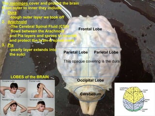
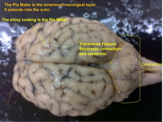
The meninges cover and protect the brain
From outer to inner they include:
1. Dura
    -tough outer layer we took off
2. Arachnoid
    -The Cerebral Spinal Fluid (CSF)
                                           Frontal Lobe
     flows between the Arachnoid
    and Pia layers and serves to nourish
    and protect the brain & spinal cord.
3. Pia
    -pearly layer extends into
                                  Parietal Lobe     Parietal Lobe
    the sulci
                                  This opaque covering is the dura


    LOBES of the BRAIN
                                          Occipital Lobe


                                           Cerebellum
Cerebellum


     Brain Stem:
      Midbrain (we won’t differentiate this)
      Pons
      Medulla
Gyrus   Sulci


Longitudinal
  Fissure
The Pia Mater is the innermost meningeal layer.
 It extends into the sulci.

The shiny coating is the Pia Mater


                                       Transverse Fissure
                                       Separates cerebellum
                                       and cerebrum

                                                              Cerebellum
Lateral View: Left Hemisphere of the
Cerebrum and cerebellum with brain
stem


                                Left Parietal Lobe

              Frontal
              Lobe                                        Transverse Fissure

                                   Temporal
                                                     Cerebellum
                                    Lobe


                                              Pons
Underside of Brain


   Brain Stem                        This usually
                                     gets pulled off
(Pons & Medulla)            Pituitarywith the dura.
                             Gland

                                                                   Olfactory


                                     Optic Chiasm
                                                    Optic Nerves
                                                                    Bulbs


            Capillary Bed
Posterior (rear) view
showing cerebellum
being pulled down to                   Occipital Lobe of Cerebrum
expose the Superior
colliculi, inferior colliculi,
and the Pineal Gland.

                                                  Pineal
                                                  Gland
                  Superior Colliculi        Inferior Colliculi


            Cerebellum
Mammillary Body
                   Optic Chiasm
Olfactory Bulb
                                                                            Pons         Medulla
                 Infundibulum
                 Stalk of the pituitary
                                          Hypothalamus
                                          Deep to the infundibulum
                                                                     Occulomotor Nerve
Corpus callosum
                      Fornix          Arbor Vitae
                                      Of Cerebellum


               Thalamus


                               Pons    Medulla
                                                      Spinal Cord
Optic Chiasm
Lateral view, hemi section of lab brain model.


                         superior


posterior                                                 anterior


                Cerebellum


                                               Pituitary Gland
                             inferior
Olfactory Tract          Pituitary Gland


                  pons
SPINAL CORD ANATOMY
                   Keep in mind that our models are HUGE. The spinal cord
                    Is really only about the thickness of your pinky finger!
                                 Posterior
                                                       Spinous Process of Vertebrae


                Posterior White Column
                                                       Central Canal: contains CSF
           Posterior Gray Horn
            Lateral Gray Horn
                                                          Contains Sensory & Interneurons
    Axons of Sensory Neurons


Dorsal Root Ganglion


                                     Anterior Gray Horn: Contains cell bodies of
                                     somatic motor neurons

                       Axons of Motor Neurons


                                   Anterior
This is the URL to a sheep dissection lab created by five students in
the bi-college (Haverford and Bryn Mawr) community: Jessica Kuhn,
Jessica Magid, Karen Revere, Elizabeth Caris, and Gray Vargas
through science education at Bryn Mawr College. It is a fantastic site
to explore the brain. Not everything you will learn will be on a test
BUT remember that educating yourself in your field is NOT about
what is going to be on the test .

Brain dissection pictures

  • 1.
    The meninges coverand protect the brain From outer to inner they include: 1. Dura -tough outer layer we took off 2. Arachnoid -The Cerebral Spinal Fluid (CSF) Frontal Lobe flows between the Arachnoid and Pia layers and serves to nourish and protect the brain & spinal cord. 3. Pia -pearly layer extends into Parietal Lobe Parietal Lobe the sulci This opaque covering is the dura LOBES of the BRAIN Occipital Lobe Cerebellum
  • 2.
    Cerebellum Brain Stem: Midbrain (we won’t differentiate this) Pons Medulla
  • 3.
    Gyrus Sulci Longitudinal Fissure
  • 4.
    The Pia Materis the innermost meningeal layer. It extends into the sulci. The shiny coating is the Pia Mater Transverse Fissure Separates cerebellum and cerebrum Cerebellum
  • 5.
    Lateral View: LeftHemisphere of the Cerebrum and cerebellum with brain stem Left Parietal Lobe Frontal Lobe Transverse Fissure Temporal Cerebellum Lobe Pons
  • 6.
    Underside of Brain Brain Stem This usually gets pulled off (Pons & Medulla) Pituitarywith the dura. Gland Olfactory Optic Chiasm Optic Nerves Bulbs Capillary Bed
  • 7.
    Posterior (rear) view showingcerebellum being pulled down to Occipital Lobe of Cerebrum expose the Superior colliculi, inferior colliculi, and the Pineal Gland. Pineal Gland Superior Colliculi Inferior Colliculi Cerebellum
  • 8.
    Mammillary Body Optic Chiasm Olfactory Bulb Pons Medulla Infundibulum Stalk of the pituitary Hypothalamus Deep to the infundibulum Occulomotor Nerve
  • 9.
    Corpus callosum Fornix Arbor Vitae Of Cerebellum Thalamus Pons Medulla Spinal Cord Optic Chiasm
  • 10.
    Lateral view, hemisection of lab brain model. superior posterior anterior Cerebellum Pituitary Gland inferior
  • 11.
    Olfactory Tract Pituitary Gland pons
  • 12.
    SPINAL CORD ANATOMY Keep in mind that our models are HUGE. The spinal cord Is really only about the thickness of your pinky finger! Posterior Spinous Process of Vertebrae Posterior White Column Central Canal: contains CSF Posterior Gray Horn Lateral Gray Horn Contains Sensory & Interneurons Axons of Sensory Neurons Dorsal Root Ganglion Anterior Gray Horn: Contains cell bodies of somatic motor neurons Axons of Motor Neurons Anterior
  • 13.
    This is theURL to a sheep dissection lab created by five students in the bi-college (Haverford and Bryn Mawr) community: Jessica Kuhn, Jessica Magid, Karen Revere, Elizabeth Caris, and Gray Vargas through science education at Bryn Mawr College. It is a fantastic site to explore the brain. Not everything you will learn will be on a test BUT remember that educating yourself in your field is NOT about what is going to be on the test .