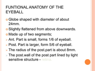
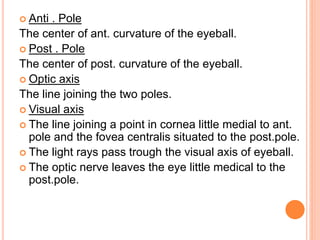
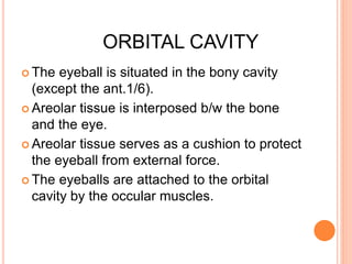
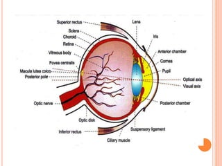
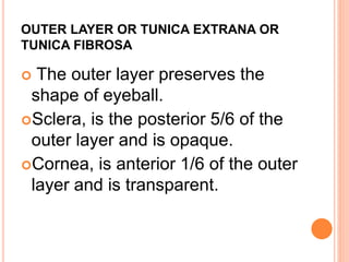
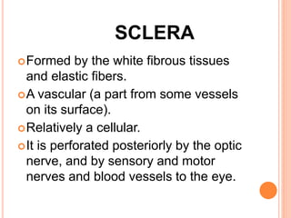
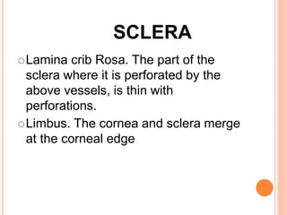
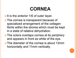
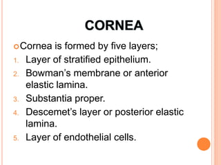
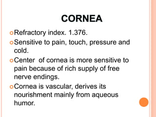
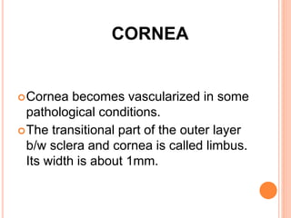
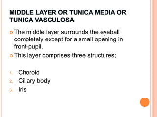
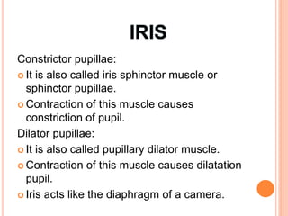
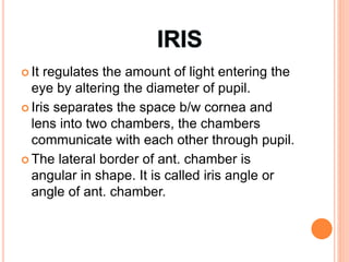
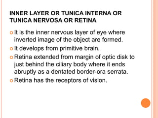
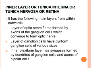
EYE/VISION
FUNTIONAL ANATOMY OF THE
EYEBALL
 Globe shaped with diameter of about
24mm.
 Slightly flattened from above downwards.
 Made up of two segments;
 Ant. Part is small, forms 1/6 of eyeball.
 Post. Part is larger, form 5/6 of eyeball.
 The radius of the post.part is about 8mm.
 The post.wall of the post part lined by light
sensitive structure - retina.
 Anti . Pole
The center of ant. curvature of the eyeball.
 Post . Pole
The center of post. curvature of the eyeball.
 Optic axis
The line joining the two poles.
 Visual axis
 The line joining a point in cornea little medial to ant.
pole and the fovea centralis situated to the post.pole.
 The light rays pass trough the visual axis of eyeball.
 The optic nerve leaves the eye little medical to the
post.pole.
ORBITAL CAVITY
 The eyeball is situated in the bony cavity
(except the ant.1/6).
 Areolar tissue is interposed b/w the bone
and the eye.
 Areolar tissue serves as a cushion to protect
the eyeball from external force.
 The eyeballs are attached to the orbital
cavity by the occular muscles.
EYELIDS
 Protects the eyeball.
 Open and close voluntarily and reflexly.
 The margins of eyelids have sensitive hairs-
cilia.
 Each cilia has a follicle, surrounded by a
nerve plexus.
 Blinking of eyelids occur when sensory
nerves (cilia) are activated.
 Prevents dust particles from reaching
eyeball.
EYELIDS
 100-150 cilia in the upper lid and about 50-
75 in the lower lid.
 Meibonian glands and some sebaceous
glands are also found in the eyelids.
 These glands open into the follicles of cilia.
 The infection of these glands leads to the
development of common eye sty.
 The opening b/w the two eyelids is called
palpebral fissure.
INNERVATION OF EYELIDS
 Sensory innervation is from the Trigeminal
(5th) cranial nerve, via the ophthalmic
division (upper lid) and maxillary division
(lower lid).
 The orbicularis oculi is innervated by the
facial (7th) nerve.
 The levator muscle in the upper lid is
supplied by the oculomoter (3rd) nerve.
BLOOD SUPPLY AND LYMPHATICS
 The eyelids are supplied by an extensive
network of blood vessels which form an
anastomosis b/w branches derived from the
external carotid artery via the face and from
the internal carotid artery via the orbit.
 Lymphatic fluid drains into the periaricular
and sub mandibular nodes.
 Periaricular lymphadenopathy is a useful
sign of infective eyelid swelling (especially
viral).
CONJUNCTIVA
 A thin mucous memb. which covers the
exposed part of the eye.
 It covers the ant. surface and also reflected
into the inner surface of eyelids.
 Bulbar portion. The part of conjunctiva
covering the eyeball.
 Palpebral portion. The part covering the
eyelids.
 The opposed portions of conjunctiva slid
over each other.
CONJUNCTIVA
The surface of conjunctiva is
lubricated by a thin film of tears
secreted by lacrimal glands.
At the upper and lower reflections b/w
eyeball and eyelid the conjunctive
forms two sacs, the superior and
inferior fornices.
LACRIMAL GLAND
 Situated in the shelter of bone, forming the
upper and outer border of wall of the eye
socket.
 Tears flow from lacrimal gland over the
surface of conjunctiva and drains into nose
via lacrimal duct, lacrimal sac and
nasolacrimal duct.
 Tear is a hypertonic fluid.
 Conjunctive is kept moist and is protected
from infection due to its continuous washing
and lubrication (by tears).
LACRIMAL GLAND
 Tears also contains lysozyme that kills bacteria.
 Secretion of tears is controlled by the
parasympathetic fiber of facial (VII cranial) nerve.
 At birth, the N/L duct may not be fully developed,
causing a watery eye.
 Acquired obstruction of the N/L duct is a common
cause of water eye in adult. It may lead to an acute
infection of the sac.
WALL OF THE EYEBALL
Compose of three layers;
1. Outer layer, which includes cornea
and sclera.
2. Middle layer; which includes choroid,
ciliary body and iris.
3. Inner layer, the retina.
OUTER LAYER OR TUNICA EXTRANA OR
TUNICA FIBROSA
 The outer layer preserves the
shape of eyeball.
Sclera, is the posterior 5/6 of the
outer layer and is opaque.
Cornea, is anterior 1/6 of the outer
layer and is transparent.
SCLERA
Formed by the white fibrous tissues
and elastic fibers.
A vascular (a part from some vessels
on its surface).
Relatively a cellular.
It is perforated posteriorly by the optic
nerve, and by sensory and motor
nerves and blood vessels to the eye.
SCLERA
Lamina crib Rosa. The part of the
sclera where it is perforated by the
above vessels, is thin with
perforations.
Limbus. The cornea and sclera merge
at the corneal edge.
CORNEA
 It is the anterior 1/6 of outer layer.
 The cornea is transparent because of
specialized arrangement of the collagen
fibrils within the stroma which must be kept
in a state of relative dehydration.
 The sclera overlaps cornea at its periphery
and appears in front as white of the eye.
 The diameter of the cornea is about 12mm
horizontally and 11mm vertically.
Cornea is formed by five layers;
1. Layer of stratified epithelium.
2. Bowman’s membrane or anterior
elastic lamina.
3. Substantia proper.
4. Descemet’s layer or posterior elastic
lamina.
5. Layer of endothelial cells.
Refractory index. 1.376.
Sensitive to pain, touch, pressure and
cold.
Center of cornea is more sensitive to
pain because of rich supply of free
nerve endings.
Cornea is vascular, derives its
nourishment mainly from aqueous
humor.
CORNEA
Cornea becomes vascularized in some
pathological conditions.
The transitional part of the outer layer
b/w sclera and cornea is called limbus.
Its width is about 1mm.
 The middle layer surrounds the eyeball
completely except for a small opening in
front-pupil.
 This layer comprises three structures;
1. Choroid
2. Ciliary body
3. Iris
CHOROID
 Forms post. 5/6 of middle layer.
 Separated from sclera by perichoroidal space.
 Antiaorly the space is limited by the insertion of
ciliary muscle into the sclera.
 Post. The space ends at short distance from
the optic nerve.
 Inner surface of choroid faces to the pigment
epithelium of retina.
 Choroid is composed of a rich capillary plexus,
numerous small arteries and veins.
CILIARY BODY
 the choroid is extended anteriorly up to the
insertion of ciliary muscle (the level of ora
serrata).
 In front of ora serrata, the middle layer is
thickened to form ciliary body.
 It is in the form of a ring.
 Its outer surface is separated from the
sclera by perichoroidal space.
 The inner surface of ciliary body faces the
vitreous body and lens.
 The ant. surface of the ciliary body faces
towards the center of cornea.
 From the surface, the iris arises.
 Ciliary body has three parts;
1. Orbiculus ciliaris.
2. Ciliary body proper.
3. Ciliary process.
I. ORBICULUS CILIARIS
 It is continuous with choroid and it forms the
posterior 2/3 of ciliary body.
 It is about 4mm broad.
II. CILIARY BODY PROPER
It is made up of two sets of ciliary
muscles;
 Outer longitudinal muscle
 Inner circular muscle
The ciliary muscles are innervated by
the parasympathetic fibers of
oculomotor nerve (6th nerve).
III. THE CILIARY PROCESS
Finger like projections from the inner
surface of the ciliary body.
There are about 70 ciliary processes.
They projects towards the central axis
of the eye to form radial fringes called
ciliaris.
IRIS
 It is the colored structure and forms the
anterior most part of the middle layer.
 It is a thin circular diaphram.
 Pupil is a circular opening in the center of
iris.
 Iris is a muscular structure and has two
muscles;
I. Constrictor pupillae, formed by circular
muscle fibers.
II. Dilator pupillae, formed by ractial muscle
fibers.
Constrictor pupillae:
 It is also called iris sphinctor muscle or
sphinctor pupillae.
 Contraction of this muscle causes
constriction of pupil.
Dilator pupillae:
 It is also called pupillary dilator muscle.
 Contraction of this muscle causes dilatation
pupil.
 Iris acts like the diaphragm of a camera.
 It regulates the amount of light entering the
eye by altering the diameter of pupil.
 Iris separates the space b/w cornea and
lens into two chambers, the chambers
communicate with each other through pupil.
 The lateral border of ant. chamber is
angular in shape. It is called iris angle or
angle of ant. chamber.
INNER LAYER OR TUNICA INTERNA OR
TUNICA NERVOSA OR RETINA
 It is the inner nervous layer of eye where
inverted image of the object are formed.
 It develops from primitive brain.
 Retina extended from margin of optic disk to
just behind the ciliary body where it ends
abruptly as a dentated border-ora serrata.
 Retina has the receptors of vision.
 It has the following main layers from within
outwards;
I. Layer of optic nerve fibres formed by
axons of the ganglion cells which
converge to form optic nerve.
II. Layer of ganglion cells have pyriform
ganglion cells of various sizes.
III. Inner plexiform layer has synapses formed
by dendrites of ganglion cells and axons of
bipolar cells.
IV. Inner nuclear layer is mainly formed by
bipolar cells which are small and oval shaped.
V. Outer plexiform layer has synapses
formed by dendrites of bipolar cells and axons
of rods and cones cells.
VI. Outer nuclear layer is formed by fibres and
nuclei of rods and cones.
VII. Rods and cones layer is the
photoreceptor layer. Rods are cylindrical,
have rhodopsin pigment
And are associated with dim light-vision (scotopic
vision). The cones have pyramidal shape and are
associated with bright light vision (photopic
vision) and colour vision.
VIII. Pigment cell layer is composed of single layer
of hexagonal epithelial cells, having dark
brown pigment (fuchsin).
 It should be noted that light has to traverse
ganglion cells and bipolar cells to reach the
photoreceptors rods and cones, where visual
impulses are set up, passed onto bipolar cells
of inner fibers pass onto cerebral cortex
(occipital lobe).
MCQ
 Choose the best answer
concerning the retina?
A. Retinal ganglion cell axons comprise the optic
nerve.
B. Cone photoreceptors fire action potentials under
conditions of bright light.
C. When light strikes a rod photoreceptor,
rhodopsin binds to sodium channels in the outer
segment membrane.
D. The transmitter released at photoreceptor
synapses is cGMP
RETINAL GANGLION CELL AXONS COMPRISE
THE OPTIC NERVE
MCQ
 Which of the following statements
about photoreceptors is true?
A. Cones detect light better than rods.
B. Both Rods and Cones hyperpolarize in response
to light.
C. Rods are enriched in the fovea.
D. Cones are more numerous than rods. E. None of
the above statements is true
BOTH RODS AND CONES HYPERPOLARIZE IN
RESPONSE TO LIGHT
THE END

Eye vision

  • 1.
  • 3.
    FUNTIONAL ANATOMY OFTHE EYEBALL  Globe shaped with diameter of about 24mm.  Slightly flattened from above downwards.  Made up of two segments;  Ant. Part is small, forms 1/6 of eyeball.  Post. Part is larger, form 5/6 of eyeball.  The radius of the post.part is about 8mm.  The post.wall of the post part lined by light sensitive structure - retina.
  • 4.
     Anti .Pole The center of ant. curvature of the eyeball.  Post . Pole The center of post. curvature of the eyeball.  Optic axis The line joining the two poles.  Visual axis  The line joining a point in cornea little medial to ant. pole and the fovea centralis situated to the post.pole.  The light rays pass trough the visual axis of eyeball.  The optic nerve leaves the eye little medical to the post.pole.
  • 5.
    ORBITAL CAVITY  Theeyeball is situated in the bony cavity (except the ant.1/6).  Areolar tissue is interposed b/w the bone and the eye.  Areolar tissue serves as a cushion to protect the eyeball from external force.  The eyeballs are attached to the orbital cavity by the occular muscles.
  • 6.
    EYELIDS  Protects theeyeball.  Open and close voluntarily and reflexly.  The margins of eyelids have sensitive hairs- cilia.  Each cilia has a follicle, surrounded by a nerve plexus.  Blinking of eyelids occur when sensory nerves (cilia) are activated.  Prevents dust particles from reaching eyeball.
  • 7.
    EYELIDS  100-150 ciliain the upper lid and about 50- 75 in the lower lid.  Meibonian glands and some sebaceous glands are also found in the eyelids.  These glands open into the follicles of cilia.  The infection of these glands leads to the development of common eye sty.  The opening b/w the two eyelids is called palpebral fissure.
  • 8.
    INNERVATION OF EYELIDS Sensory innervation is from the Trigeminal (5th) cranial nerve, via the ophthalmic division (upper lid) and maxillary division (lower lid).  The orbicularis oculi is innervated by the facial (7th) nerve.  The levator muscle in the upper lid is supplied by the oculomoter (3rd) nerve.
  • 9.
    BLOOD SUPPLY ANDLYMPHATICS  The eyelids are supplied by an extensive network of blood vessels which form an anastomosis b/w branches derived from the external carotid artery via the face and from the internal carotid artery via the orbit.  Lymphatic fluid drains into the periaricular and sub mandibular nodes.  Periaricular lymphadenopathy is a useful sign of infective eyelid swelling (especially viral).
  • 10.
    CONJUNCTIVA  A thinmucous memb. which covers the exposed part of the eye.  It covers the ant. surface and also reflected into the inner surface of eyelids.  Bulbar portion. The part of conjunctiva covering the eyeball.  Palpebral portion. The part covering the eyelids.  The opposed portions of conjunctiva slid over each other.
  • 11.
    CONJUNCTIVA The surface ofconjunctiva is lubricated by a thin film of tears secreted by lacrimal glands. At the upper and lower reflections b/w eyeball and eyelid the conjunctive forms two sacs, the superior and inferior fornices.
  • 12.
    LACRIMAL GLAND  Situatedin the shelter of bone, forming the upper and outer border of wall of the eye socket.  Tears flow from lacrimal gland over the surface of conjunctiva and drains into nose via lacrimal duct, lacrimal sac and nasolacrimal duct.  Tear is a hypertonic fluid.  Conjunctive is kept moist and is protected from infection due to its continuous washing and lubrication (by tears).
  • 13.
    LACRIMAL GLAND  Tearsalso contains lysozyme that kills bacteria.  Secretion of tears is controlled by the parasympathetic fiber of facial (VII cranial) nerve.  At birth, the N/L duct may not be fully developed, causing a watery eye.  Acquired obstruction of the N/L duct is a common cause of water eye in adult. It may lead to an acute infection of the sac.
  • 14.
    WALL OF THEEYEBALL Compose of three layers; 1. Outer layer, which includes cornea and sclera. 2. Middle layer; which includes choroid, ciliary body and iris. 3. Inner layer, the retina.
  • 15.
    OUTER LAYER ORTUNICA EXTRANA OR TUNICA FIBROSA  The outer layer preserves the shape of eyeball. Sclera, is the posterior 5/6 of the outer layer and is opaque. Cornea, is anterior 1/6 of the outer layer and is transparent.
  • 16.
    SCLERA Formed by thewhite fibrous tissues and elastic fibers. A vascular (a part from some vessels on its surface). Relatively a cellular. It is perforated posteriorly by the optic nerve, and by sensory and motor nerves and blood vessels to the eye.
  • 17.
    SCLERA Lamina crib Rosa.The part of the sclera where it is perforated by the above vessels, is thin with perforations. Limbus. The cornea and sclera merge at the corneal edge.
  • 18.
    CORNEA  It isthe anterior 1/6 of outer layer.  The cornea is transparent because of specialized arrangement of the collagen fibrils within the stroma which must be kept in a state of relative dehydration.  The sclera overlaps cornea at its periphery and appears in front as white of the eye.  The diameter of the cornea is about 12mm horizontally and 11mm vertically.
  • 19.
    Cornea is formedby five layers; 1. Layer of stratified epithelium. 2. Bowman’s membrane or anterior elastic lamina. 3. Substantia proper. 4. Descemet’s layer or posterior elastic lamina. 5. Layer of endothelial cells.
  • 20.
    Refractory index. 1.376. Sensitiveto pain, touch, pressure and cold. Center of cornea is more sensitive to pain because of rich supply of free nerve endings. Cornea is vascular, derives its nourishment mainly from aqueous humor.
  • 21.
    CORNEA Cornea becomes vascularizedin some pathological conditions. The transitional part of the outer layer b/w sclera and cornea is called limbus. Its width is about 1mm.
  • 22.
     The middlelayer surrounds the eyeball completely except for a small opening in front-pupil.  This layer comprises three structures; 1. Choroid 2. Ciliary body 3. Iris
  • 23.
    CHOROID  Forms post.5/6 of middle layer.  Separated from sclera by perichoroidal space.  Antiaorly the space is limited by the insertion of ciliary muscle into the sclera.  Post. The space ends at short distance from the optic nerve.  Inner surface of choroid faces to the pigment epithelium of retina.  Choroid is composed of a rich capillary plexus, numerous small arteries and veins.
  • 24.
    CILIARY BODY  thechoroid is extended anteriorly up to the insertion of ciliary muscle (the level of ora serrata).  In front of ora serrata, the middle layer is thickened to form ciliary body.  It is in the form of a ring.  Its outer surface is separated from the sclera by perichoroidal space.  The inner surface of ciliary body faces the vitreous body and lens.
  • 25.
     The ant.surface of the ciliary body faces towards the center of cornea.  From the surface, the iris arises.  Ciliary body has three parts; 1. Orbiculus ciliaris. 2. Ciliary body proper. 3. Ciliary process.
  • 26.
    I. ORBICULUS CILIARIS It is continuous with choroid and it forms the posterior 2/3 of ciliary body.  It is about 4mm broad.
  • 27.
    II. CILIARY BODYPROPER It is made up of two sets of ciliary muscles;  Outer longitudinal muscle  Inner circular muscle The ciliary muscles are innervated by the parasympathetic fibers of oculomotor nerve (6th nerve).
  • 28.
    III. THE CILIARYPROCESS Finger like projections from the inner surface of the ciliary body. There are about 70 ciliary processes. They projects towards the central axis of the eye to form radial fringes called ciliaris.
  • 30.
    IRIS  It isthe colored structure and forms the anterior most part of the middle layer.  It is a thin circular diaphram.  Pupil is a circular opening in the center of iris.  Iris is a muscular structure and has two muscles; I. Constrictor pupillae, formed by circular muscle fibers. II. Dilator pupillae, formed by ractial muscle fibers.
  • 31.
    Constrictor pupillae:  Itis also called iris sphinctor muscle or sphinctor pupillae.  Contraction of this muscle causes constriction of pupil. Dilator pupillae:  It is also called pupillary dilator muscle.  Contraction of this muscle causes dilatation pupil.  Iris acts like the diaphragm of a camera.
  • 32.
     It regulatesthe amount of light entering the eye by altering the diameter of pupil.  Iris separates the space b/w cornea and lens into two chambers, the chambers communicate with each other through pupil.  The lateral border of ant. chamber is angular in shape. It is called iris angle or angle of ant. chamber.
  • 33.
    INNER LAYER ORTUNICA INTERNA OR TUNICA NERVOSA OR RETINA  It is the inner nervous layer of eye where inverted image of the object are formed.  It develops from primitive brain.  Retina extended from margin of optic disk to just behind the ciliary body where it ends abruptly as a dentated border-ora serrata.  Retina has the receptors of vision.
  • 34.
     It hasthe following main layers from within outwards; I. Layer of optic nerve fibres formed by axons of the ganglion cells which converge to form optic nerve. II. Layer of ganglion cells have pyriform ganglion cells of various sizes. III. Inner plexiform layer has synapses formed by dendrites of ganglion cells and axons of bipolar cells.
  • 35.
    IV. Inner nuclearlayer is mainly formed by bipolar cells which are small and oval shaped. V. Outer plexiform layer has synapses formed by dendrites of bipolar cells and axons of rods and cones cells. VI. Outer nuclear layer is formed by fibres and nuclei of rods and cones. VII. Rods and cones layer is the photoreceptor layer. Rods are cylindrical, have rhodopsin pigment
  • 36.
    And are associatedwith dim light-vision (scotopic vision). The cones have pyramidal shape and are associated with bright light vision (photopic vision) and colour vision. VIII. Pigment cell layer is composed of single layer of hexagonal epithelial cells, having dark brown pigment (fuchsin).  It should be noted that light has to traverse ganglion cells and bipolar cells to reach the photoreceptors rods and cones, where visual impulses are set up, passed onto bipolar cells of inner fibers pass onto cerebral cortex (occipital lobe).
  • 38.
    MCQ  Choose thebest answer concerning the retina? A. Retinal ganglion cell axons comprise the optic nerve. B. Cone photoreceptors fire action potentials under conditions of bright light. C. When light strikes a rod photoreceptor, rhodopsin binds to sodium channels in the outer segment membrane. D. The transmitter released at photoreceptor synapses is cGMP
  • 39.
    RETINAL GANGLION CELLAXONS COMPRISE THE OPTIC NERVE
  • 40.
    MCQ  Which ofthe following statements about photoreceptors is true? A. Cones detect light better than rods. B. Both Rods and Cones hyperpolarize in response to light. C. Rods are enriched in the fovea. D. Cones are more numerous than rods. E. None of the above statements is true
  • 41.
    BOTH RODS ANDCONES HYPERPOLARIZE IN RESPONSE TO LIGHT
  • 42.