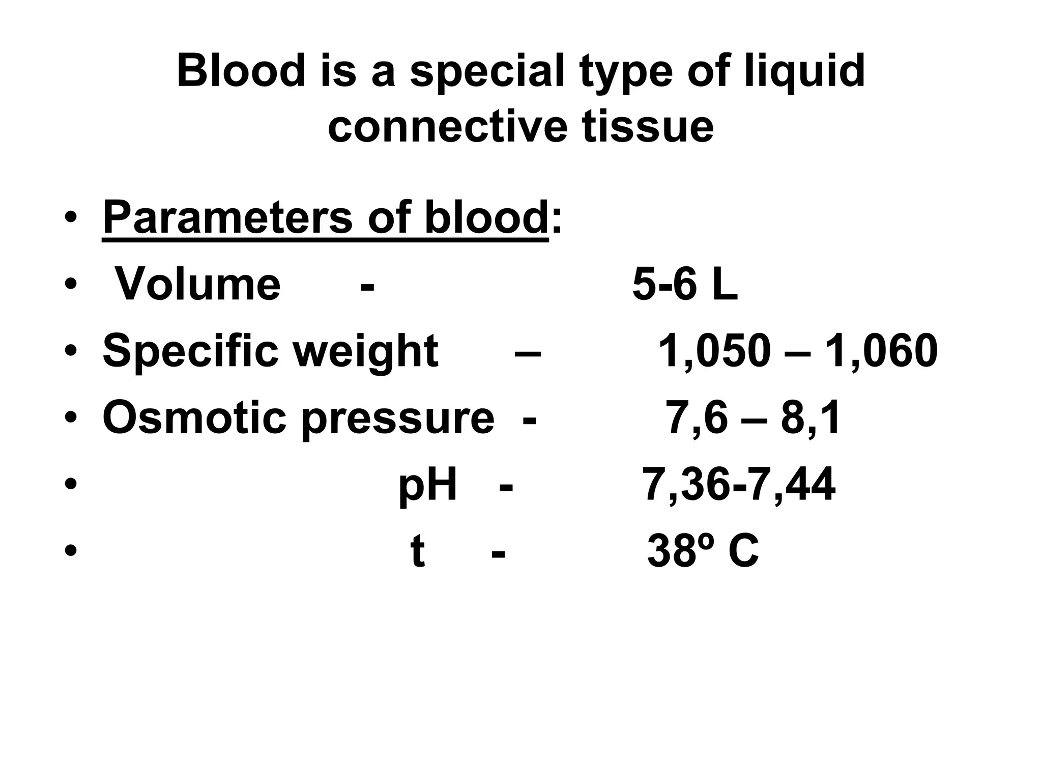
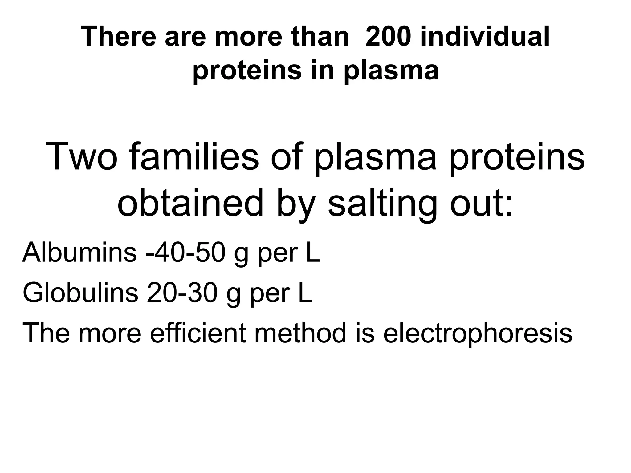
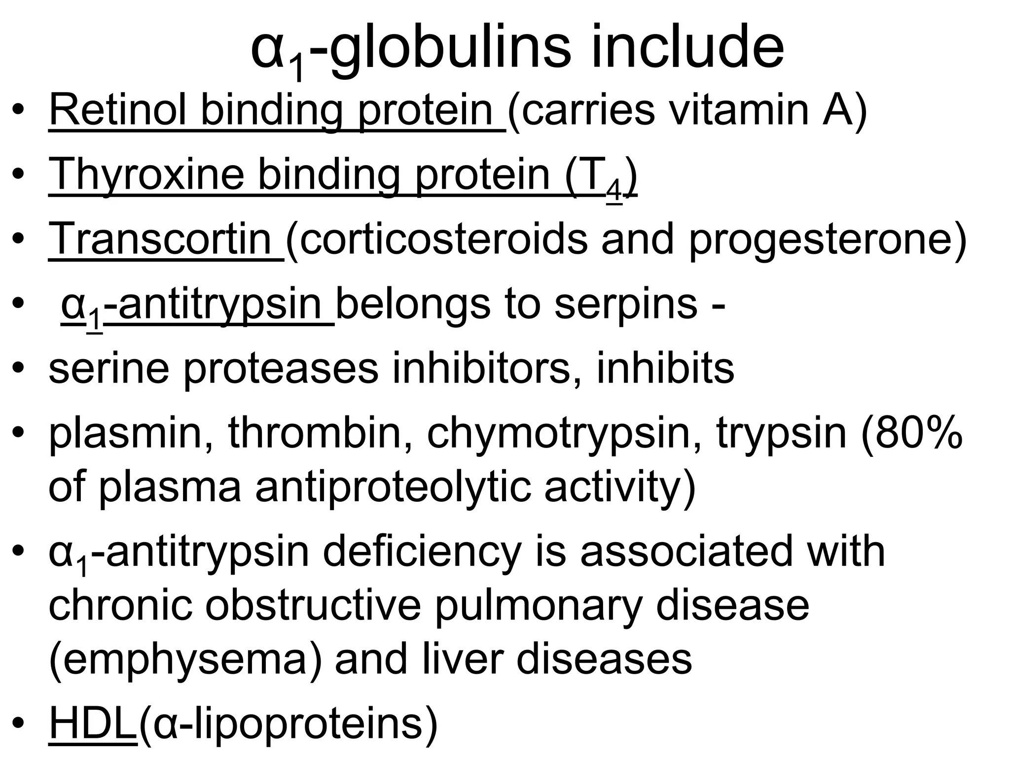
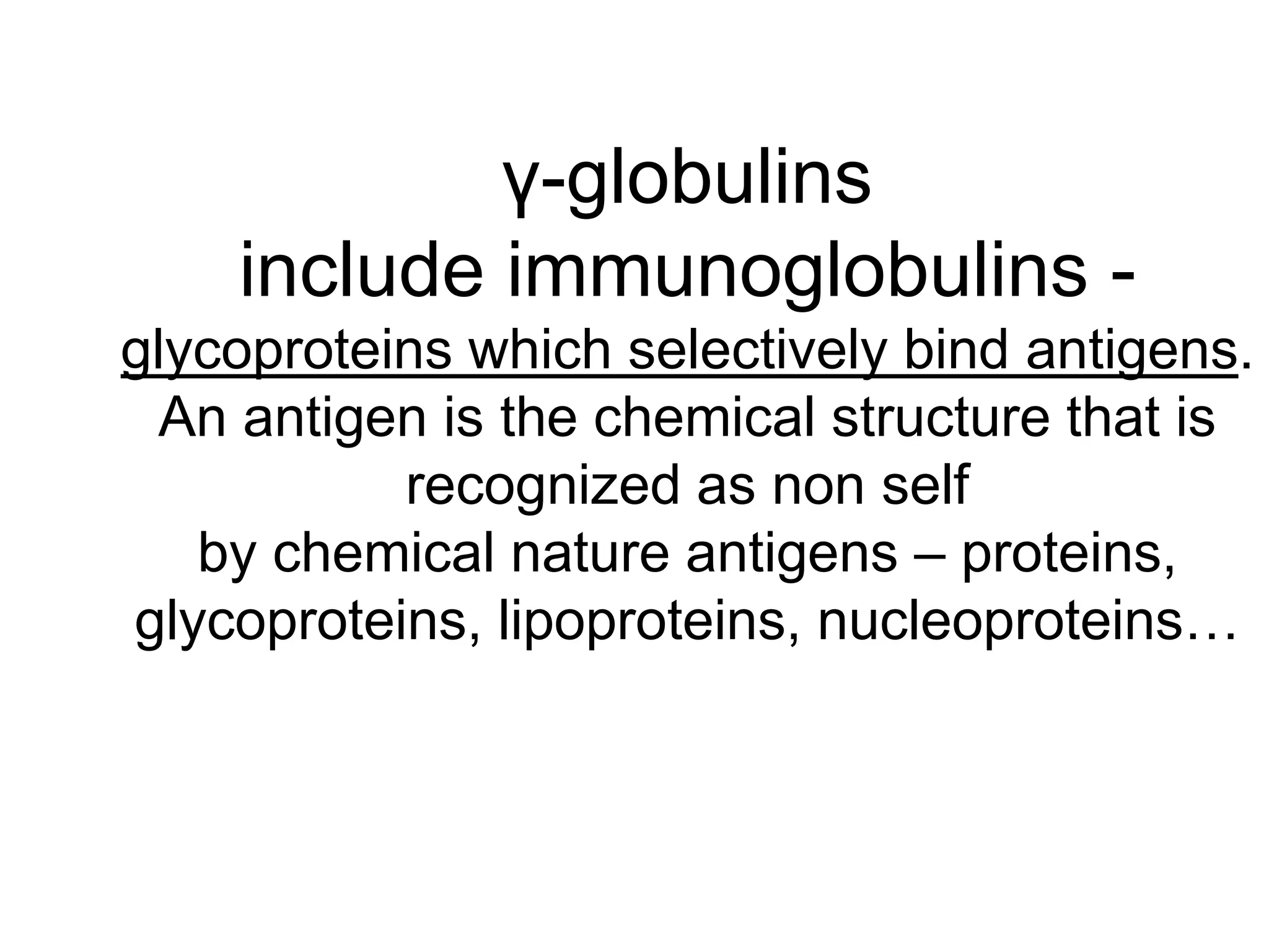
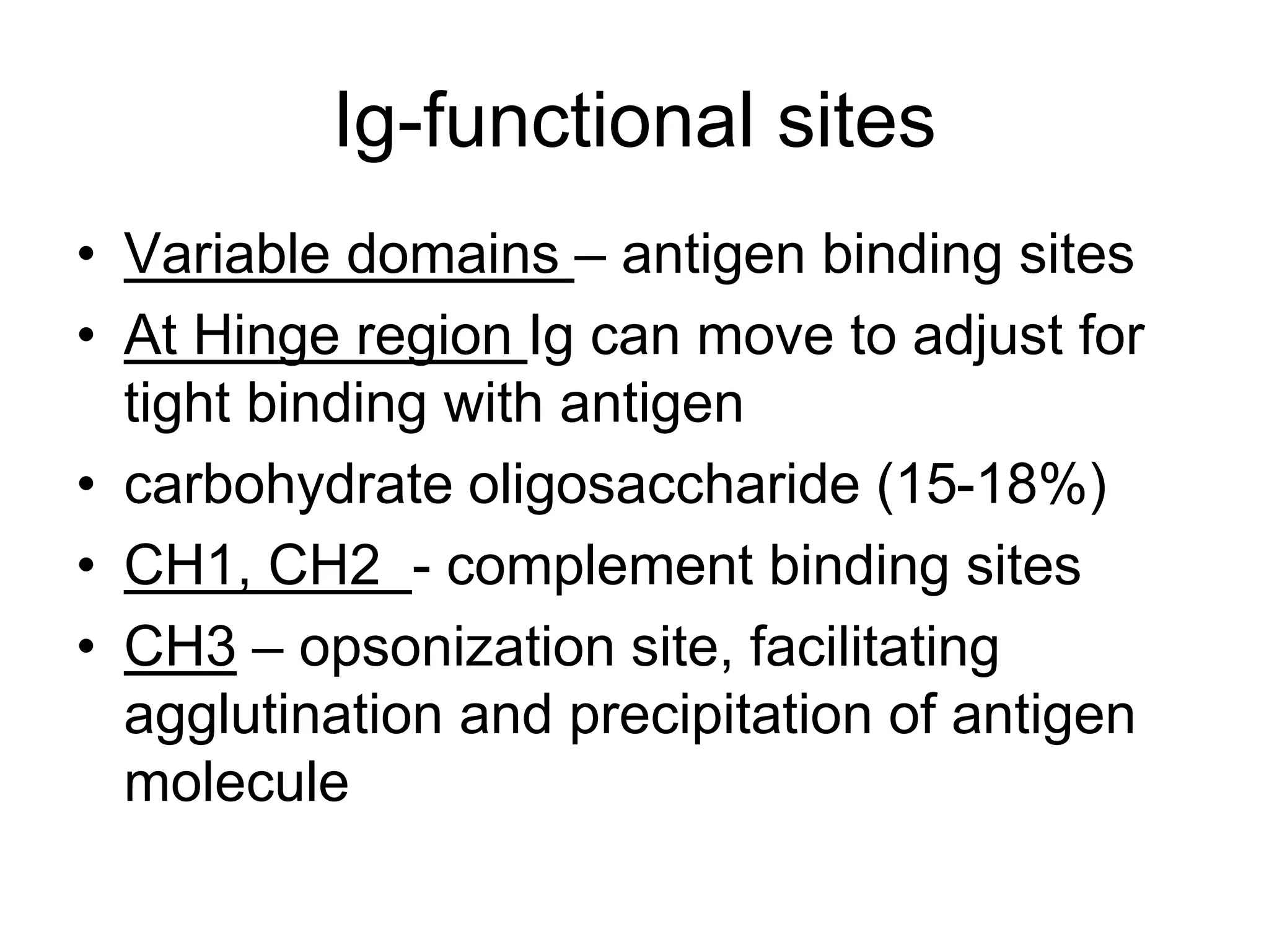
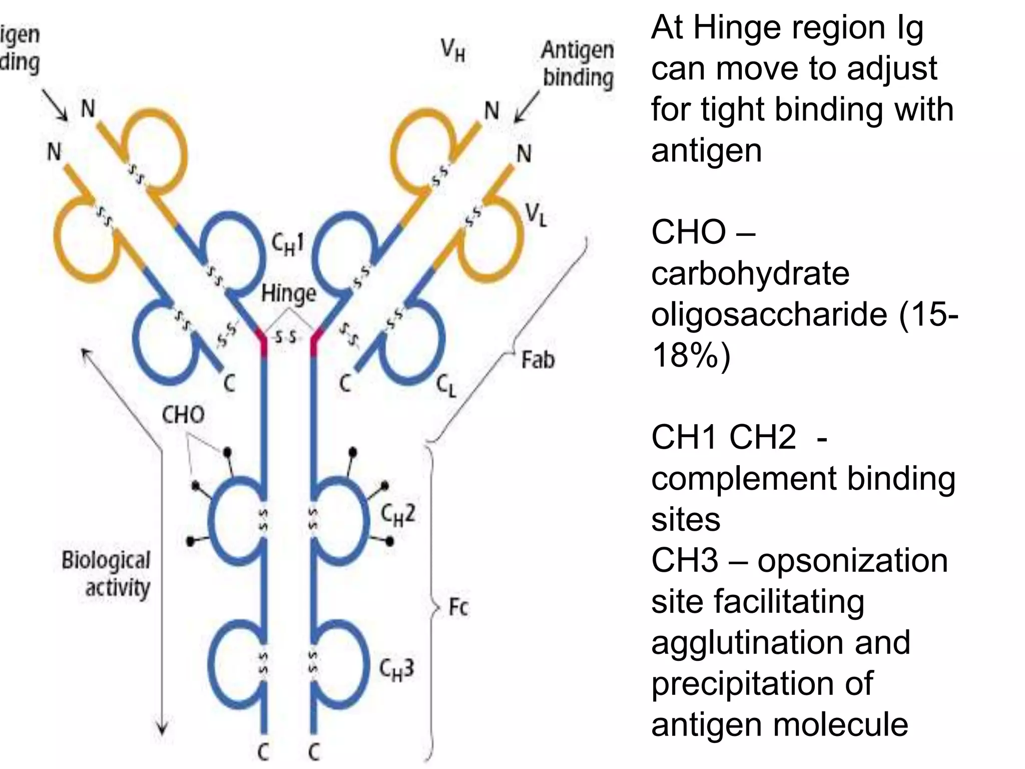
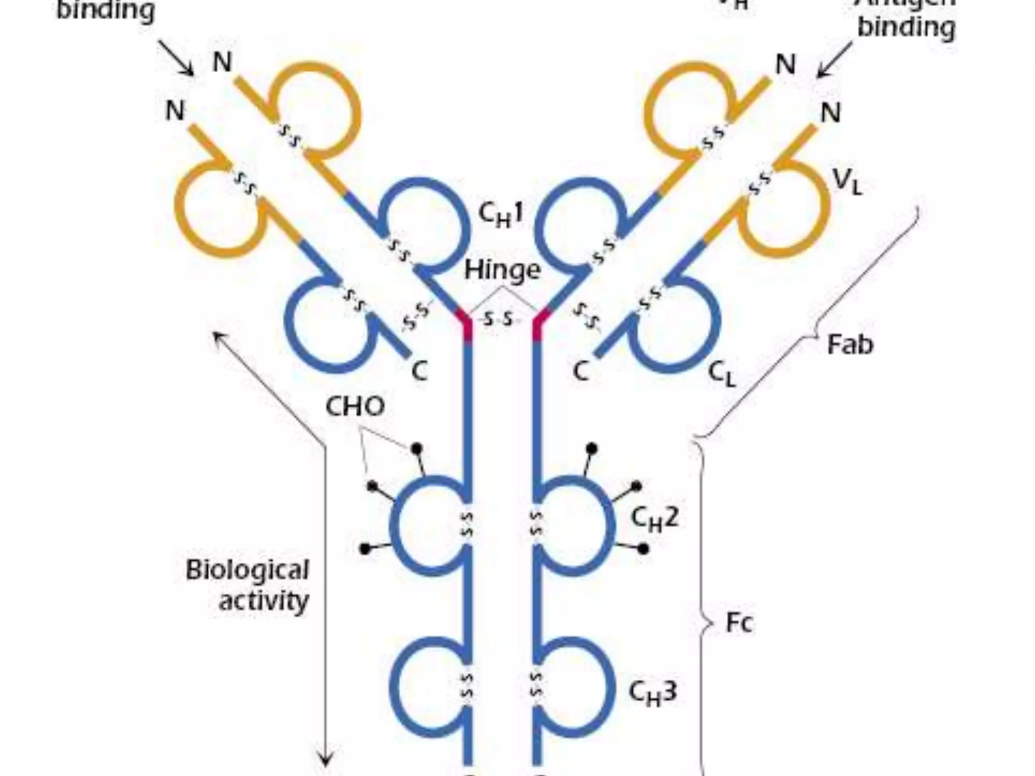
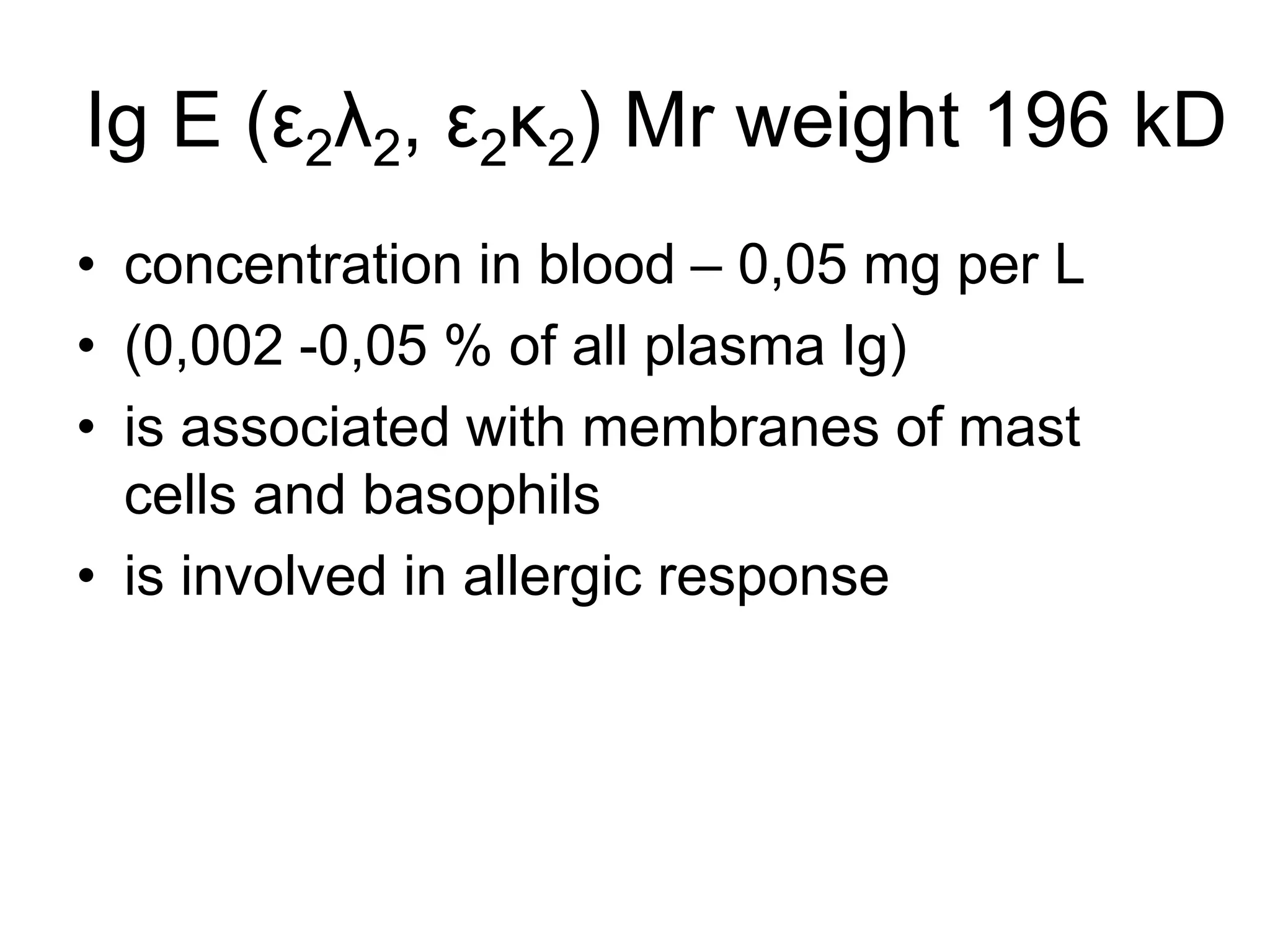
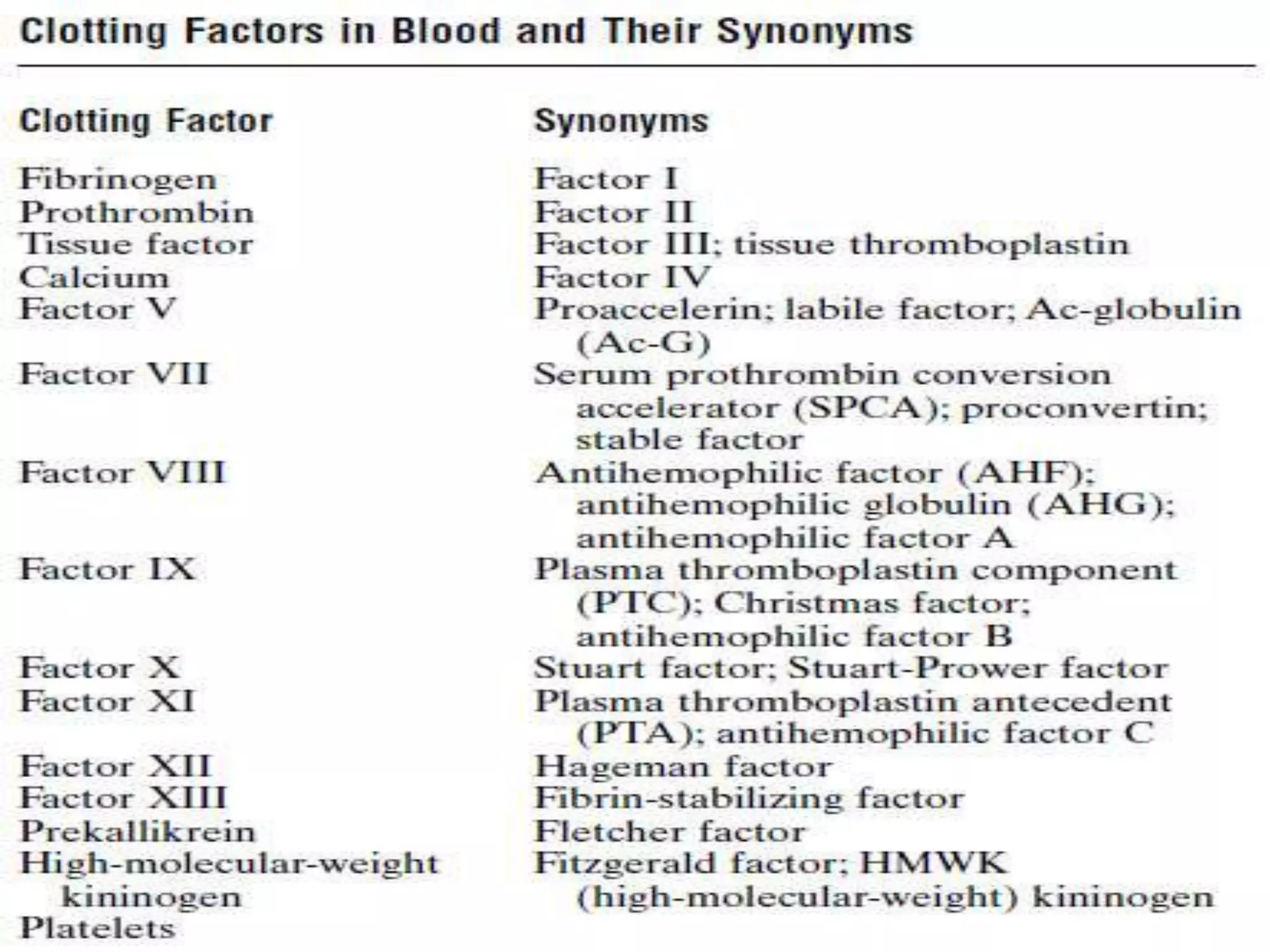
ФБОУ БГМУ ВО БГМУ
BLOOD PLASMA PROTEINS
Blood is a special type of liquid
connective tissue
• Parameters of blood:
• Volume - 5-6 L
• Specific weight – 1,050 – 1,060
• Osmotic pressure - 7,6 – 8,1
• pH - 7,36-7,44
• t - 38º C
Functions of blood
• 1. Transport
• gases - O2, CO2 respiratory function
• amino acids, fatty acids, glucose – nutritive function
• hormones – regulatory function
• waste product – excretory function
• 2. Protection, immune response
• leucocytes, antibodies, acute phase proteins
• 3. Thermoregulation
• 4. Homeostasis (maintenance of pH,
osmotic pressure…)
Composition of blood
Plasma
(55% of whole blood)
Ratio: plasma-cellular part
• Up to 95% of plasma volume is water. The
major solute is heterogeneous proteins (6-
8% by weight)
• (Total plasma protein – 65-85 g per L
• (estimated by biuret method)
• 4% of plasma protein is fibrinogen
• concentration in plasma – 3,5-4 g per L
• Molecular weight – 340 kD
• Glycoprotein
• Fibrinogen → fibrin → clot
• Serum is defibrinated plasma
There are more than 200 individual
proteins in plasma
Two families of plasma proteins
obtained by salting out:
Albumins -40-50 g per L
Globulins 20-30 g per L
The more efficient method is electrophoresis
Types of Electrophoresis
depending supporting medium:
• 1. filter paper electrophoresis(takes 16-18
hours, 5 bands are obtained)
• 2. Agar gel electrophoresis (1-2 h-s, 5
bands)
• 3. PAGE (polyacrylamide gel EP)
combines electrophoresis and sieving
effect, 20 fractions
• 4. immune electrophoresis followed by
antigen-antibody reaction, 40 fractions
Agar gel electrophoresis is
commonly used in clinical laboratory
Technique
• 1. Electrophoretic run is carried at pH 8,6.
Proteins are charged negatively and move
toward anode
• 2. After run is completed proteins are fixed
by methanol
• 3. Then stained using dyes
• 4. Proteins are quantified by densitometry
Normal proteinogram
In agar gel (or paper) electrophoresis
more than 200 serum proteins are
separated into 5 bands:
Albumins - 55-65 %
α1 –globulins - 4-7%
α2-globulins - 5-9%
β-globulins - 7-14%
γ-globulins - 14-22%
Proteinogram of patient with
myeloma (peak in γ-globulins)
Inflammatory (left -peak in α- and β-
globulins) in and
normal(right)proteinogram
α1-antitrypsin deficiency(left) and
immunodeficiency(right) state
Proteinogram abnormalities
Liver
diseases
Nephrotic
syndrome
Acute inflammation
Myeloma
Immunodeficiency
state
Heterogeneous plasma proteins
depending the function are
classified as:
1. transport proteins
• 2. proteins involved in hemostasis
• 3. antiproteases
• 4. proteins of immune system
• 5. APP - acute phase proteins
• (include proteins of classes 1-4)
Albumins - molecular weight – 60-70 kD
• half-life - 20 days
• Produced by liver (25% of protein synthesis in
liver, 12 g per day, used as a liver function test)
• Functions
• 1. provide of 80% of blood oncotic pressure
• 2. transport of fatty acids, bilirubin, Ca, 10% of
plasma Cu, bile acids, sulfa-drugs, antibiotics…
• 3. depot of amino acids
• liver↔ blood proteins ↔ tissues
• Therapeutic use – in burns, hemorrhages…
α1-globulins include
• Retinol binding protein (carries vitamin A)
• Thyroxine binding protein (T4)
• Transcortin (corticosteroids and progesterone)
• α1-antitrypsin belongs to serpins -
• serine proteases inhibitors, inhibits
• plasmin, thrombin, chymotrypsin, trypsin (80%
of plasma antiproteolytic activity)
• α1-antitrypsin deficiency is associated with
chronic obstructive pulmonary disease
(emphysema) and liver diseases
• HDL(α-lipoproteins)
α2 –globulins include
• Haptoglobin binds Hb released in RBC
hemolysis, preventing the loss of iron in urine
• α2 –macroglobulin – 10% of blood antiprotease
activity
• ceruloplasmin - blue due to presence of Cu –
0,32 %, 6-8 atoms of Cu per molecule, 90% 0f
plasma Cu
• An antioxidant
• ferroxidase: Fe2+→Fe3+ (incorpor. in transferrin)
• Concentration decreases in Wilson”s
disease(hepatocellular degeneration, defect of
β-globulins include
• LDL (β-lipoproteins)
• Transferrin presents 3% of total plasma protein
• specific iron binding protein
• 1/3 of transferrin is saturated with Fe3+(in iron
deficient anemia % of saturation lowers)
• half-life is 7 days (the better indicator of protein
turnover in liver than albumin
• fibrinogen
• CRP – C-reactive protein reacts with C-
polysaccharide of pneumococci, produced by
liver. Functions – activation of complement,
APP - acute phase proteins -
• Concentration may increase in 5 to 1000 folds
in inflammatory and neoplastic conditions
• Positive APP (increase in inflammation)
• – CRP, haptoglobin, fibrinogen ceruloplasmin,
serum amyloid A (more than 30 plasma
proteins)
• Negative APP (decrease in inflammation) –
albumin, thyroxine binding protein, transferrin
γ-globulins
include immunoglobulins -
glycoproteins which selectively bind antigens.
An antigen is the chemical structure that is
recognized as non self
by chemical nature antigens – proteins,
glycoproteins, lipoproteins, nucleoproteins…
Ig-functional sites
• Variable domains – antigen binding sites
• At Hinge region Ig can move to adjust for
tight binding with antigen
• carbohydrate oligosaccharide (15-18%)
• CH1, CH2 - complement binding sites
• CH3 – opsonization site, facilitating
agglutination and precipitation of antigen
molecule
At Hinge region Ig
can move to adjust
for tight binding with
antigen
CHO –
carbohydrate
oligosaccharide (15-
18%)
CH1 CH2 -
complement binding
sites
CH3 – opsonization
site facilitating
agglutination and
precipitation of
antigen molecule
There are 5 classes of Ig –
A,D,E,G,M
• Light chains (220 amino acid long) are
identical in all Ig (two types of L-chains - λ
and κ )
• Heavy chains (440 -700 amino acid long)
are specific for different classes of Ig
IgA (α2λ2, α2κ2) Mr weight is 160kD
• concentration in plasma – 0,8-4 g per L (20% of
plasma Ig)
• The major Ig of exocrine secretions (saliva,
milk, tears…)
• In exocrine secretions is found as a dimer
Dimer is more stable and
protected from proteolysis
IgG(γ2λ2 and γ2κ2)
Mr weight 170kD
• Concentration in blood – 8-18 g per L,
the major plasma Ig (75% of all plasma
Ig)
• provides passive immunity, the only
class of Ig are traversed across
placenta
• Functions are opsonization,
complement activation, activation of
phagocytosis
IgM (μ2λ2 and μ2κ2) Mr weight 170kD
• Concentration in blood – 0,6-2,8 g per L, (7%
of all plasma Ig)
• Is the first antibody to appear in the first
contact with an antigen (the primary
respond)
• The largest Ig due to formation of pentamers
Ig M pentamer
Ig D (δ2λ2, δ2κ2) Mr weight 185 kD
• concentration in blood – 0,03-0,15 g per L
(0,25 % of all plasma Ig)
• represents 1% of plasma membrane
proteins in immature B-lymphocytes)
• function is to participate in antigen
dependent differentiation of B-cells
Ig E (ε2λ2, ε2κ2) Mr weight 196 kD
• concentration in blood – 0,05 mg per L
• (0,002 -0,05 % of all plasma Ig)
• is associated with membranes of mast
cells and basophils
• is involved in allergic response
Plasma enzymes:
-Secretory (normal components of plasma –
enzymes of blood clotting, lipoprotein lipase,
pseudo choline esterase…)
-Indicatory (intracellular enzymes entering plasma
in corresponding tissue damage - AlAT, AsAT,
amylase, isozymes of LDH, Creatine Kinase )
-Excretory (are normally excreted into the bile,
increased activity in plasma signals cholestasis –
alkaline phosphatase, γ-GGT (glutamyl
transferase), leucine aminopeptidase)
Complement system
Is composed of about 2O plasma proteins
that act in sequential cascade
Functions:
1)Lysis of foreign cells by rupturing their
membranes
2)Opsonization – activation of phagocytosis
3)Activation of inflammation
Classical pathway of
complement activation
Связывание железа в активном центре тренсферрина
Biochemistry of blood.ppt

Biochemistry of blood.ppt

  • 1.
    ФБОУ БГМУ ВОБГМУ BLOOD PLASMA PROTEINS
  • 2.
    Blood is aspecial type of liquid connective tissue • Parameters of blood: • Volume - 5-6 L • Specific weight – 1,050 – 1,060 • Osmotic pressure - 7,6 – 8,1 • pH - 7,36-7,44 • t - 38º C
  • 3.
    Functions of blood •1. Transport • gases - O2, CO2 respiratory function • amino acids, fatty acids, glucose – nutritive function • hormones – regulatory function • waste product – excretory function • 2. Protection, immune response • leucocytes, antibodies, acute phase proteins • 3. Thermoregulation • 4. Homeostasis (maintenance of pH, osmotic pressure…)
  • 4.
  • 5.
  • 6.
    • Up to95% of plasma volume is water. The major solute is heterogeneous proteins (6- 8% by weight) • (Total plasma protein – 65-85 g per L • (estimated by biuret method) • 4% of plasma protein is fibrinogen • concentration in plasma – 3,5-4 g per L • Molecular weight – 340 kD • Glycoprotein • Fibrinogen → fibrin → clot • Serum is defibrinated plasma
  • 7.
    There are morethan 200 individual proteins in plasma Two families of plasma proteins obtained by salting out: Albumins -40-50 g per L Globulins 20-30 g per L The more efficient method is electrophoresis
  • 8.
    Types of Electrophoresis dependingsupporting medium: • 1. filter paper electrophoresis(takes 16-18 hours, 5 bands are obtained) • 2. Agar gel electrophoresis (1-2 h-s, 5 bands) • 3. PAGE (polyacrylamide gel EP) combines electrophoresis and sieving effect, 20 fractions • 4. immune electrophoresis followed by antigen-antibody reaction, 40 fractions
  • 9.
    Agar gel electrophoresisis commonly used in clinical laboratory
  • 10.
    Technique • 1. Electrophoreticrun is carried at pH 8,6. Proteins are charged negatively and move toward anode • 2. After run is completed proteins are fixed by methanol • 3. Then stained using dyes • 4. Proteins are quantified by densitometry
  • 11.
  • 12.
    In agar gel(or paper) electrophoresis more than 200 serum proteins are separated into 5 bands: Albumins - 55-65 % α1 –globulins - 4-7% α2-globulins - 5-9% β-globulins - 7-14% γ-globulins - 14-22%
  • 13.
    Proteinogram of patientwith myeloma (peak in γ-globulins)
  • 14.
    Inflammatory (left -peakin α- and β- globulins) in and normal(right)proteinogram
  • 15.
  • 16.
  • 17.
    Heterogeneous plasma proteins dependingthe function are classified as: 1. transport proteins • 2. proteins involved in hemostasis • 3. antiproteases • 4. proteins of immune system • 5. APP - acute phase proteins • (include proteins of classes 1-4)
  • 18.
    Albumins - molecularweight – 60-70 kD • half-life - 20 days • Produced by liver (25% of protein synthesis in liver, 12 g per day, used as a liver function test) • Functions • 1. provide of 80% of blood oncotic pressure • 2. transport of fatty acids, bilirubin, Ca, 10% of plasma Cu, bile acids, sulfa-drugs, antibiotics… • 3. depot of amino acids • liver↔ blood proteins ↔ tissues • Therapeutic use – in burns, hemorrhages…
  • 19.
    α1-globulins include • Retinolbinding protein (carries vitamin A) • Thyroxine binding protein (T4) • Transcortin (corticosteroids and progesterone) • α1-antitrypsin belongs to serpins - • serine proteases inhibitors, inhibits • plasmin, thrombin, chymotrypsin, trypsin (80% of plasma antiproteolytic activity) • α1-antitrypsin deficiency is associated with chronic obstructive pulmonary disease (emphysema) and liver diseases • HDL(α-lipoproteins)
  • 20.
    α2 –globulins include •Haptoglobin binds Hb released in RBC hemolysis, preventing the loss of iron in urine • α2 –macroglobulin – 10% of blood antiprotease activity • ceruloplasmin - blue due to presence of Cu – 0,32 %, 6-8 atoms of Cu per molecule, 90% 0f plasma Cu • An antioxidant • ferroxidase: Fe2+→Fe3+ (incorpor. in transferrin) • Concentration decreases in Wilson”s disease(hepatocellular degeneration, defect of
  • 21.
    β-globulins include • LDL(β-lipoproteins) • Transferrin presents 3% of total plasma protein • specific iron binding protein • 1/3 of transferrin is saturated with Fe3+(in iron deficient anemia % of saturation lowers) • half-life is 7 days (the better indicator of protein turnover in liver than albumin • fibrinogen • CRP – C-reactive protein reacts with C- polysaccharide of pneumococci, produced by liver. Functions – activation of complement,
  • 22.
    APP - acutephase proteins - • Concentration may increase in 5 to 1000 folds in inflammatory and neoplastic conditions • Positive APP (increase in inflammation) • – CRP, haptoglobin, fibrinogen ceruloplasmin, serum amyloid A (more than 30 plasma proteins) • Negative APP (decrease in inflammation) – albumin, thyroxine binding protein, transferrin
  • 23.
    γ-globulins include immunoglobulins - glycoproteinswhich selectively bind antigens. An antigen is the chemical structure that is recognized as non self by chemical nature antigens – proteins, glycoproteins, lipoproteins, nucleoproteins…
  • 25.
    Ig-functional sites • Variabledomains – antigen binding sites • At Hinge region Ig can move to adjust for tight binding with antigen • carbohydrate oligosaccharide (15-18%) • CH1, CH2 - complement binding sites • CH3 – opsonization site, facilitating agglutination and precipitation of antigen molecule
  • 26.
    At Hinge regionIg can move to adjust for tight binding with antigen CHO – carbohydrate oligosaccharide (15- 18%) CH1 CH2 - complement binding sites CH3 – opsonization site facilitating agglutination and precipitation of antigen molecule
  • 29.
    There are 5classes of Ig – A,D,E,G,M • Light chains (220 amino acid long) are identical in all Ig (two types of L-chains - λ and κ ) • Heavy chains (440 -700 amino acid long) are specific for different classes of Ig
  • 30.
    IgA (α2λ2, α2κ2)Mr weight is 160kD • concentration in plasma – 0,8-4 g per L (20% of plasma Ig) • The major Ig of exocrine secretions (saliva, milk, tears…) • In exocrine secretions is found as a dimer
  • 31.
    Dimer is morestable and protected from proteolysis
  • 32.
    IgG(γ2λ2 and γ2κ2) Mrweight 170kD • Concentration in blood – 8-18 g per L, the major plasma Ig (75% of all plasma Ig) • provides passive immunity, the only class of Ig are traversed across placenta • Functions are opsonization, complement activation, activation of phagocytosis
  • 33.
    IgM (μ2λ2 andμ2κ2) Mr weight 170kD • Concentration in blood – 0,6-2,8 g per L, (7% of all plasma Ig) • Is the first antibody to appear in the first contact with an antigen (the primary respond) • The largest Ig due to formation of pentamers
  • 34.
  • 35.
    Ig D (δ2λ2,δ2κ2) Mr weight 185 kD • concentration in blood – 0,03-0,15 g per L (0,25 % of all plasma Ig) • represents 1% of plasma membrane proteins in immature B-lymphocytes) • function is to participate in antigen dependent differentiation of B-cells
  • 36.
    Ig E (ε2λ2,ε2κ2) Mr weight 196 kD • concentration in blood – 0,05 mg per L • (0,002 -0,05 % of all plasma Ig) • is associated with membranes of mast cells and basophils • is involved in allergic response
  • 37.
    Plasma enzymes: -Secretory (normalcomponents of plasma – enzymes of blood clotting, lipoprotein lipase, pseudo choline esterase…) -Indicatory (intracellular enzymes entering plasma in corresponding tissue damage - AlAT, AsAT, amylase, isozymes of LDH, Creatine Kinase ) -Excretory (are normally excreted into the bile, increased activity in plasma signals cholestasis – alkaline phosphatase, γ-GGT (glutamyl transferase), leucine aminopeptidase)
  • 38.
    Complement system Is composedof about 2O plasma proteins that act in sequential cascade Functions: 1)Lysis of foreign cells by rupturing their membranes 2)Opsonization – activation of phagocytosis 3)Activation of inflammation
  • 39.
  • 40.
    Связывание железа вактивном центре тренсферрина