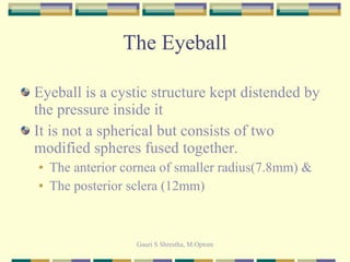
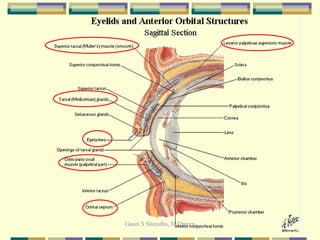
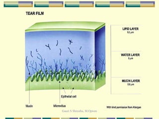
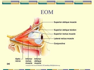
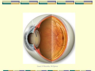
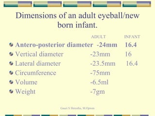
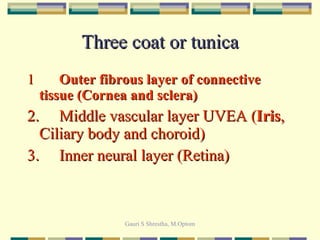
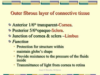
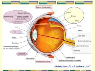
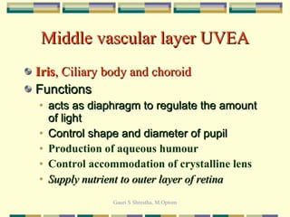
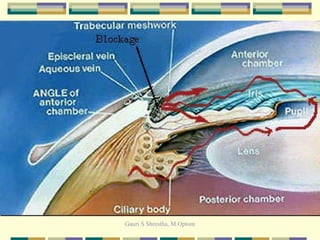
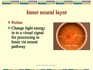
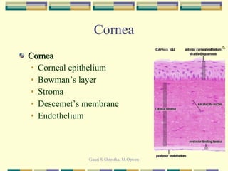
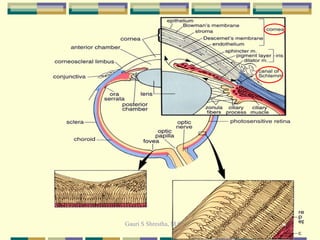
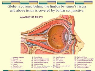
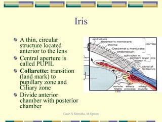
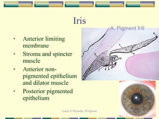
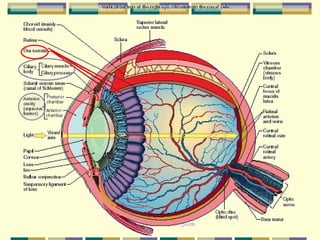
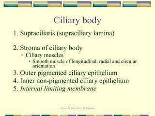
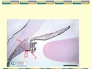
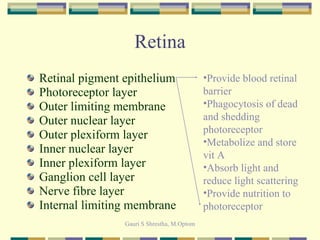
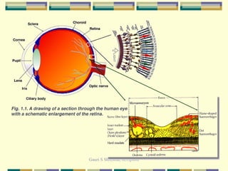
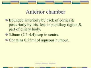
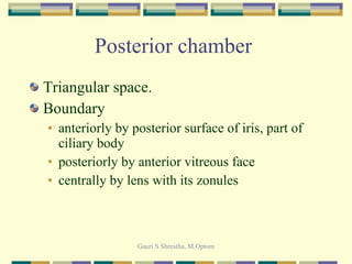
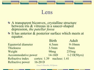
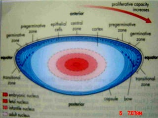
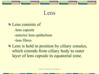
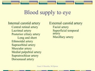
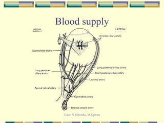
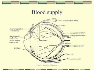
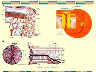
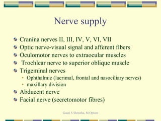
Anatomy of Eye GS Shrestha, M. Optom Teaching Assistant
Learning Objective Describe dimensions and gross anatomical structure of eye Describe different coats of eye Describe the spaces in eye Describe the functions of eye
The Eyeball Eyeball is a cystic structure kept distended by the pressure inside it It is not a spherical but consists of two modified spheres fused together.  The anterior cornea of smaller radius(7.8mm) &  The posterior sclera (12mm)
 
 
EOM
 
Dimensions of an adult eyeball/new born infant.   ADULT   INFANT Antero-posterior diameter   - 24mm   16.4 Vertical diameter   -23mm   16 Lateral diameter   -23.5mm  16.4 Circumference   -75mm Volume   -6.5ml Weight   -7gm
Three coat or tunica 1 Outer fibrous layer of connective tissue (Cornea and sclera) 2.  Middle vascular layer UVEA ( Iris , Ciliary body and choroid) 3. Inner neural layer (Retina)
Outer fibrous layer of connective tissue Anterior 1/6 th  transparent- Cornea . Posterior 5/6 th opaque- Sclera . Junction of cornea & sclera – Limbus Function Protection for structure within maintain globe’s shape Provide resistance to the pressure of the fluids inside Transmittance of light from cornea to retina
 
Middle vascular layer UVEA Iris , Ciliary body and choroid Functions acts as diaphragm to regulate the amount of light Control shape and diameter of pupil Production of aqueous humour Control accommodation of crystalline lens Supply nutrient to outer layer of retina
 
Inner neural layer Retina Change light energy in to a visual signal for processing in brain via neural pathway
Cornea Cornea Corneal epithelium Bowman’s layer Stroma Descemet’s membrane Endothelium
Sclera It opaque and porcelain white Consists of indistinct 3 layers: outer episclera to inner layer scleral stroma Thinnest 0.3mm at insertion of rectus muscles, thickest 1.0mm at post pole, 0.4-0.5mm at equator
 
Limbus  Transition zone between peripheral cornea & anterior sclera 1.5mm-horizontal & 2mm at vertical meridian Structures included   Conjunctiva Tenon’s capsule Episclera Corneoscleral stroma Aqueous outflow apparatus
Globe is covered behind the limbus by tenon’s fascia and above tenon is covered by bulbar conjunctiva
Iris A thin, circular structure located anterior to the lens Central aperture is called PUPIL Collarette:  transition (land mark) to pupillary zone and Ciliary zone Divide anterior chamber with posterior chamber
Iris Anterior limiting membrane Stroma and spincter muscle Anterior non-pigmented epithelium and dilator muscle Posterior pigmented epithelium A. Pigment frill
Ciliary body Ring shaped structure Width 5.9mm nasal side 6.7mm on temporal side Extend from root of iris at scleral spur to ora serrata Divided in to two parts Pars plicata containing ciliary processes Pars plana: a transition b/w pars plicata and ora serrata
 
Ciliary body 1. Supraciliaris (supraciliary lamina) 2. Stroma of ciliary body Ciliary muscles Smooth muscle of longitudinal, radial and circular orientation 3. Outer pigmented ciliary epithelium  4. Inner non-pigmented ciliary epithelium 5.  Internal limiting membrane
1 2 3 4  5
Choroid Extend from ora serrata to optic disc Primarily made up of blood vessels and covered both side by connective tissue Layers Suprachoroidal Lamina Choroidal stroma Choriocapillaries Bruch’s membrane
Retina
Retina Inner most and transparent layer Extend from optic disc to ora serrata Two basic layers:  pigment layer  and  neural layer Function Transformation of light energy to neural signal by photoreceptor, bipolar cells and ganglion cells Its thickness at posterior pole in peripapillary region is approx 0.5mm, at equator 0.18-0.2mm & at ora serrata approx 0.1mm
 
Retina Retinal pigment epithelium Photoreceptor layer Outer limiting membrane Outer nuclear layer Outer plexiform layer Inner nuclear layer Inner plexiform layer Ganglion cell layer Nerve fibre layer Internal limiting membrane Provide blood retinal barrier Phagocytosis of dead and shedding photoreceptor Metabolize and store vit A Absorb light and reduce light scattering Provide nutrition to photoreceptor
 
Anterior chamber Bounded anteriorly by back of cornea & posteriorly by iris, lens in pupillary region & part of ciliary body. 3.0mm (2.5-4.4)deep in centre. Contains 0.25ml of aqueous humour.
Posterior chamber  Triangular space. Boundary anteriorly by posterior surface of iris, part of ciliary body posteriorly by anterior vitreous face centrally by lens with its zonules
Lens A transparent biconvex, crystalline structure between iris & vitreous in a saucer-shaped depression,  the patellar fossa It has anterior & posterior surface which meets at equator.   Birth  Adult Equatorial diameter  6.5mm  9-10mm Thickness   3.5mm    5mm Weight   90mg  255mg Accommodative power  14-16D  1-2 D(50yrs) Refractive index  cortex: 1.39  nucleus: 1.41 Refractive power  16-20 D
 
Lens Lens consists of  -lens capsule -anterior lens epithelium -lens fibres Lens is hold in position by ciliary zonules, which extends from ciliary body to outer layer of lens capsule in equatorial zone.
 
Vitreous humour  It is an inert, transparent, colourless, jellylike structure. Vitreous cavity is bounded anteriorly by lens & ciliary body ; posteriorly by retina. Space of berger Wieger’s ligament Weight-4gm
Vitreous humour Vitreous attached firmly to retina at 4 sites: At ora serrata At peripapillary region At fovea At retinal vasculatures.
Blood supply to eye Internal carotid artery Central retinal artery Lacrimal artery Posterior ciliary artery Long and short Ethmoidal artery Supraorbital artery Muscular artery Medial palpebral artery Supratrochlear artery Dorsonasal artery External carotid artery Facial artery Superficial temporal artery Maxillary artery
Blood supply
Blood supply
 
Vein Superior ophthalmic vein Angular vein Supraorbital vein Ethmoidal vein Muscular vein Lacrimal vein Anterior ciliary vein Central retinal vein Upper vortex vein Inferior ophthalmic vein lower eyelids,  lateral muscles,  inferior conjunctiva,  lacrimal sac,  lower vortex veins pterygoid venous plexus
Nerve supply Cranina nerves II, III, IV, V, VI, VII Optic nerve-visual signal and afferent fibers  Oculomotor nerves to extraocular muscles Trochlear nerve to superior oblique muscle Trigeminal nerves Ophthalmic (lacrimal, frontal and nasociliary nerves) maxillary division Abducent nerve  Facial nerve (secretomotor fibres)
THANK YOU

Anatomy for bn

  • 1.
    Anatomy of EyeGS Shrestha, M. Optom Teaching Assistant
  • 2.
    Learning Objective Describedimensions and gross anatomical structure of eye Describe different coats of eye Describe the spaces in eye Describe the functions of eye
  • 3.
    The Eyeball Eyeballis a cystic structure kept distended by the pressure inside it It is not a spherical but consists of two modified spheres fused together. The anterior cornea of smaller radius(7.8mm) & The posterior sclera (12mm)
  • 4.
  • 5.
  • 6.
  • 7.
  • 8.
    Dimensions of anadult eyeball/new born infant. ADULT INFANT Antero-posterior diameter - 24mm 16.4 Vertical diameter -23mm 16 Lateral diameter -23.5mm 16.4 Circumference -75mm Volume -6.5ml Weight -7gm
  • 9.
    Three coat ortunica 1 Outer fibrous layer of connective tissue (Cornea and sclera) 2. Middle vascular layer UVEA ( Iris , Ciliary body and choroid) 3. Inner neural layer (Retina)
  • 10.
    Outer fibrous layerof connective tissue Anterior 1/6 th transparent- Cornea . Posterior 5/6 th opaque- Sclera . Junction of cornea & sclera – Limbus Function Protection for structure within maintain globe’s shape Provide resistance to the pressure of the fluids inside Transmittance of light from cornea to retina
  • 11.
  • 12.
    Middle vascular layerUVEA Iris , Ciliary body and choroid Functions acts as diaphragm to regulate the amount of light Control shape and diameter of pupil Production of aqueous humour Control accommodation of crystalline lens Supply nutrient to outer layer of retina
  • 13.
  • 14.
    Inner neural layerRetina Change light energy in to a visual signal for processing in brain via neural pathway
  • 15.
    Cornea Cornea Cornealepithelium Bowman’s layer Stroma Descemet’s membrane Endothelium
  • 16.
    Sclera It opaqueand porcelain white Consists of indistinct 3 layers: outer episclera to inner layer scleral stroma Thinnest 0.3mm at insertion of rectus muscles, thickest 1.0mm at post pole, 0.4-0.5mm at equator
  • 17.
  • 18.
    Limbus Transitionzone between peripheral cornea & anterior sclera 1.5mm-horizontal & 2mm at vertical meridian Structures included Conjunctiva Tenon’s capsule Episclera Corneoscleral stroma Aqueous outflow apparatus
  • 19.
    Globe is coveredbehind the limbus by tenon’s fascia and above tenon is covered by bulbar conjunctiva
  • 20.
    Iris A thin,circular structure located anterior to the lens Central aperture is called PUPIL Collarette: transition (land mark) to pupillary zone and Ciliary zone Divide anterior chamber with posterior chamber
  • 21.
    Iris Anterior limitingmembrane Stroma and spincter muscle Anterior non-pigmented epithelium and dilator muscle Posterior pigmented epithelium A. Pigment frill
  • 22.
    Ciliary body Ringshaped structure Width 5.9mm nasal side 6.7mm on temporal side Extend from root of iris at scleral spur to ora serrata Divided in to two parts Pars plicata containing ciliary processes Pars plana: a transition b/w pars plicata and ora serrata
  • 23.
  • 24.
    Ciliary body 1.Supraciliaris (supraciliary lamina) 2. Stroma of ciliary body Ciliary muscles Smooth muscle of longitudinal, radial and circular orientation 3. Outer pigmented ciliary epithelium 4. Inner non-pigmented ciliary epithelium 5. Internal limiting membrane
  • 25.
    1 2 34 5
  • 26.
    Choroid Extend fromora serrata to optic disc Primarily made up of blood vessels and covered both side by connective tissue Layers Suprachoroidal Lamina Choroidal stroma Choriocapillaries Bruch’s membrane
  • 27.
  • 28.
    Retina Inner mostand transparent layer Extend from optic disc to ora serrata Two basic layers: pigment layer and neural layer Function Transformation of light energy to neural signal by photoreceptor, bipolar cells and ganglion cells Its thickness at posterior pole in peripapillary region is approx 0.5mm, at equator 0.18-0.2mm & at ora serrata approx 0.1mm
  • 29.
  • 30.
    Retina Retinal pigmentepithelium Photoreceptor layer Outer limiting membrane Outer nuclear layer Outer plexiform layer Inner nuclear layer Inner plexiform layer Ganglion cell layer Nerve fibre layer Internal limiting membrane Provide blood retinal barrier Phagocytosis of dead and shedding photoreceptor Metabolize and store vit A Absorb light and reduce light scattering Provide nutrition to photoreceptor
  • 31.
  • 32.
    Anterior chamber Boundedanteriorly by back of cornea & posteriorly by iris, lens in pupillary region & part of ciliary body. 3.0mm (2.5-4.4)deep in centre. Contains 0.25ml of aqueous humour.
  • 33.
    Posterior chamber Triangular space. Boundary anteriorly by posterior surface of iris, part of ciliary body posteriorly by anterior vitreous face centrally by lens with its zonules
  • 34.
    Lens A transparentbiconvex, crystalline structure between iris & vitreous in a saucer-shaped depression, the patellar fossa It has anterior & posterior surface which meets at equator. Birth Adult Equatorial diameter 6.5mm 9-10mm Thickness 3.5mm 5mm Weight 90mg 255mg Accommodative power 14-16D 1-2 D(50yrs) Refractive index cortex: 1.39 nucleus: 1.41 Refractive power 16-20 D
  • 35.
  • 36.
    Lens Lens consistsof -lens capsule -anterior lens epithelium -lens fibres Lens is hold in position by ciliary zonules, which extends from ciliary body to outer layer of lens capsule in equatorial zone.
  • 37.
  • 38.
    Vitreous humour It is an inert, transparent, colourless, jellylike structure. Vitreous cavity is bounded anteriorly by lens & ciliary body ; posteriorly by retina. Space of berger Wieger’s ligament Weight-4gm
  • 39.
    Vitreous humour Vitreousattached firmly to retina at 4 sites: At ora serrata At peripapillary region At fovea At retinal vasculatures.
  • 40.
    Blood supply toeye Internal carotid artery Central retinal artery Lacrimal artery Posterior ciliary artery Long and short Ethmoidal artery Supraorbital artery Muscular artery Medial palpebral artery Supratrochlear artery Dorsonasal artery External carotid artery Facial artery Superficial temporal artery Maxillary artery
  • 41.
  • 42.
  • 43.
  • 44.
    Vein Superior ophthalmicvein Angular vein Supraorbital vein Ethmoidal vein Muscular vein Lacrimal vein Anterior ciliary vein Central retinal vein Upper vortex vein Inferior ophthalmic vein lower eyelids, lateral muscles, inferior conjunctiva, lacrimal sac, lower vortex veins pterygoid venous plexus
  • 45.
    Nerve supply Craninanerves II, III, IV, V, VI, VII Optic nerve-visual signal and afferent fibers Oculomotor nerves to extraocular muscles Trochlear nerve to superior oblique muscle Trigeminal nerves Ophthalmic (lacrimal, frontal and nasociliary nerves) maxillary division Abducent nerve Facial nerve (secretomotor fibres)
  • 46.

Editor's Notes

  • #6 Lubrication, hydration, optical integration and defense mechanism
  • #7 Symmetrical co-ordinated eye movement is governed by six extra ocular muscles
  • #19 Limbal vein to episcleral vessels then drain to anterior ciliary vein.
  • #39 Hyaloid artery system