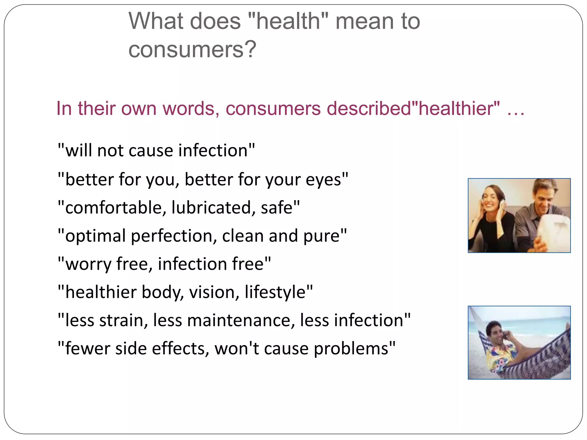
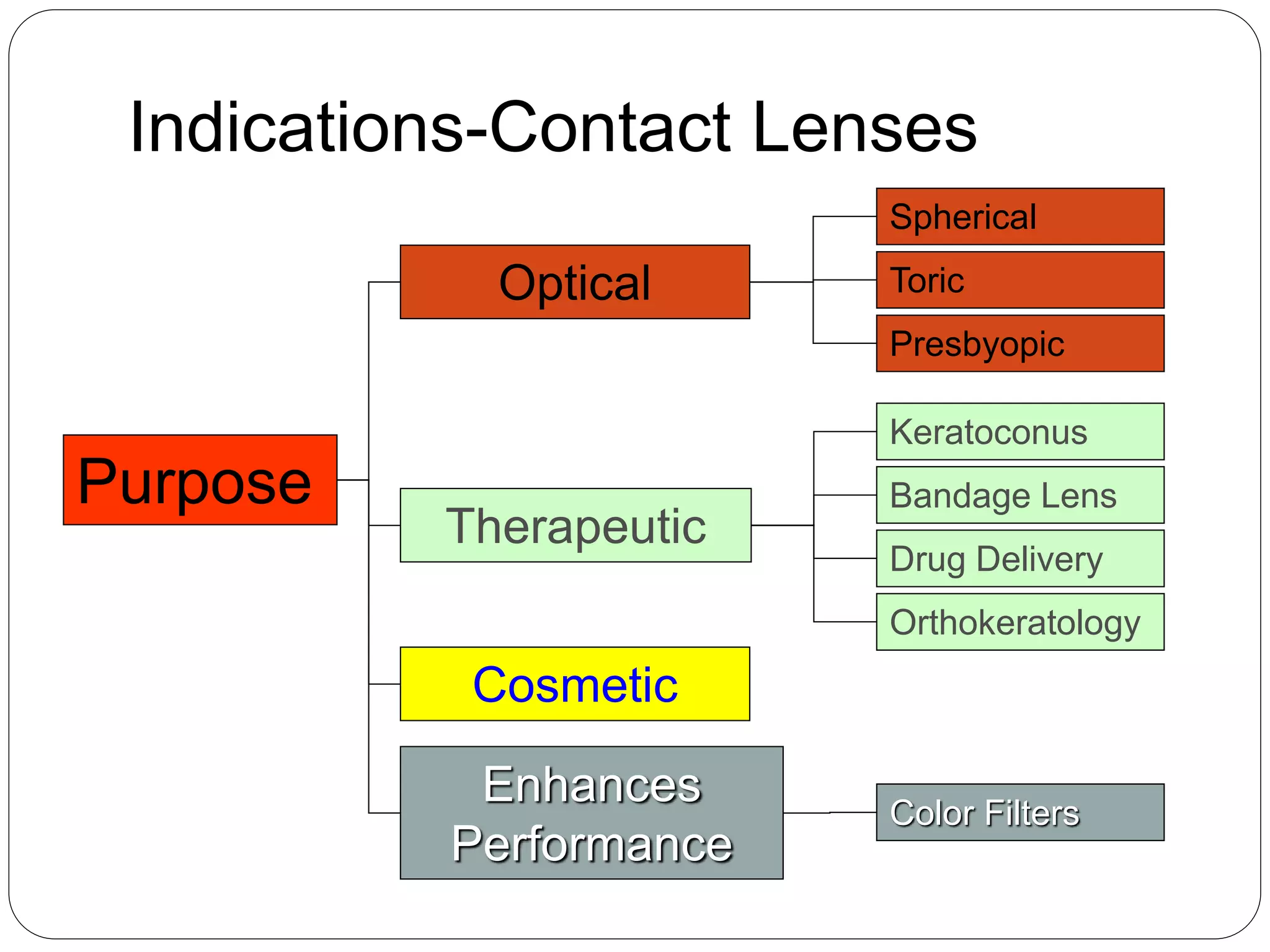
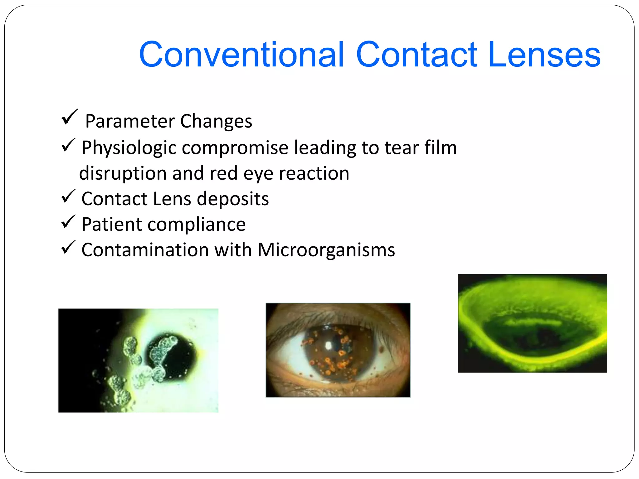
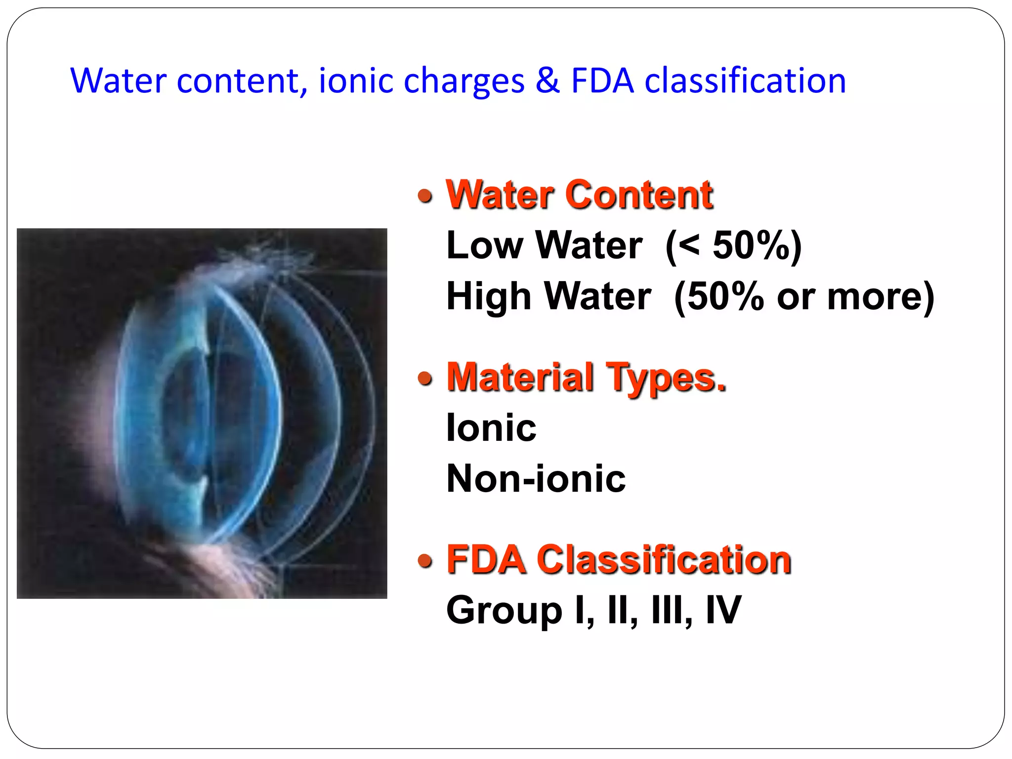
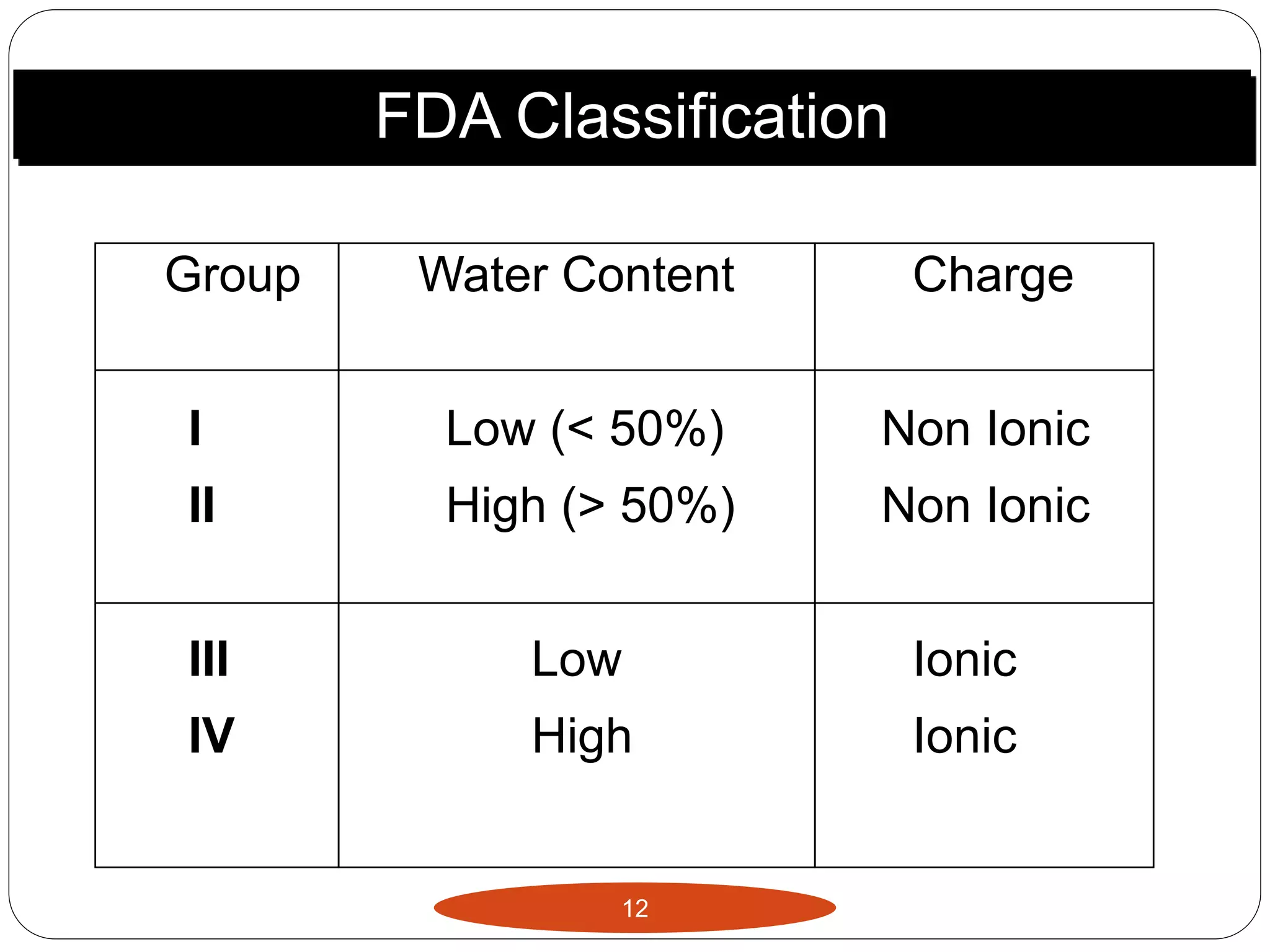
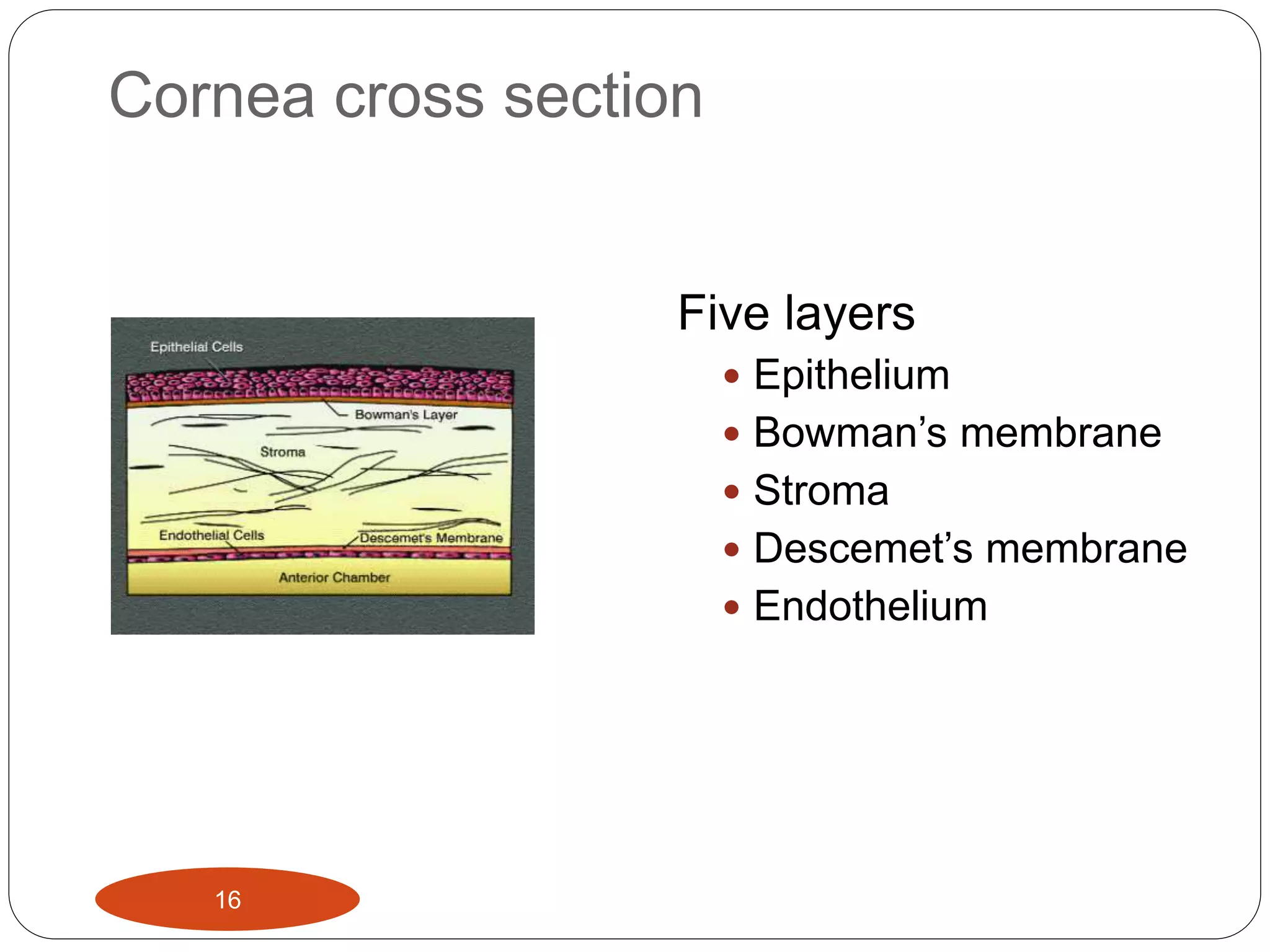
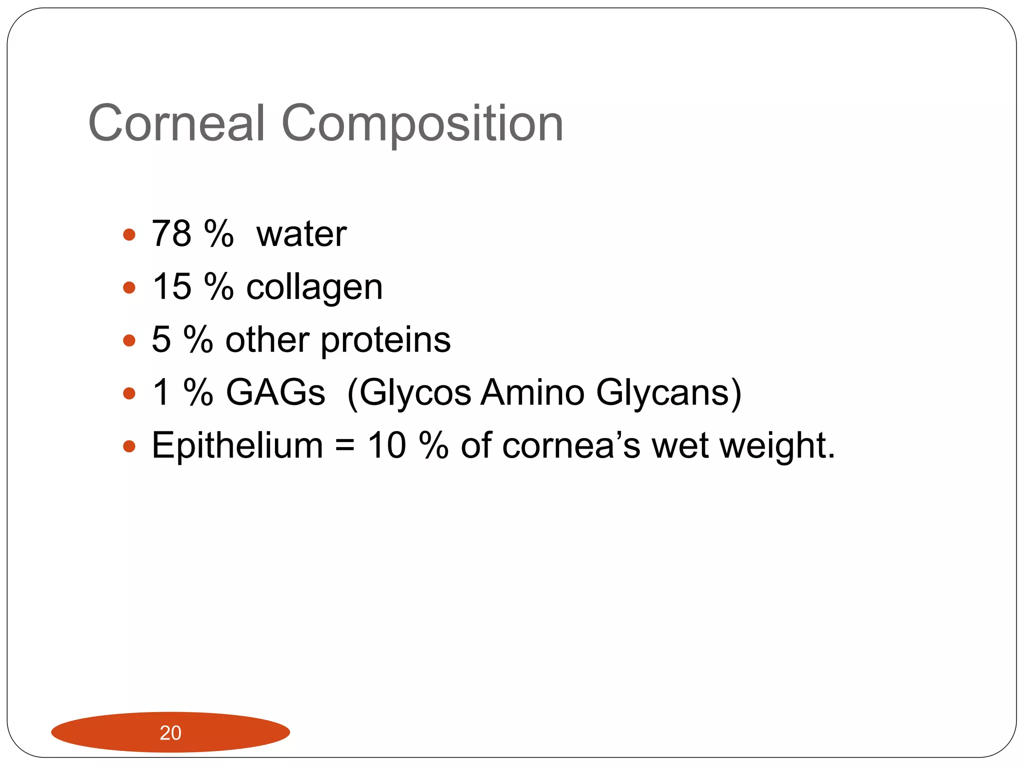
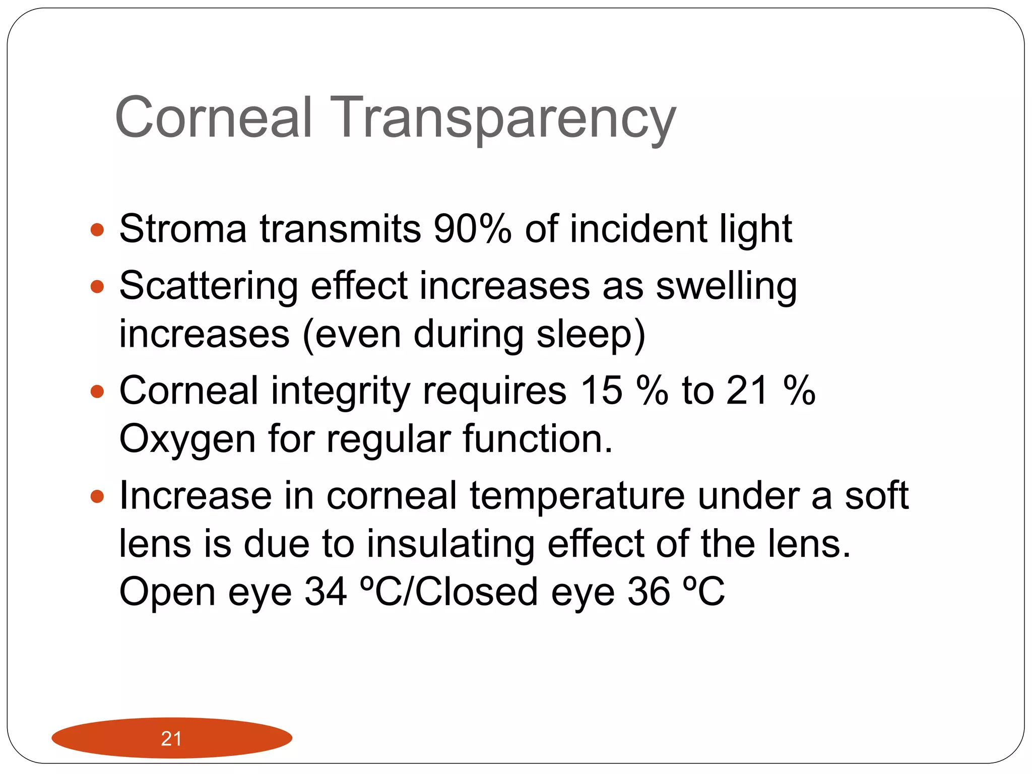
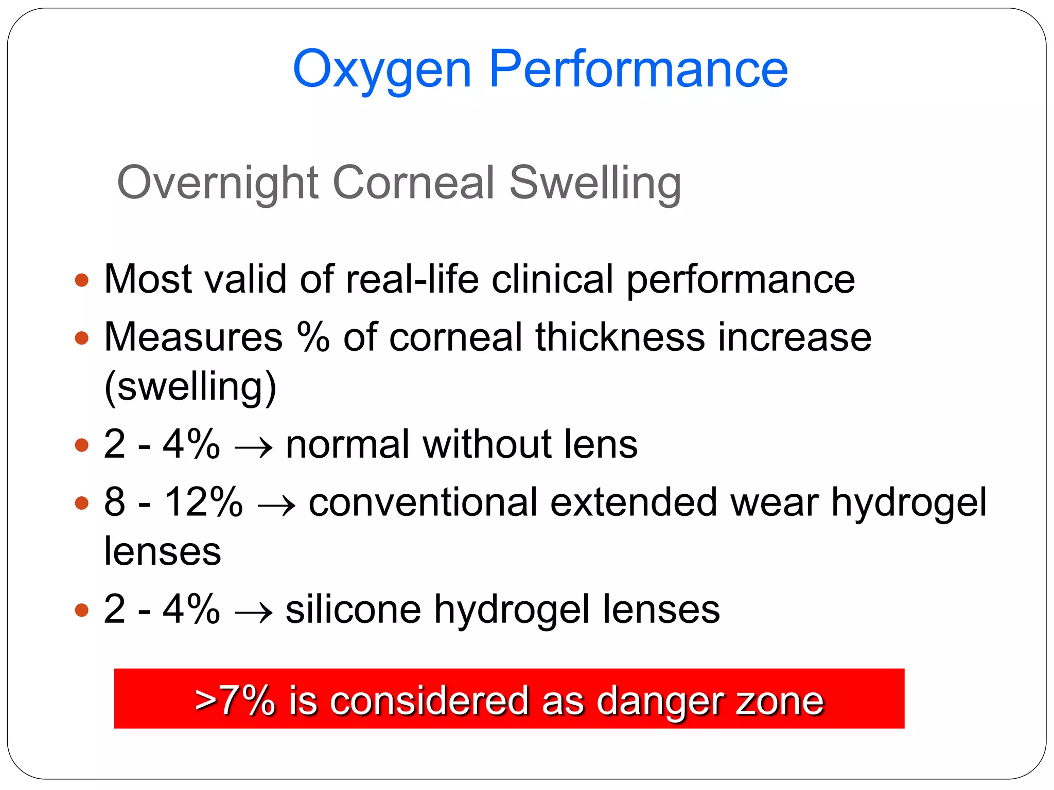
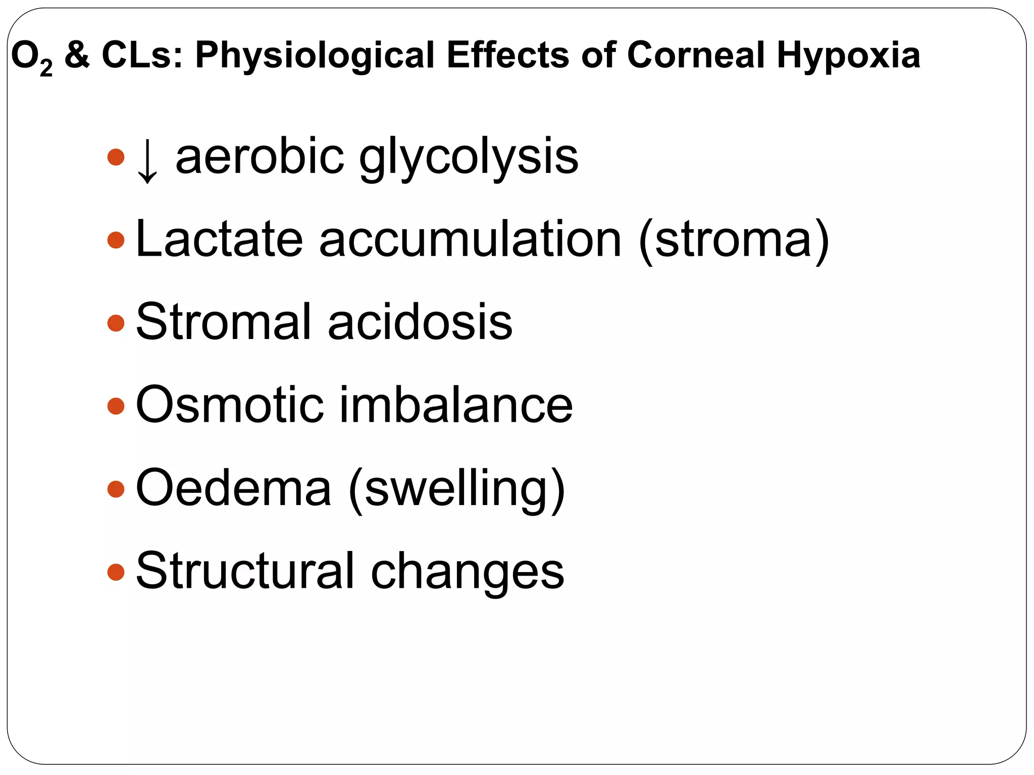
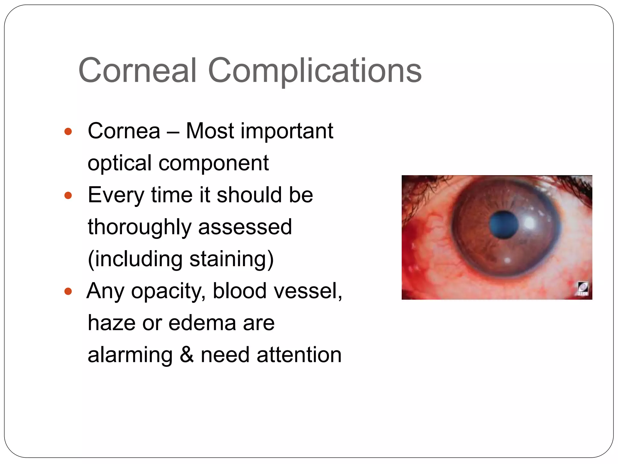
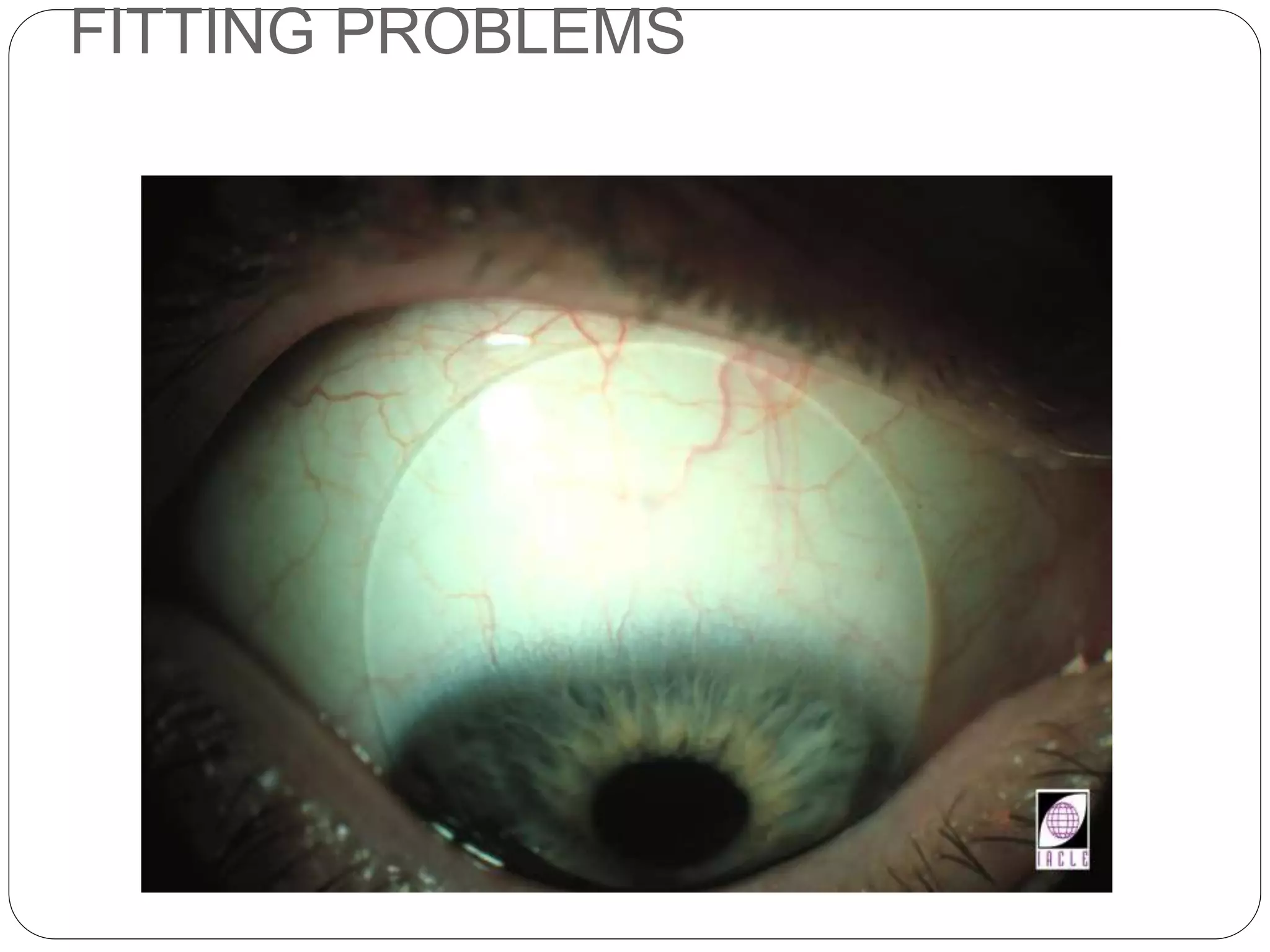
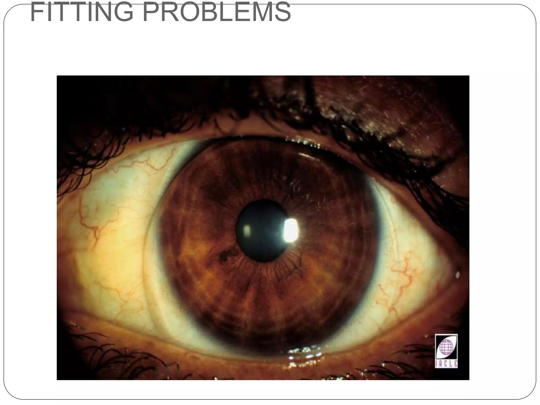
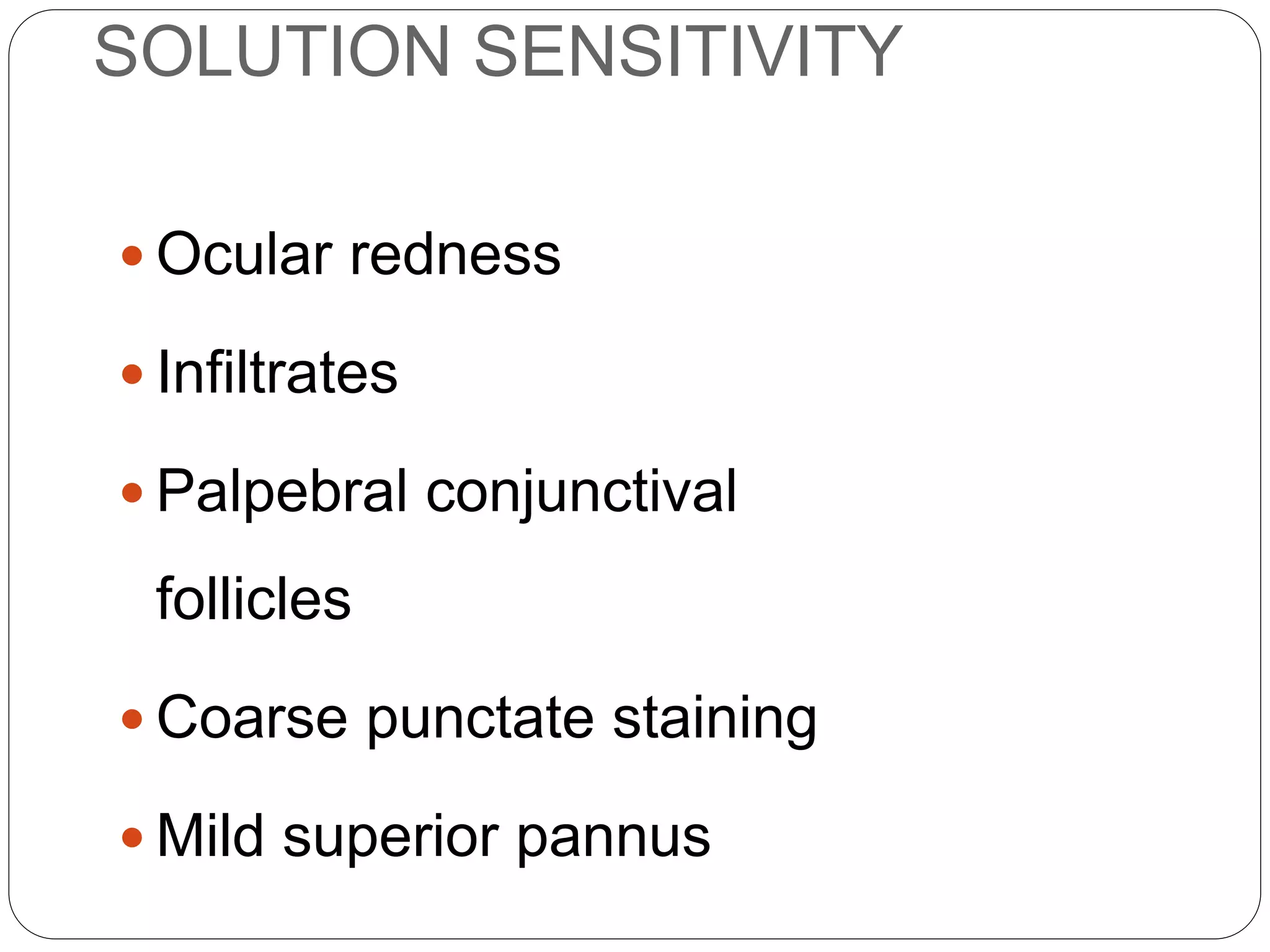
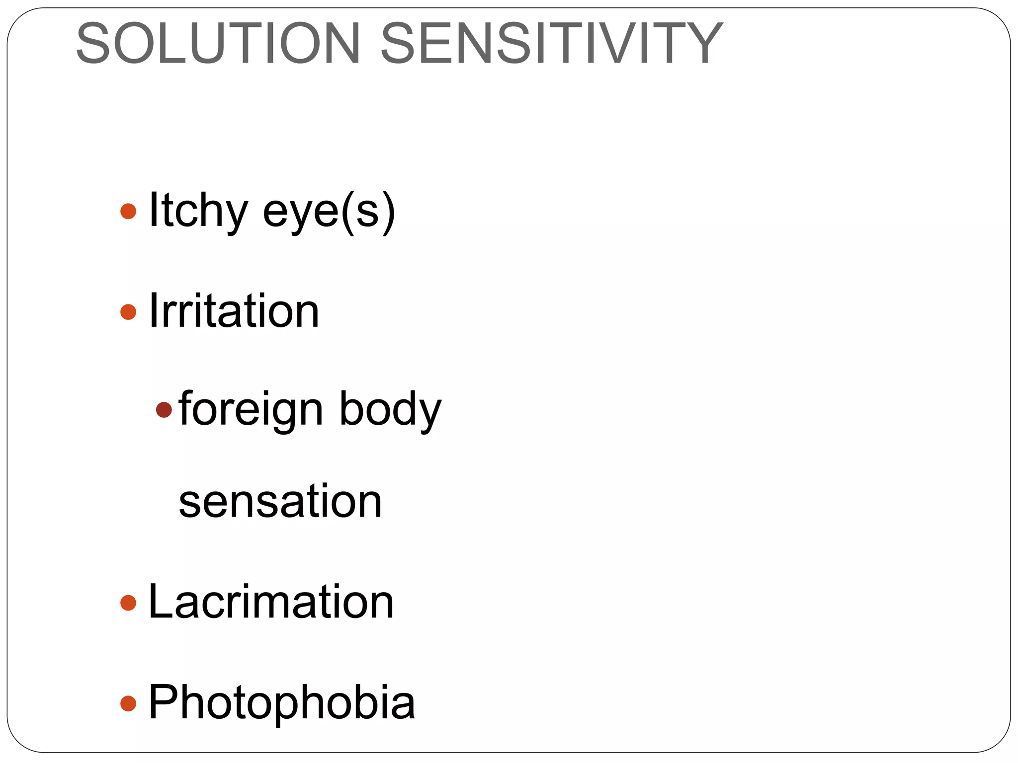
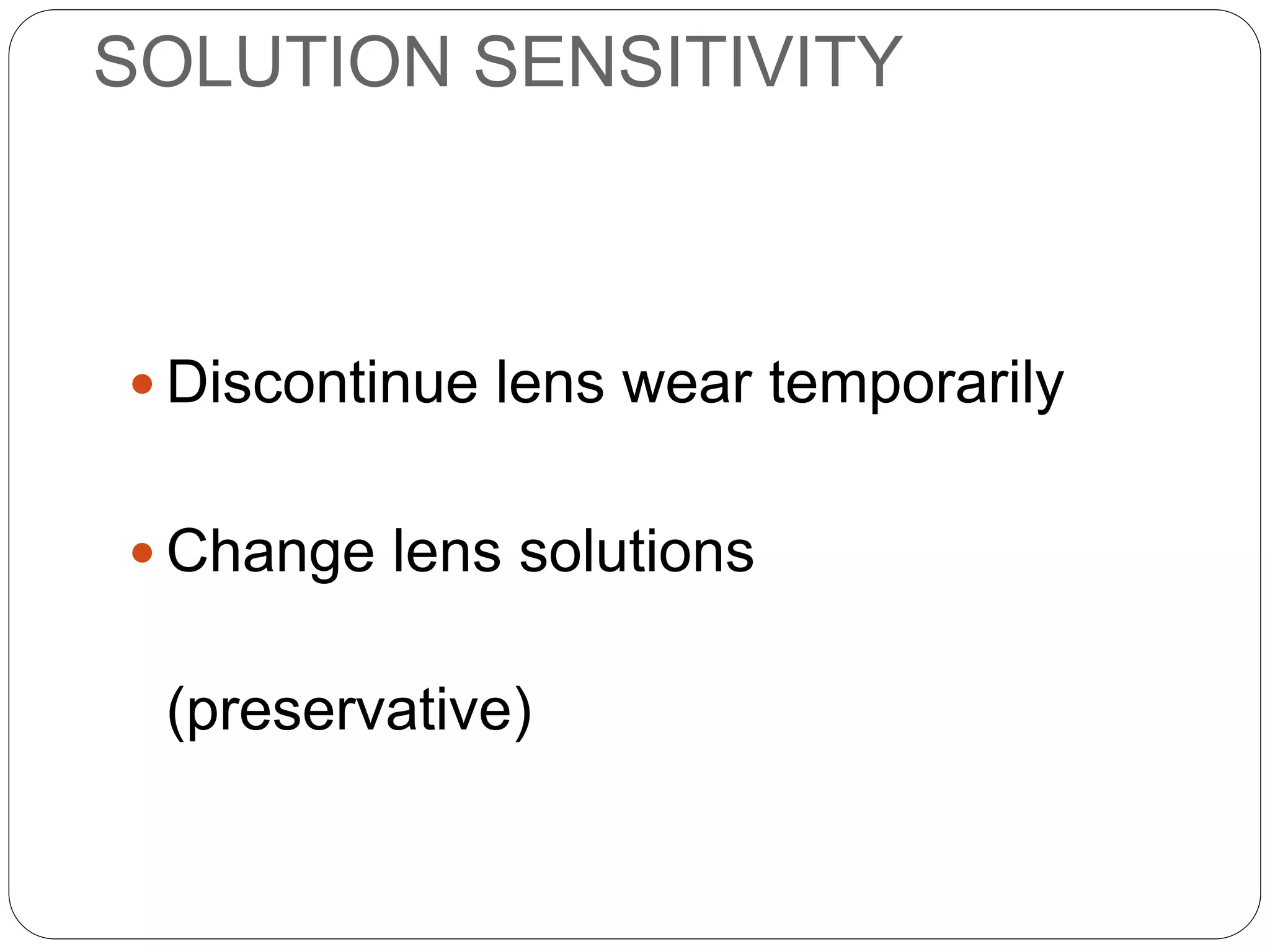
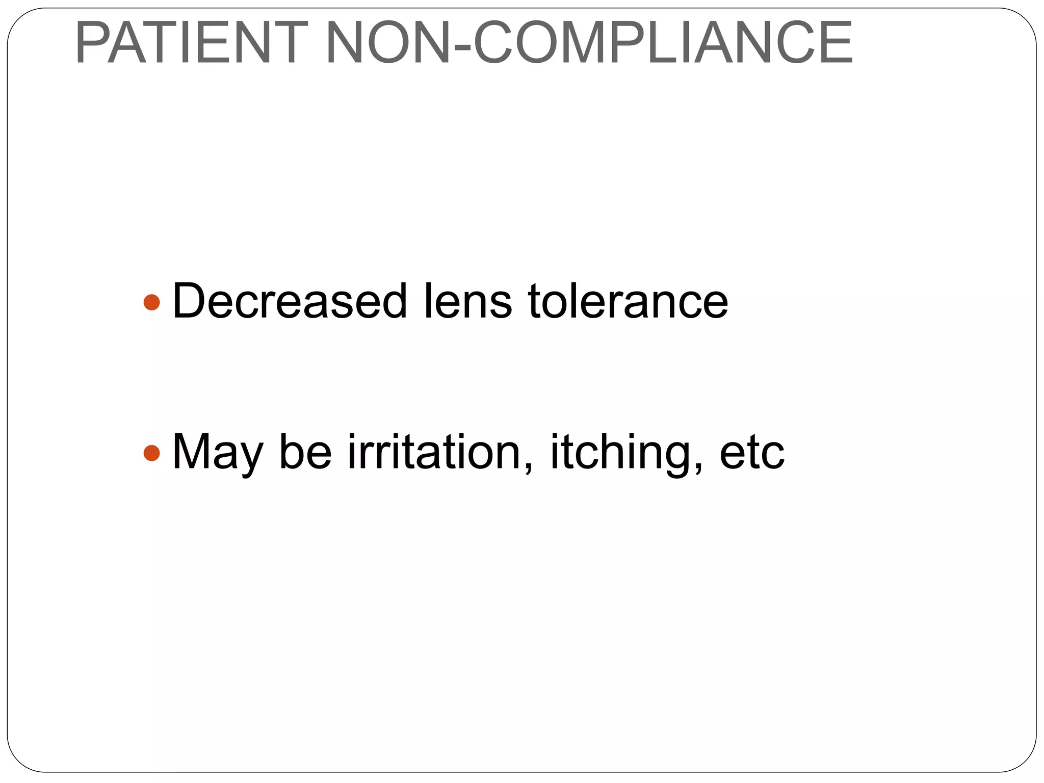
Cornea in Contact Lens
wear
Mr.Vishakh G. Nair
M.Optom FIACLE
Faculty Member
Doctor of Optometry(OD) Programme
Ministry Of Higher Education -KSA
Bharati Vidyapeeth University
Medical College
School of Optometry- PUNE
Success criteria of contact lens
wear……
12 hrs of comfortable, continuous
contact lens wear
Lenses should be comfortable
almost immediately after insertion
in the eye
End of Day comfort
Vision should be good throughout
Convenienc
e
 Health
 Quality
 Value
Consumers indicate that a "healthier
contact lens" is the most important
need
What does "health" mean to
consumers?
In their own words, consumers described"healthier" …
"will not cause infection"
"better for you, better for your eyes"
"comfortable, lubricated, safe"
"optimal perfection, clean and pure"
"worry free, infection free"
"healthier body, vision, lifestyle"
"less strain, less maintenance, less infection"
"fewer side effects, won't cause problems"
Indications-Contact Lenses
Purpose
Cosmetic
Spherical
Toric
Presbyopic
Optical
Keratoconus
Bandage Lens
Drug Delivery
Orthokeratology
Therapeutic
Enhances
Performance
Color Filters
Hydrogel CL Modalities
Soft Contact
Lenses
Traditional
Planned
Replacement/
Disposable
Daily (Disp)
Bi-weekly
Monthly
Quarterly
7
 Daily Wear:
 Extended Wear
(Flexible Wear):
 Continuous Wear:
- Wear during wake hours;
Requires daily removal & care.
- Wear up to 7 days/ 6 nights,
without daily removal & care*.
- Wear up to 30 days/ 29 nights,
without daily removal & care*.
*Silicone Hydrogels
Classification based on
Wear time
 Parameter Changes
 Physiologic compromise leading to tear film
disruption and red eye reaction
 Contact Lens deposits
 Patient compliance
 Contamination with Microorganisms
Conventional Contact Lenses
How do protein deposits get
denatured?
Native Protein
• Transparent
• Natural part of tears
• Beneficial to eyes
(anti-microbial activity)
Denatured Protein
• Translucent/opaque
• Strongly binds to lens
surface
• Harmful to eyes
Environmental
Factors:
• Heat
• Drying
• Oxidation
• UV light
Silicone Hydrogels – Awesome Twosome
The SILICONE phase facilitates oxygen
transmission
The HYDROGEL phase allows good lens movement
 Water Content
Low Water (< 50%)
High Water (50% or more)
 Material Types.
Ionic
Non-ionic
 FDA Classification
Group I, II, III, IV
Water content, ionic charges & FDA classification
12
Group Water Content Charge
I Low (< 50%) Non Ionic
II High (> 50%) Non Ionic
III Low Ionic
IV High Ionic
FDA Classification
 Less corneal edema
 Less microcyst response
 Less neovascularization
 Less limbal hyperemia
 Less infiltrative keretitis
 Less infectious keretitis
 Safe, clear and comfortable lens wear
Improved oxygen transmission with silicone
hydrogel lenses
Cornea
 Window of eye – watch
glass
 Transparent
 Important optical
element (43 D)
 No blood vessels
 Oxygen from
atmosphere
 Very sensitive
14
CORNEAL SHAPE
15
 Aspheric, flattens towards periphery
 Front apical radius = 7.8 mm
 Back apical radius = 6.5 mm
 Average refractive index 1.376
 Center thickness = 0.52 mm
 Peripheral thickness = 0.67 mm
 Horizontal diameter = 12 mm
 Vertical diameter = 11 mm
Cornea cross section
Five layers
 Epithelium
 Bowman’s membrane
 Stroma
 Descemet’s membrane
 Endothelium
16
Dua’s Layer
18
19
Corneal Composition
20
 78 % water
 15 % collagen
 5 % other proteins
 1 % GAGs (Glycos Amino Glycans)
 Epithelium = 10 % of cornea’s wet weight.
Corneal Transparency
21
 Stroma transmits 90% of incident light
 Scattering effect increases as swelling
increases (even during sleep)
 Corneal integrity requires 15 % to 21 %
Oxygen for regular function.
 Increase in corneal temperature under a soft
lens is due to insulating effect of the lens.
Open eye 34 ºC/Closed eye 36 ºC
Oxygen supply to Cornea
22
 Atmosphere via Tear film
 Epithelium (21 %)
 Capillaries of limbal region
 Aqueous humor via corneal endothelium
 Endothelium (7.4%)
 Capillaries of palpebral conjunctiva
 Carbon-di-oxide is thrown out
Dk - Oxygen Permeability.
Amount of O2 material
allow through
Dk/t - Oxygen
Transmissibility.
Amount of O2 material of
certain thickness allow
through
Holden - Mertz Criteria
Dk/t
required for
DW = 24 x
10-9
Dk/t
required for
EW = 87 x
10-9
Oxygen Performance
Overnight Corneal Swelling
 Most valid of real-life clinical performance
 Measures % of corneal thickness increase
(swelling)
 2 - 4%  normal without lens
 8 - 12%  conventional extended wear hydrogel
lenses
 2 - 4%  silicone hydrogel lenses
>7% is considered as danger zone
Oxygen Performance
↓ aerobic glycolysis
Lactate accumulation (stroma)
Stromal acidosis
Osmotic imbalance
Oedema (swelling)
Structural changes
O2 & CLs: Physiological Effects of Corneal Hypoxia
EFFECTS OF TISSUE ACIDOSIS
Ocular Examination
 With Contact Lenses
 Visual Acuity
 Over refraction
 Lens surface quality
 Lens fitting assessment
Ocular Examination
 Without Contact Lenses
 Slit-lamp examination
 Tear film assessment
With Contact Lenses
 Lens fit assessment
 Check whether the lens
fit is same as it was
during dispensing the
lens
 If not, find out the cause
and plan for changing
the lens parameter
Check-up without lenses
 Repeat retinoscopy & subjective acceptance after 30
minutes of removal
 Amount of corneal edema ?
 Amount of change in refractive error ?
 Compare findings with base line results
 Degree of change indicative of cause !!
Check-up without lenses
 Immediately on lens removal
 Slit Lamp exam for Corneal integrity
 Evert eye lid, Upper Tarsal Conjunctiva
 Keratometry – mires, reading
 Effect of lens wear on Cornea
 Retinoscopy & subjective refraction
 Switched contact lenses ?
 Amount of Corneal changes
Possible causes of problems
 First find out whether problems are due to Contact
Lens or the Eye
 Symptoms which stay even after lens removal are possibly
due to eye, other-wise lens
 Fresh trial lens which does not give problems even at end
of full day wear indicates the patient’s earlier lenses are
damaged
 Non-compliance to instructions !
Identify & Rectify problems
 Find out the problem
and its cause
 Myths and
misconceptions !
 Sensitivity to Lens care
system ?
 Recommend solution to
problem
Review of Lens Care system
 Lens handling – insertion & removal
 Ask patient to demonstrate
 Use of solutions and steps
 Name of solutions, their use
 Proper steps and sequence involved
 Change Lens care system if required to balance efficacy & economy
 Patient and Lens Hygiene
 Wash hands before handling lenses
 Trim finger nails, remove cream on hands
 Clean lens storage case once a week
Components of Care and Maintenance
ü Daily cleaner
ü Rinsing solution
ü Disinfecting solution
ü Protein removers
ü Lubricating/ rewetting
solution
ü Lens storage case
Wash hands Either Rub + Rinse
Or Rinse OnlyDisinfect
Daily Lens Care Procedures
 Failure to prescribe proper
lens care regimen
 Failure of compliance
 Reduced comfort
 Reduced vision
 Increased risk of contamination 
complications/ infections
 Dissatisfied patient
Failure of
CL wear
Signs vs Symptoms
 Sign
 Structural change that the practitioner observes during
examination (what you see)
 Symptom
 patient complaint such as irritation, dryness, pain or
visual disturbances (what they feel)
Normal CL Adaptive
Symptom
 Variable vision
 Mild discomfort
 Tearing
 Redness
 Frequent blinking
 Intermittent dryness
 Sensitivity to light
 Minor discharge on the eyelids
When to change Lenses ?
 How long to use same
contact lens ?
 Life depends on
 Wear tear
 Patient tolerance
 Amount of lens care
 CL material
 After or before problems
occur ?
What to change in CL?
 Parameter changes
 Change Contact Lens raw material
 Ocular response to old lenses
 Wearing schedule changes
 Change replacement modality
 Shorter is better to avoid complications
Cornea in Contact Lens wear
 Reduced oxygen supply (Hypoxia)
 Increased accummulation of carbon dioxide
(Hypercapnia)
 Reduced tear exchange
 Increased corneal temperature
 Increased risk of infections and inflammations
Corneal Complications
 Cornea – Most important
optical component
 Every time it should be
thoroughly assessed
(including staining)
 Any opacity, blood vessel,
haze or edema are
alarming & need attention
Common Etiological Factors Causing
Corneal Complications
 Hypoxia and Hypercapnia
 Allergy and Toxicity
 Mechanical effects
 Osmotic effects
Silicon Hydrogel
 Almost eliminates hypoxia
 Reduces dehydration
 Provides good wetting
 Reduces protein deposition
 Enhances on eye comfort
COMPLICATIONS: AETIOLOGIES
 Physical, including lens fit
 Lens back vertex power (BVP)
 Lens dehydration
 Surface deposits/poor wettability
 Altered blinking
 Hypoxia
COMPLICATIONS: AETIOLOGIES
 Pre-existing systemic or ocular disease
 Environmental
 Chemical
 Microbiological
 Immunological
 Patient non-compliance
COMPLICATIONS:
PHYSICAL FACTORS
 Lens:
fit
surface properties
lens condition
lens shape (design)
 Blinking:
completeness
lid tonus
COMPLICATIONS:
VISUAL FACTORS
 Accuracy of Rx prescribed
 Reproducibility/optical quality
 Accuracy of Rx dispensed
 Binocular vision a complication?
 Incipient presbyopia
 Distance vs near (esp. torics)
COMPLICATIONS:
PHYSIOLOGICAL FACTORS
 Dk/t
 Water content
factors affecting water content
 Blinking
frequency
completeness
 Environment
COMPLICATIONS:
PATHOLOGICAL FACTORS
 Condition of lenses
micro-organisms
immunological issues
 Chemical
 Environmental
 Pre-existing ocular pathology
COMPLICATIONS:
WEARER-RELATED
 Non-compliance:
misunderstanding
ignorance and carelessness
deliberate deviation
little or no care of lenses
 Failure to attend after-care visits
 Swapped lenses
 Poor personal hygiene
PREVENTING COMPLICATIONS
 Patient selection
 Lens selection
 Patient education
 preventative measures
 signs, symptoms, action
 After-care and appropriate intervention
CORNEAL OEDEMA:
INTRODUCTION
 Mild oedema is a natural consequence of sleep
 Recognized as contact lens-induced since
introduction of CLs (1888)
 Measured indirectly by corneal swelling (pachometry)
 SCL-induced oedema involves the whole of the
cornea and is diffuse
CORNEAL OEDEMA
 Diffuse
 Structural and functional
changes
involve all layers
subtle to obvious
 More central than peripheral
SIGNS
CORNEAL OEDEMA
SIGNS
TOPOGRAPHICAL SWELLING
CORNEAL SWELLING (%)
CORNEAL LOCATION (mm)
SOFLENS 04
Temporal Nasal
– 9.00 D
– 6.00 D
– 1.25 D
0
5 5 64 43 32 21 10
5
10
15
20
CORNEALSWELLING(%)
(Holden et al., 1985)
CORNEAL OEDEMA: STRIAE
INTRODUCTION
 Appear in the posterior stroma
 Occur in:
inflammatory conditions
traumatic conditions
degenerative conditions
CL wear (hypoxia & other CL effects)
 Usually vertically oriented
1-3 mm in length
whispy white lines
usually thicker than nerve fibres
CORNEAL OEDEMA: STRIAE
SIGNS AND AETIOLOGY
STROMAL STRIAE
 Corneal hypoxia causes stromal
oedema
 Minimum of 5% corneal swelling
required
 Separation of lamellae
AETIOLOGY
STROMAL STRIAE
 Intervention is warranted
 Increase lens Dk/t
 Reduce wearing time
MANAGEMENT
CORNEAL OEDEMA: FOLDS & BLACK
LINES - INTRODUCTION
 Terms used somewhat interchangeably
 Buckling of the component layers resulting from
corneal oedema
 Limbus constrains lateral corneal expansion
 Require 7 to 12+% corneal swelling
black lines result from swelling at the
higher end of this range
CORNEAL OEDEMA: FOLDS AND
BLACK LINES: SIGNS
CORNEAL OEDEMA
 Generally asymptomatic unless corneal
swelling is significant
 Reduced vision:
diffusive ‘spectacle’ blur
haziness, haloes, coloured haloes
little or no Rx change detectable
SYMPTOMS
CORNEAL OEDEMA
 Normal eye closure (sleep)
 Anterior eye hypoxia
 Hypotonicity of the tears
 Reduced tear film evaporation
 Stromal pH changes
 Reduced endothelial pump
efficacy
 Increased temperature under
lenses
 Trauma
AETIOLOGY
CORNEAL OEDEMA
 Minimize contact lens effects:
 maximize lens Dk/t (priority)
 optimize lens fit (less significant)
 fit siloxane hydrogels
 Decrease lens wear
MANAGEMENT
CORNEAL EXHAUSTION SYNDROME:
INTRODUCTION
 Result of long-term wear of lenses with no, or low,
O2 transmissibility
 PMMA (most likely)
 low water, toric SCLs
 low water, high BVP spherical SCLs
 Sudden development of lens intolerance
 All layers of the cornea affected
CORNEAL EXHAUSTION
SYNDROME
 Altered/irregular refraction
 Distorted keratometer mires
 Acute oedema
 Posterior stromal haze/opacities
 Endothelial changes
polymegethism
distortion/bumpiness
SIGNS
CORNEAL EXHAUSTION
SYNDROME
 Reduced lens comfort
 Decreased wearing time
 Blurred or fluctuating vision
SYMPTOMS
CORNEAL EXHAUSTION
SYNDROME
 Long term wear of low Dk/t
lenses
 Chronic hypoxia
 Acidosis
 Endothelial function is
compromised
AETIOLOGY
CORNEAL EXHAUSTION
SYNDROME
 High Dk/t lens to prevent occurrence
 Refit with lenses of much higher Dk/t :
siloxane hydrogels
RGPs
MANAGEMENT
CORNEAL VASCULARIZATION:
INTRODUCTION
 Sometimes called neovascularization (new blood
vessels)
 Vessels observed are often not ‘new’
 All lenses make limbal vasculature more obvious,
RGP lenses and siloxane hydrogels produce the
least obvious alterations
 Need to distinguish between ‘new’ vessels and
filled ghost vessels
CORNEAL VASCULARIZATION
 Early signs are vessel spikes
 Branching capillaries from limbal arcade
episcleral vessels
(anterior ciliary artery)
 Anterior or deep vessels
level of stimulating pathology
SIGNS
CORNEAL VASCULARIZATION
 Limbal versus clear corneal
involvement
 Transudate from limbal capillaries
 Lipid deposits from deep vessels
 Ghost vessels
SIGNS
ANTERIOR EYE VASCULATURE
CORNEAL VASCULARIZATION
 Description
 Documentation
extent of penetration
location
depth
severity
 Assessment by quadrant
SIGNS
CORNEAL VASCULARIZATION
SIGNS
CORNEAL VASCULARIZATION
SIGNS
CORNEAL VASCULARIZATION
 Asymptomatic
 Vascularization may
accompany painful anterior
segment disease
SYMPTOMS
CORNEAL VASCULARIZATION
 Vaso-proliferative stimuli include:
angiogenic factors (existing
vessels)
tight-fitting lenses
lactic acid build-up
oedema (not in isolation)
inflammatory mediators
trauma/disease
keratocytes and inflammatory cells
neural control
POSSIBLE AETIOLOGIES
CORNEAL VASCULARIZATION
 Individual susceptibility
 Long-term contact lens-induced hypoxia
 more common in extended wear
 Latent period
AETIOLOGY
CORNEAL VASCULARIZATION
 Acceptable level of vessel growth
 budding
 limbal or clear cornea
 Optimize lens fitting
 Increase lens Dk/t
MANAGEMENT
CORNEAL VASCULARIZATION
 Change lens solutions
 Patient education and follow-
up
 Optimize fitting
characteristics
 Decrease wearing time
 Refit with siloxane hydrogel
MANAGEMENT
OEDEMA-INDUCED REFRACTIVE
CHANGES: INTRODUCTION
 Often neglected aspect of CL practice as
‘physiology’ issues are pursued
 Changes are usually small with SCLs
spectacle blur uncommon
 Changes are usually myopic
 Changes are usually slow (weeks,
months)
OEDEMA-INDUCED REFRACTIVE
CHANGES: SIGNS
 Corneal oedema
 Decreased acuity on testing
 Usually, corneal curvature also altered
may be steeper or flatter
meridians of one eye can change in
opposite directions
eccentricity may also be altered
 Correlation between changes in curvature
and Rx often poor
OEDEMA-INDUCED REFRACTIVE
CHANGES: SIGNS
Refractive changes (greater in EW):
 Determine after lens removal
 Maximum spherical change <–0.50D (<0.1mm)
hyperopic shifts have been reported
 Maximum cylindrical change <0.33D (<0.06mm)
flattening initially, steepening eventually
 Irregular changes uncommon
 Over-Rx should agree with change measured after
lens removal
 Recovery may be:
rapid after lens removal
nil
OEDEMA-INDUCED REFRACTIVE
CHANGES: SYMPTOMS
 Reduced vision:
 diffusive blur
 refractive blur
 Often, none – change is uncovered during a
consultation or after-care visit
OEDEMA-INDUCED REFRACTIVE
CHANGES: AETIOLOGY
 Poor lens physiological performance
corneal hypoxia
greater with thick lenses (low Dk/t &
rigidity)
 Uneven corneal swelling:
different local Dk/ts under lens
lens design factors
differing BVPs
 Corneal ‘softening’
corneal molding (individual
characteristic)
OEDEMA-INDUCED REFRACTIVE
CHANGES: MANAGEMENT
 Try to determine if Rx change is lens-induced
 Refit with lenses with superior physiological
characteristics
use thinner lenses
fit siloxane hydrogels from the start
use lenses with lower rigidity
 Monitor eyes exhibiting change more closely
 Treat larger than acceptable changes more
seriously (set limits for what is ‘acceptable’)
OCULAR DISCOMFORT:
INTRODUCTION
 Probably the most common contact lens-induced
condition
 May be accompanied by redness and corneal
staining
 Must consider the possibility that it is not contact
lens related
 Various descriptions may be used
 Descriptions may be misleading
DISCOMFORT
 Bulbar and/or palpebral
conjunctival redness
 Staining (with sodium
fluorescein, Rose Bengal,
Lissamine Green)
 May be none
SIGNS
• Surface drying
• Surface deposits
• Excessive lens movement
• Defect or damage
LENS SIGNS
DISCOMFORT
DISCOMFORT
 Mild to painful
 Vague to specific
can indicate cause
SYMPTOMS
DISCOMFORT
SYMPTOMS
 Dryness
 Itchiness
 Grittiness (scratchiness)
 Watery
 Stinging/burning
 Pain
DISCOMFORT
 Multiple aetiologies
physical
physiological
psychological
 Careful questioning of the
patient
AETIOLOGY
ACUTE DISCOMFORT
AETIOLOGY
 Mechanical
foreign body (debris trapped under
lens)
ocular surface damage
 Toxicity
solution mediated
 Lens defect
surface
edge
 Edge: shape & thickness
 Lens fit (too tight, too loose)
 Infection/inflammation
CHRONIC DISCOMFORT
AETIOLOGY
 Drying of the lens front surface
 Drying of the exposed conjunctiva
 Depletion of the post-lens tear film
 Reduced lubricity of the lens front surface
 Tightening of the lens fit
CHRONIC DISCOMFORT
AETIOLOGY
 Front & back surface deposits
 lens movement
 Limbal/conjunctival hyperaemia
 Hypoxia & retention of metabolic by-products
 Hypersensitivity to preserved solutions
 Altered conjunctival sensitivity
DISCOMFORT
 Can be difficult to alleviate
 Optimize lens:
fitting
care
replacement schedule
 Change:
material (to higher or lower water
content)
change lens design
 Use dehydration-resistant lens
MANAGEMENT
OCULAR REDNESS:
INTRODUCTION
 Subject to individual variation
 Pre-lens wear assessment is essential
 Need to assess:
location of redness
extent of redness
depth of vessels involved
type (bulbar, tarsal, limbal)
OCULAR REDNESS
 Location may be:
localized
sector
diffuse
bulbar and/or limbal
 Deep and/or superficial
vessels
SIGNS
OCULAR REDNESS
SIGNS
OCULAR REDNESS
SIGNS
OCULAR REDNESS
SIGNS
OCULAR REDNESS
SIGNS
OCULAR REDNESS
SIGNS
OCULAR REDNESS
SIGNS
OCULAR REDNESS
SIGNS
OCULAR REDNESS
 Asymptomatic to a hot/burning
sensation
 Typical symptoms include:
irritation
dryness
lens intolerance
SYMPTOMS
OCULAR REDNESS
 Multifactorial
 Lens effects on:
tear film
blinking
AETIOLOGY
OCULAR REDNESS
AETIOLOGY
 Tear deficiency
 Mechanical (physical)
 Toxicity/solution incompatibility
 CLPC
 Allergy
 Infective keratitis
 ‘Sterile’ keratitis
 Hypoxia
after Munro & Covey, 1998
OCULAR REDNESS
 Based on cause that needs to be eliminated, or at
least reduced
 Individual basis
 Minimize dehydration of:
ocular surface
lens surface
MANAGEMENT
CONTACT LENS-INDUCED
PAPILLARY CONJUNCTIVITIS
 CLPC or earlier, Giant Papillary Conjunctivitis
(GPC)
 Mainly a result of SCL wear
 Usually bilateral
 Incidence: 0.8% and 12%
 Onset: 2 months to 14 years
 Induced by RGP lenses less commonly
CONTACT LENS PAPILLARY
CONJUNCTIVITIS: SIGNS
 Enlarged papillae
 Roughened appearance
(irregular specular reflection)
 Palpebral redness
 Tissue oedema
 Precursor to Giant Papillary
Conjunctivitis (GPC)
CONTACT LENS PAPILLARY
CONJUNCTIVITIS: SIGNS
CONTACT LENS PAPILLARY
CONJUNCTIVITIS: SIGNS
CONJUNCTIVA: ZONES/AREAS
CLOSED EYE
Marginal
Tarsal
Tarsal
Limbal
Bulbar
Fornix
Orbital
Marginal
Orbital
Fornix
Bulbar
Limbal
Cornea
Tarsal plate
(tarsus)
Tear fluid
CONTACT LENS PAPILLARY
CONJUNCTIVITIS: SIGNS
CONTACT LENS PAPILLARY
CONJUNCTIVITIS: SIGNS
CONTACT LENS PAPILLARY
CONJUNCTIVITIS: SIGNS
PAPILLAE
CHARACTERISTICS
 Raised, cobblestone-like
 Walls perpendicular to tarsal plate
 Occurs in allergic diseases
 Vessels at base & as central vascular tuft
 Associated with allergy/allergic response
 Eventually, apex may appear as a white point
 Mainly inflammatory cells
 0.3 – 0.9 mm diameter
 May be accompanied by strands of mucus
 Seen in the normal conjunctiva
CONTACT LENS PAPILLARY
CONJUNCTIVITIS: SIGNS
CONTACT LENS PAPILLARY
CONJUNCTIVITIS: SIGNS
CONTACT LENS PAPILLARY
CONJUNCTIVITIS:SIGNS
CONTACT LENS PAPILLARY
CONJUNCTIVITIS: SIGNS
CONTACT LENS PAPILLARY
CONJUNCTIVITIS: SYMPTOMS
 Most likely asymptomatic in early stages
 In moderate/advanced stages:
 increased lens awareness
 increased lens movement
 increased mucus affecting:
– drying of lens surface
– lens deposits
– decreased or fluctuating vision
 itching
 lens intolerance
CONTACT LENS PAPILLARY
CONJUNCTIVITIS: AETIOLOGY
 Lens front surface deposits
mechanical irritation
immune response
drying of lens surface
CONTACT LENS PAPILLARY
CONJUNCTIVITIS: MANAGEMENT
 Modify lens wear
 Change lens design
 Frequent lens replacement
 Optimize lens care and
maintenance
CONTACT LENS PAPILLARY
CONJUNCTIVITIS: MANAGEMENT
 RGP lenses
 Pharmacological therapy
 Patient education
 Complete resolution is
unlikely
CLARE: INTRODUCTION
 An acute inflammatory response usually
associated with SCL EW
 Sudden onset, usually early AM
 Presentation is dramatic
 5% - 30% incidence in SCL EW, 4% in disposable
SCL EW
 More likely in first 3 months of lens wear
 More common in female wearers
CLARE SIGNS
CLARE
 Moderate to severe ocular redness
may have 360° conjunctival
redness
 Diffuse infiltrates (2-3 mm from
limbus)
may also have focal zones of
infiltrates
 Minimal or no staining
 Lacrimation, often profuse
SIGNS
CLARE SIGNS
CLARE
 Patient woken (often early AM) by a
painful eye
 Photophobia
 Lacrimation
 Ocular irritation
SYMPTOMS
CLARE
 Extended wear (closed-eye
hypoxia)
 Lens binding overnight
entrapped debris & deposits
 Gram-negative bacteria
 Sensitivity to lens care products
 Debilitated general health
 Some seasonal variation
AETIOLOGY
CLARE
 Temporary discontinuation of
wear
 Palliative therapy
saline rinse (sterile)
lubrication
 Regular lens replacement
 Low toxicity lens care products
MANGEMENT
CLARE
 Monitor recovery
complete resolution of infiltrates
about 1-3 weeks
 Re-start with DW initially
 Caution with continued EW
optimize fitting
change lens type
change lens care products
 Monitor for recurrence
MANGEMENT
FITTING PROBLEMS
SIGNS
• Decentration
• Corneal coverage
- inadequate
- limbal exposure
• Movement on
blinking
- limited
- excessive
FITTING PROBLEMS
SIGNS
• Lag on eye
movement
- horizontal gaze
- vertical gaze
• Tightness
- push up test
- excessive
- loose, inadequate
- recentration speed
FITTING PROBLEMS
SIGNS
• Retinoscopy reflex
- central shadow (steep)
- peripheral distortion (flat)
• Over refraction
- variable
• Keratometer mires
- irregular
- improve on blinking
(steep)
- change on blinking (flat)
FITTING PROBLEMS
SIGNS
• Edge stand-off
- flat fit
- inside-out lens
• Ocular changes
- redness
- staining
— cornea
— conjunctiva
- palpebral conjunctiva
FITTING PROBLEMS
SIGNS
FITTING PROBLEMS
SIGNS
FITTING PROBLEMS
SIGNS
FITTING PROBLEMS
SIGNS
FITTING PROBLEMS
 Poor or variable vision
 Discomfort
 Decreased tolerance to lens wear
reduced wearing time
early onset of symptoms
 Lost lens
 Uneasiness about lens staying on the
eye
 Comfort not a reliable indicator of fit
SYMPTOMS
FITTING PROBLEMS
 Inappropriate lens parameters
poor selection
 Limited lens choice
stock (compromise too
great)
 Change in parameters over
time
 Lens spoilage
 Manufacturer’s tolerances too
lax
AETIOLOGY
FITTING PROBLEMS
 After-care evaluation
 Refitting as required
change parameters/material
stock lens with a compromise?
choose alternative lens
custom design
 Change manufacturers
 Revise office systems to trap
errors
MANAGEMENT
‘SMILE’ STAINING:
INTRODUCTION
 Staining in the inferior cornea
 Pattern similar to ‘smiling face’
 Severity subject to individual variation
 Worst with high water, ultrathin SCLs
Smile Smile Desiccation
More central
‘SMILE’ STAINING: SIGNS
 Punctate staining in the inferior quadrant
 Staining may coalesce
 Stained area isolated from the limbus
 Severity ranges from diffuse to severe
 Severe cases may also have lighter staining
superiorly
 Staining area (the ‘mouth’) may vary from small to
almost semi-circular
‘SMILE’ STAINING: SIGNS
‘SMILE’ STAINING: SYMPTOMS
 Most are asymptomatic
Symptoms reported include:
 Dryness
 Itchiness
 Grittiness
 Lens awareness
Usually, pain is not reported
‘SMILE’ STAINING: AETIOLOGY
 Pervaporation leads to:
 depletion of the aqueous phase of the post-lens tear
film
 desiccation of corneal epithelium
 lens adherence (?)
 Potentially, corneal area not covered by the upper
eyelid can be affected
 Desiccated area stains with sodium fluorescein
‘SMILE’ STAINING:
MANAGEMENT
 Try to ascertain fundamental cause(s)
 If pervaporation:
 increase centre thickness
 lower water content
 alter lens design, e.g. FOZD
 some combination of the above
 If stock lens, try another lens series or a different
manufacturer
SOLUTION SENSITIVITY
 Ocular redness
 Infiltrates
 Palpebral conjunctival
follicles
 Coarse punctate staining
 Mild superior pannus
SIGNS
SOLUTION SENSITIVITY
 Itchy eye(s)
 Irritation
foreign body
sensation
 Lacrimation
 Photophobia
SYMPTOMS
SOLUTION SENSITIVITY
 Accumulation of preservatives from lens care
products, especially storage solutions
 Delayed hypersensitivity to the preservatives in lens
care products
 Predisposition to sensitivities
AETIOLOGY
SOLUTION SENSITIVITY
 Discontinue lens wear temporarily
 Change lens solutions
(preservative)
MANAGEMENT
PATIENT NON-COMPLIANCE
INTRODUCTION
 Non-compliance is common in health care
 Contact lenses are no exception
 Main aspects include:
lens care procedures
lens care products
lens wear schedules
lens replacement schedules
PATIENT NON-COMPLIANCE
 Specific to type of non-compliance
 Lens related
 deposits
 discoloration
 altered physical properties
 shape distortion
 Ocular
 redness (eyeball and lids)
 epithelial staining
 corneal oedema
 papillae (follicles if toxicity an issue)
SIGNS
PATIENT NON-COMPLIANCE
 Decreased lens tolerance
 May be irritation, itching, etc
SYMPTOMS
PATIENT NON-COMPLIANCE
 Little or no contact lens education
 Lack of information reinforcement
 Poor/no after-care scheduling
 Failure to appreciate their own role
in successful contact lens wear
 Human nature
AETIOLOGY
PATIENT NON-COMPLIANCE
 Careful questioning and observation
 After-care evaluation
 Strong emphasis on patient education
 Discontinuation from lens wear
MANAGEMENT
LENS CASE CONTAMINATION
 Dirty lens case
 Discoloured appearance
 Association with corneal
inflammation and infection
SIGNS
LENS CASE CONTAMINATION
SIGNS
LENS CASE CONTAMINATION
 Ocular irritation
 Infection associated
with:
pain
lacrimation
photophobia
SYMPTOMS
LENS CASE CONTAMINATION
 Poor hygiene
 Neglect
 Infrequent
replacement
 Biofilm formation
AETIOLOGY
LENS CASE CONTAMINATION
 Patient education
 Routine case cleaning
 Regular case replacement
MANAGENT
LENS DEPOSITS
 Front and back surface build-up
irregular specular reflection
allow lens to dehydrate
move lens to differentiate deposits from
debris
 Palpebral conjunctival inflammation
 Corneal staining
SIGNS
LENS DEPOSITS
SIGNS
LENS DEPOSITS
SIGNS
LENS DEPOSITS
SIGNS
LENS DEPOSITS
SIGNS
LENS DEPOSITS
SIGNS
LENS DEPOSITS
 Discomfort
 Lens awareness
 Reduced vision
SYMPTOMS
LENS DEPOSITS
 Interaction between:
lens material
tear film/ocular surface
environment
 Proteins denature when attached to the
lens
AETIOLOGY
LENS DEPOSITS
 Optimize care and maintenance
solutions
regimen
 Frequent lens replacement
daily disposal
MANAGEMENT
VISION PROBLEMS
 Reasonably frequent symptom but
less so than discomfort/dryness,
and redness
 Establish cause, or association
 Record vision data:
at fitting visit
on delivery
at all subsequent visits
VISION PROBLEMS
 Decreased habitual vision
high contrast
low contrast
distance and/or near
 Decreased contrast
sensitivity
 Unilateral or bilateral
SIGNS
VISION PROBLEMS
SIGNS
VISION PROBLEMS
 Blurred or variable vision
 Problems with binocular
vision
 Poorer vision at night
 Glare
 Haze
 Fluctuations of the foregoing
 Asthenopia
 Alteration(s)
during lens wear
after lens wear
SYMPTOMS
VISION PROBLEMS
 Blur may be:
refractive
diffusive
 Incorrect BVP
 Poor fitting
 Surface deposits
 Myopic creep
extended wear
AETIOLOGY
VISION PROBLEMS
 Optimize lens fit
 Accurate over-refraction
 Adjust spherical and/or cylindrical
power as indicated
 Change lens material
 Lens care issues
MANAGEMENT
LENS WRINKLING
 Corrugated central or mid-peripheral
lens area
 Does not change with a blink
 Lens wrinkling can cause corneal
SIGNS
LENS WRINKLING
SIGNS
LENS WRINKLING
SIGNS
LENS WRINKLING
SIGNS
LENS WRINKLING
SIGNS
LENS WRINKLING
Severe vision reduction if wrinkling
is in the pupil zone
Onset may be rapid (minutes) to
months
Recovery: ‘prompt’ to ‘hours after’
SYMPTOMS
LENS WRINKLING
Usually a combination of factors:
• High minus BVP
• Low water material
• Ultra-thin central lens design
• Thick mid-peripheral zone
• Lid forces on blinking
AETIOLOGY (Minus Lenses)
AETIOLOGY (Plus Lenses)
LENS WRINKLING
 Usually a combination of factors:
higher plus BVP
low water material
thin peripheral lens design
lid forces on blinking
LENS WRINKLING
 Change lens design
 higher water content
 thicker design
 Consider a more rigid material
 siloxane hydrogel
 Consider RGP lenses
MANAGEMENT
CHANGES IN REFRACTION
 Decreased VA
 Does over-Rx (with) or ‘best’ Rx (without):
 improve VA (to baseline, < baseline)
 fail to improve VA
 If YES, refraction has altered
 myopic creep
 lens-induced change (regular)
 true change in Rx
 If NO, other issues involved:
 change in corneal topography (irregular)
 distorted lens
 poor lens condition (e.g. deposits)
SIGNS: WITH & WITHOUT LENSES
CHANGES IN REFRACTION
 Blurred vision
distance
intermediate
near
all distances
 Asthenopia
SYMPTOMS
CHANGES IN REFRACTION
 Natural
growth/development
 Normal age changes
 Onset of presbyopia
 Lens-induced
chronic hypoxia
bearing pressure
 Irregular astigmatism
 Onset of keratoconus
AETIOLOGY
CHANGES IN REFRACTION
 Accurate refraction
 Assess VA carefully
 Adjust lens BVP
 sphere, cyl, axis
 Optimize fitting
 Use high Dk/t material
MANAGEMENT
RESIDUAL REFRACTIVE ERROR (RRE)
INTRODUCTION
 Refractive error remaining with primary vision
correction in place
errors & omissions excluded
 May be spherical and/or cylindrical
 Corneal or ‘internal’ cause(s)
regular or irregular
 Other: disease, senile changes
retinal, lenticular, etc
RESIDUAL REFRACTIVE ERROR
SIGNS
 VA less than expected
 VA not responsive to Rx changes
 Pinhole may improve VA but….
problem may not be all refractive
required Rx may be impossible to
determine
Rx changes may be to no avail
 Usually, reduced VA mirrors RRE magnitude
RESIDUAL REFRACTIVE ERROR
SYMPTOMS
 Sub-optimal vision
apparent at trial fitting
apparent at dispensing
apparent once wear commences
does not improve with time
 Even small reductions can be symptomatic
RESIDUAL REFRACTIVE ERROR
AETIOLOGY
 Degree of lens conformance (to cornea)
 Usually, anatomical
if astigmatic, usually Against-the-Rule
 If toric, lens orientation may vary with fixation
distance, esp. at near distances
 RRE may not be sphero-cylindrical
  full correction is difficult or
impossible
RESIDUAL REFRACTIVE ERROR
MANAGEMENT
 Do spectacles or CLs provide best
VA?
 Supply best correction possible
 If toric SCL orientation varies:
try another design (thinner?)
alter head position/body posture
optimize Rx for preferred distance
 Complete solution unlikely
compromises are probably required
Moral of the Story..
Experience is like a comb that life gives
you when you have lost all your hair
PRACTITIONER SKILL
Ability to:
investigate
deduce
analyse
diagnose
solve
problems
educate
PRACTITIONER SKILL
Employ SOAPE:
 Subjective
 Objective
 Assessment
 Plan
 Education
Clear Cornea
Clear Vision
Clean Practice
THANK YOU
vishakhnair@rediffmail.com

Cornea in Contact Lens wear

  • 1.
    Cornea in ContactLens wear Mr.Vishakh G. Nair M.Optom FIACLE Faculty Member Doctor of Optometry(OD) Programme Ministry Of Higher Education -KSA Bharati Vidyapeeth University Medical College School of Optometry- PUNE
  • 2.
    Success criteria ofcontact lens wear…… 12 hrs of comfortable, continuous contact lens wear Lenses should be comfortable almost immediately after insertion in the eye End of Day comfort Vision should be good throughout
  • 3.
    Convenienc e  Health  Quality Value Consumers indicate that a "healthier contact lens" is the most important need
  • 4.
    What does "health"mean to consumers? In their own words, consumers described"healthier" … "will not cause infection" "better for you, better for your eyes" "comfortable, lubricated, safe" "optimal perfection, clean and pure" "worry free, infection free" "healthier body, vision, lifestyle" "less strain, less maintenance, less infection" "fewer side effects, won't cause problems"
  • 5.
    Indications-Contact Lenses Purpose Cosmetic Spherical Toric Presbyopic Optical Keratoconus Bandage Lens DrugDelivery Orthokeratology Therapeutic Enhances Performance Color Filters
  • 6.
    Hydrogel CL Modalities SoftContact Lenses Traditional Planned Replacement/ Disposable Daily (Disp) Bi-weekly Monthly Quarterly
  • 7.
    7  Daily Wear: Extended Wear (Flexible Wear):  Continuous Wear: - Wear during wake hours; Requires daily removal & care. - Wear up to 7 days/ 6 nights, without daily removal & care*. - Wear up to 30 days/ 29 nights, without daily removal & care*. *Silicone Hydrogels Classification based on Wear time
  • 8.
     Parameter Changes Physiologic compromise leading to tear film disruption and red eye reaction  Contact Lens deposits  Patient compliance  Contamination with Microorganisms Conventional Contact Lenses
  • 9.
    How do proteindeposits get denatured? Native Protein • Transparent • Natural part of tears • Beneficial to eyes (anti-microbial activity) Denatured Protein • Translucent/opaque • Strongly binds to lens surface • Harmful to eyes Environmental Factors: • Heat • Drying • Oxidation • UV light
  • 10.
    Silicone Hydrogels –Awesome Twosome The SILICONE phase facilitates oxygen transmission The HYDROGEL phase allows good lens movement
  • 11.
     Water Content LowWater (< 50%) High Water (50% or more)  Material Types. Ionic Non-ionic  FDA Classification Group I, II, III, IV Water content, ionic charges & FDA classification
  • 12.
    12 Group Water ContentCharge I Low (< 50%) Non Ionic II High (> 50%) Non Ionic III Low Ionic IV High Ionic FDA Classification
  • 13.
     Less cornealedema  Less microcyst response  Less neovascularization  Less limbal hyperemia  Less infiltrative keretitis  Less infectious keretitis  Safe, clear and comfortable lens wear Improved oxygen transmission with silicone hydrogel lenses
  • 14.
    Cornea  Window ofeye – watch glass  Transparent  Important optical element (43 D)  No blood vessels  Oxygen from atmosphere  Very sensitive 14
  • 15.
    CORNEAL SHAPE 15  Aspheric,flattens towards periphery  Front apical radius = 7.8 mm  Back apical radius = 6.5 mm  Average refractive index 1.376  Center thickness = 0.52 mm  Peripheral thickness = 0.67 mm  Horizontal diameter = 12 mm  Vertical diameter = 11 mm
  • 16.
    Cornea cross section Fivelayers  Epithelium  Bowman’s membrane  Stroma  Descemet’s membrane  Endothelium 16
  • 17.
  • 18.
  • 19.
  • 20.
    Corneal Composition 20  78% water  15 % collagen  5 % other proteins  1 % GAGs (Glycos Amino Glycans)  Epithelium = 10 % of cornea’s wet weight.
  • 21.
    Corneal Transparency 21  Stromatransmits 90% of incident light  Scattering effect increases as swelling increases (even during sleep)  Corneal integrity requires 15 % to 21 % Oxygen for regular function.  Increase in corneal temperature under a soft lens is due to insulating effect of the lens. Open eye 34 ºC/Closed eye 36 ºC
  • 22.
    Oxygen supply toCornea 22  Atmosphere via Tear film  Epithelium (21 %)  Capillaries of limbal region  Aqueous humor via corneal endothelium  Endothelium (7.4%)  Capillaries of palpebral conjunctiva  Carbon-di-oxide is thrown out
  • 24.
    Dk - OxygenPermeability. Amount of O2 material allow through Dk/t - Oxygen Transmissibility. Amount of O2 material of certain thickness allow through Holden - Mertz Criteria Dk/t required for DW = 24 x 10-9 Dk/t required for EW = 87 x 10-9 Oxygen Performance
  • 25.
    Overnight Corneal Swelling Most valid of real-life clinical performance  Measures % of corneal thickness increase (swelling)  2 - 4%  normal without lens  8 - 12%  conventional extended wear hydrogel lenses  2 - 4%  silicone hydrogel lenses >7% is considered as danger zone Oxygen Performance
  • 26.
    ↓ aerobic glycolysis Lactateaccumulation (stroma) Stromal acidosis Osmotic imbalance Oedema (swelling) Structural changes O2 & CLs: Physiological Effects of Corneal Hypoxia
  • 27.
  • 28.
    Ocular Examination  WithContact Lenses  Visual Acuity  Over refraction  Lens surface quality  Lens fitting assessment
  • 29.
    Ocular Examination  WithoutContact Lenses  Slit-lamp examination  Tear film assessment
  • 30.
    With Contact Lenses Lens fit assessment  Check whether the lens fit is same as it was during dispensing the lens  If not, find out the cause and plan for changing the lens parameter
  • 31.
    Check-up without lenses Repeat retinoscopy & subjective acceptance after 30 minutes of removal  Amount of corneal edema ?  Amount of change in refractive error ?  Compare findings with base line results  Degree of change indicative of cause !!
  • 32.
    Check-up without lenses Immediately on lens removal  Slit Lamp exam for Corneal integrity  Evert eye lid, Upper Tarsal Conjunctiva  Keratometry – mires, reading  Effect of lens wear on Cornea  Retinoscopy & subjective refraction  Switched contact lenses ?  Amount of Corneal changes
  • 33.
    Possible causes ofproblems  First find out whether problems are due to Contact Lens or the Eye  Symptoms which stay even after lens removal are possibly due to eye, other-wise lens  Fresh trial lens which does not give problems even at end of full day wear indicates the patient’s earlier lenses are damaged  Non-compliance to instructions !
  • 34.
    Identify & Rectifyproblems  Find out the problem and its cause  Myths and misconceptions !  Sensitivity to Lens care system ?  Recommend solution to problem
  • 35.
    Review of LensCare system  Lens handling – insertion & removal  Ask patient to demonstrate  Use of solutions and steps  Name of solutions, their use  Proper steps and sequence involved  Change Lens care system if required to balance efficacy & economy  Patient and Lens Hygiene  Wash hands before handling lenses  Trim finger nails, remove cream on hands  Clean lens storage case once a week
  • 36.
    Components of Careand Maintenance ü Daily cleaner ü Rinsing solution ü Disinfecting solution ü Protein removers ü Lubricating/ rewetting solution ü Lens storage case
  • 37.
    Wash hands EitherRub + Rinse Or Rinse OnlyDisinfect Daily Lens Care Procedures
  • 38.
     Failure toprescribe proper lens care regimen  Failure of compliance  Reduced comfort  Reduced vision  Increased risk of contamination  complications/ infections  Dissatisfied patient Failure of CL wear
  • 39.
    Signs vs Symptoms Sign  Structural change that the practitioner observes during examination (what you see)  Symptom  patient complaint such as irritation, dryness, pain or visual disturbances (what they feel)
  • 40.
    Normal CL Adaptive Symptom Variable vision  Mild discomfort  Tearing  Redness  Frequent blinking  Intermittent dryness  Sensitivity to light  Minor discharge on the eyelids
  • 41.
    When to changeLenses ?  How long to use same contact lens ?  Life depends on  Wear tear  Patient tolerance  Amount of lens care  CL material  After or before problems occur ?
  • 42.
    What to changein CL?  Parameter changes  Change Contact Lens raw material  Ocular response to old lenses  Wearing schedule changes  Change replacement modality  Shorter is better to avoid complications
  • 43.
    Cornea in ContactLens wear  Reduced oxygen supply (Hypoxia)  Increased accummulation of carbon dioxide (Hypercapnia)  Reduced tear exchange  Increased corneal temperature  Increased risk of infections and inflammations
  • 44.
    Corneal Complications  Cornea– Most important optical component  Every time it should be thoroughly assessed (including staining)  Any opacity, blood vessel, haze or edema are alarming & need attention
  • 45.
    Common Etiological FactorsCausing Corneal Complications  Hypoxia and Hypercapnia  Allergy and Toxicity  Mechanical effects  Osmotic effects
  • 46.
    Silicon Hydrogel  Almosteliminates hypoxia  Reduces dehydration  Provides good wetting  Reduces protein deposition  Enhances on eye comfort
  • 47.
    COMPLICATIONS: AETIOLOGIES  Physical,including lens fit  Lens back vertex power (BVP)  Lens dehydration  Surface deposits/poor wettability  Altered blinking  Hypoxia
  • 48.
    COMPLICATIONS: AETIOLOGIES  Pre-existingsystemic or ocular disease  Environmental  Chemical  Microbiological  Immunological  Patient non-compliance
  • 49.
    COMPLICATIONS: PHYSICAL FACTORS  Lens: fit surfaceproperties lens condition lens shape (design)  Blinking: completeness lid tonus
  • 50.
    COMPLICATIONS: VISUAL FACTORS  Accuracyof Rx prescribed  Reproducibility/optical quality  Accuracy of Rx dispensed  Binocular vision a complication?  Incipient presbyopia  Distance vs near (esp. torics)
  • 51.
    COMPLICATIONS: PHYSIOLOGICAL FACTORS  Dk/t Water content factors affecting water content  Blinking frequency completeness  Environment
  • 52.
    COMPLICATIONS: PATHOLOGICAL FACTORS  Conditionof lenses micro-organisms immunological issues  Chemical  Environmental  Pre-existing ocular pathology
  • 53.
    COMPLICATIONS: WEARER-RELATED  Non-compliance: misunderstanding ignorance andcarelessness deliberate deviation little or no care of lenses  Failure to attend after-care visits  Swapped lenses  Poor personal hygiene
  • 54.
    PREVENTING COMPLICATIONS  Patientselection  Lens selection  Patient education  preventative measures  signs, symptoms, action  After-care and appropriate intervention
  • 55.
    CORNEAL OEDEMA: INTRODUCTION  Mildoedema is a natural consequence of sleep  Recognized as contact lens-induced since introduction of CLs (1888)  Measured indirectly by corneal swelling (pachometry)  SCL-induced oedema involves the whole of the cornea and is diffuse
  • 56.
    CORNEAL OEDEMA  Diffuse Structural and functional changes involve all layers subtle to obvious  More central than peripheral SIGNS
  • 57.
  • 58.
    TOPOGRAPHICAL SWELLING CORNEAL SWELLING(%) CORNEAL LOCATION (mm) SOFLENS 04 Temporal Nasal – 9.00 D – 6.00 D – 1.25 D 0 5 5 64 43 32 21 10 5 10 15 20 CORNEALSWELLING(%) (Holden et al., 1985)
  • 59.
    CORNEAL OEDEMA: STRIAE INTRODUCTION Appear in the posterior stroma  Occur in: inflammatory conditions traumatic conditions degenerative conditions CL wear (hypoxia & other CL effects)  Usually vertically oriented 1-3 mm in length whispy white lines usually thicker than nerve fibres
  • 60.
  • 61.
    STROMAL STRIAE  Cornealhypoxia causes stromal oedema  Minimum of 5% corneal swelling required  Separation of lamellae AETIOLOGY
  • 62.
    STROMAL STRIAE  Interventionis warranted  Increase lens Dk/t  Reduce wearing time MANAGEMENT
  • 63.
    CORNEAL OEDEMA: FOLDS& BLACK LINES - INTRODUCTION  Terms used somewhat interchangeably  Buckling of the component layers resulting from corneal oedema  Limbus constrains lateral corneal expansion  Require 7 to 12+% corneal swelling black lines result from swelling at the higher end of this range
  • 64.
    CORNEAL OEDEMA: FOLDSAND BLACK LINES: SIGNS
  • 65.
    CORNEAL OEDEMA  Generallyasymptomatic unless corneal swelling is significant  Reduced vision: diffusive ‘spectacle’ blur haziness, haloes, coloured haloes little or no Rx change detectable SYMPTOMS
  • 66.
    CORNEAL OEDEMA  Normaleye closure (sleep)  Anterior eye hypoxia  Hypotonicity of the tears  Reduced tear film evaporation  Stromal pH changes  Reduced endothelial pump efficacy  Increased temperature under lenses  Trauma AETIOLOGY
  • 67.
    CORNEAL OEDEMA  Minimizecontact lens effects:  maximize lens Dk/t (priority)  optimize lens fit (less significant)  fit siloxane hydrogels  Decrease lens wear MANAGEMENT
  • 68.
    CORNEAL EXHAUSTION SYNDROME: INTRODUCTION Result of long-term wear of lenses with no, or low, O2 transmissibility  PMMA (most likely)  low water, toric SCLs  low water, high BVP spherical SCLs  Sudden development of lens intolerance  All layers of the cornea affected
  • 69.
    CORNEAL EXHAUSTION SYNDROME  Altered/irregularrefraction  Distorted keratometer mires  Acute oedema  Posterior stromal haze/opacities  Endothelial changes polymegethism distortion/bumpiness SIGNS
  • 70.
    CORNEAL EXHAUSTION SYNDROME  Reducedlens comfort  Decreased wearing time  Blurred or fluctuating vision SYMPTOMS
  • 71.
    CORNEAL EXHAUSTION SYNDROME  Longterm wear of low Dk/t lenses  Chronic hypoxia  Acidosis  Endothelial function is compromised AETIOLOGY
  • 72.
    CORNEAL EXHAUSTION SYNDROME  HighDk/t lens to prevent occurrence  Refit with lenses of much higher Dk/t : siloxane hydrogels RGPs MANAGEMENT
  • 73.
    CORNEAL VASCULARIZATION: INTRODUCTION  Sometimescalled neovascularization (new blood vessels)  Vessels observed are often not ‘new’  All lenses make limbal vasculature more obvious, RGP lenses and siloxane hydrogels produce the least obvious alterations  Need to distinguish between ‘new’ vessels and filled ghost vessels
  • 74.
    CORNEAL VASCULARIZATION  Earlysigns are vessel spikes  Branching capillaries from limbal arcade episcleral vessels (anterior ciliary artery)  Anterior or deep vessels level of stimulating pathology SIGNS
  • 75.
    CORNEAL VASCULARIZATION  Limbalversus clear corneal involvement  Transudate from limbal capillaries  Lipid deposits from deep vessels  Ghost vessels SIGNS
  • 76.
  • 77.
    CORNEAL VASCULARIZATION  Description Documentation extent of penetration location depth severity  Assessment by quadrant SIGNS
  • 78.
  • 79.
  • 80.
    CORNEAL VASCULARIZATION  Asymptomatic Vascularization may accompany painful anterior segment disease SYMPTOMS
  • 81.
    CORNEAL VASCULARIZATION  Vaso-proliferativestimuli include: angiogenic factors (existing vessels) tight-fitting lenses lactic acid build-up oedema (not in isolation) inflammatory mediators trauma/disease keratocytes and inflammatory cells neural control POSSIBLE AETIOLOGIES
  • 82.
    CORNEAL VASCULARIZATION  Individualsusceptibility  Long-term contact lens-induced hypoxia  more common in extended wear  Latent period AETIOLOGY
  • 83.
    CORNEAL VASCULARIZATION  Acceptablelevel of vessel growth  budding  limbal or clear cornea  Optimize lens fitting  Increase lens Dk/t MANAGEMENT
  • 84.
    CORNEAL VASCULARIZATION  Changelens solutions  Patient education and follow- up  Optimize fitting characteristics  Decrease wearing time  Refit with siloxane hydrogel MANAGEMENT
  • 85.
    OEDEMA-INDUCED REFRACTIVE CHANGES: INTRODUCTION Often neglected aspect of CL practice as ‘physiology’ issues are pursued  Changes are usually small with SCLs spectacle blur uncommon  Changes are usually myopic  Changes are usually slow (weeks, months)
  • 86.
    OEDEMA-INDUCED REFRACTIVE CHANGES: SIGNS Corneal oedema  Decreased acuity on testing  Usually, corneal curvature also altered may be steeper or flatter meridians of one eye can change in opposite directions eccentricity may also be altered  Correlation between changes in curvature and Rx often poor
  • 87.
    OEDEMA-INDUCED REFRACTIVE CHANGES: SIGNS Refractivechanges (greater in EW):  Determine after lens removal  Maximum spherical change <–0.50D (<0.1mm) hyperopic shifts have been reported  Maximum cylindrical change <0.33D (<0.06mm) flattening initially, steepening eventually  Irregular changes uncommon  Over-Rx should agree with change measured after lens removal  Recovery may be: rapid after lens removal nil
  • 88.
    OEDEMA-INDUCED REFRACTIVE CHANGES: SYMPTOMS Reduced vision:  diffusive blur  refractive blur  Often, none – change is uncovered during a consultation or after-care visit
  • 89.
    OEDEMA-INDUCED REFRACTIVE CHANGES: AETIOLOGY Poor lens physiological performance corneal hypoxia greater with thick lenses (low Dk/t & rigidity)  Uneven corneal swelling: different local Dk/ts under lens lens design factors differing BVPs  Corneal ‘softening’ corneal molding (individual characteristic)
  • 90.
    OEDEMA-INDUCED REFRACTIVE CHANGES: MANAGEMENT Try to determine if Rx change is lens-induced  Refit with lenses with superior physiological characteristics use thinner lenses fit siloxane hydrogels from the start use lenses with lower rigidity  Monitor eyes exhibiting change more closely  Treat larger than acceptable changes more seriously (set limits for what is ‘acceptable’)
  • 91.
    OCULAR DISCOMFORT: INTRODUCTION  Probablythe most common contact lens-induced condition  May be accompanied by redness and corneal staining  Must consider the possibility that it is not contact lens related  Various descriptions may be used  Descriptions may be misleading
  • 92.
    DISCOMFORT  Bulbar and/orpalpebral conjunctival redness  Staining (with sodium fluorescein, Rose Bengal, Lissamine Green)  May be none SIGNS
  • 93.
    • Surface drying •Surface deposits • Excessive lens movement • Defect or damage LENS SIGNS DISCOMFORT
  • 94.
    DISCOMFORT  Mild topainful  Vague to specific can indicate cause SYMPTOMS
  • 95.
    DISCOMFORT SYMPTOMS  Dryness  Itchiness Grittiness (scratchiness)  Watery  Stinging/burning  Pain
  • 96.
  • 97.
    ACUTE DISCOMFORT AETIOLOGY  Mechanical foreignbody (debris trapped under lens) ocular surface damage  Toxicity solution mediated  Lens defect surface edge  Edge: shape & thickness  Lens fit (too tight, too loose)  Infection/inflammation
  • 98.
    CHRONIC DISCOMFORT AETIOLOGY  Dryingof the lens front surface  Drying of the exposed conjunctiva  Depletion of the post-lens tear film  Reduced lubricity of the lens front surface  Tightening of the lens fit
  • 99.
    CHRONIC DISCOMFORT AETIOLOGY  Front& back surface deposits  lens movement  Limbal/conjunctival hyperaemia  Hypoxia & retention of metabolic by-products  Hypersensitivity to preserved solutions  Altered conjunctival sensitivity
  • 100.
    DISCOMFORT  Can bedifficult to alleviate  Optimize lens: fitting care replacement schedule  Change: material (to higher or lower water content) change lens design  Use dehydration-resistant lens MANAGEMENT
  • 101.
    OCULAR REDNESS: INTRODUCTION  Subjectto individual variation  Pre-lens wear assessment is essential  Need to assess: location of redness extent of redness depth of vessels involved type (bulbar, tarsal, limbal)
  • 102.
    OCULAR REDNESS  Locationmay be: localized sector diffuse bulbar and/or limbal  Deep and/or superficial vessels SIGNS
  • 103.
  • 104.
  • 105.
  • 106.
  • 107.
  • 108.
  • 109.
  • 110.
    OCULAR REDNESS  Asymptomaticto a hot/burning sensation  Typical symptoms include: irritation dryness lens intolerance SYMPTOMS
  • 111.
    OCULAR REDNESS  Multifactorial Lens effects on: tear film blinking AETIOLOGY
  • 112.
    OCULAR REDNESS AETIOLOGY  Teardeficiency  Mechanical (physical)  Toxicity/solution incompatibility  CLPC  Allergy  Infective keratitis  ‘Sterile’ keratitis  Hypoxia after Munro & Covey, 1998
  • 113.
    OCULAR REDNESS  Basedon cause that needs to be eliminated, or at least reduced  Individual basis  Minimize dehydration of: ocular surface lens surface MANAGEMENT
  • 114.
    CONTACT LENS-INDUCED PAPILLARY CONJUNCTIVITIS CLPC or earlier, Giant Papillary Conjunctivitis (GPC)  Mainly a result of SCL wear  Usually bilateral  Incidence: 0.8% and 12%  Onset: 2 months to 14 years  Induced by RGP lenses less commonly
  • 115.
    CONTACT LENS PAPILLARY CONJUNCTIVITIS:SIGNS  Enlarged papillae  Roughened appearance (irregular specular reflection)  Palpebral redness  Tissue oedema  Precursor to Giant Papillary Conjunctivitis (GPC)
  • 116.
  • 117.
  • 118.
  • 119.
  • 120.
  • 121.
  • 122.
    PAPILLAE CHARACTERISTICS  Raised, cobblestone-like Walls perpendicular to tarsal plate  Occurs in allergic diseases  Vessels at base & as central vascular tuft  Associated with allergy/allergic response  Eventually, apex may appear as a white point  Mainly inflammatory cells  0.3 – 0.9 mm diameter  May be accompanied by strands of mucus  Seen in the normal conjunctiva
  • 123.
  • 124.
  • 125.
  • 126.
  • 127.
    CONTACT LENS PAPILLARY CONJUNCTIVITIS:SYMPTOMS  Most likely asymptomatic in early stages  In moderate/advanced stages:  increased lens awareness  increased lens movement  increased mucus affecting: – drying of lens surface – lens deposits – decreased or fluctuating vision  itching  lens intolerance
  • 128.
    CONTACT LENS PAPILLARY CONJUNCTIVITIS:AETIOLOGY  Lens front surface deposits mechanical irritation immune response drying of lens surface
  • 129.
    CONTACT LENS PAPILLARY CONJUNCTIVITIS:MANAGEMENT  Modify lens wear  Change lens design  Frequent lens replacement  Optimize lens care and maintenance
  • 130.
    CONTACT LENS PAPILLARY CONJUNCTIVITIS:MANAGEMENT  RGP lenses  Pharmacological therapy  Patient education  Complete resolution is unlikely
  • 131.
    CLARE: INTRODUCTION  Anacute inflammatory response usually associated with SCL EW  Sudden onset, usually early AM  Presentation is dramatic  5% - 30% incidence in SCL EW, 4% in disposable SCL EW  More likely in first 3 months of lens wear  More common in female wearers
  • 132.
  • 133.
    CLARE  Moderate tosevere ocular redness may have 360° conjunctival redness  Diffuse infiltrates (2-3 mm from limbus) may also have focal zones of infiltrates  Minimal or no staining  Lacrimation, often profuse SIGNS
  • 134.
  • 135.
    CLARE  Patient woken(often early AM) by a painful eye  Photophobia  Lacrimation  Ocular irritation SYMPTOMS
  • 136.
    CLARE  Extended wear(closed-eye hypoxia)  Lens binding overnight entrapped debris & deposits  Gram-negative bacteria  Sensitivity to lens care products  Debilitated general health  Some seasonal variation AETIOLOGY
  • 137.
    CLARE  Temporary discontinuationof wear  Palliative therapy saline rinse (sterile) lubrication  Regular lens replacement  Low toxicity lens care products MANGEMENT
  • 138.
    CLARE  Monitor recovery completeresolution of infiltrates about 1-3 weeks  Re-start with DW initially  Caution with continued EW optimize fitting change lens type change lens care products  Monitor for recurrence MANGEMENT
  • 139.
    FITTING PROBLEMS SIGNS • Decentration •Corneal coverage - inadequate - limbal exposure • Movement on blinking - limited - excessive
  • 140.
    FITTING PROBLEMS SIGNS • Lagon eye movement - horizontal gaze - vertical gaze • Tightness - push up test - excessive - loose, inadequate - recentration speed
  • 141.
    FITTING PROBLEMS SIGNS • Retinoscopyreflex - central shadow (steep) - peripheral distortion (flat) • Over refraction - variable • Keratometer mires - irregular - improve on blinking (steep) - change on blinking (flat)
  • 142.
    FITTING PROBLEMS SIGNS • Edgestand-off - flat fit - inside-out lens • Ocular changes - redness - staining — cornea — conjunctiva - palpebral conjunctiva
  • 143.
  • 144.
  • 145.
  • 146.
  • 147.
    FITTING PROBLEMS  Pooror variable vision  Discomfort  Decreased tolerance to lens wear reduced wearing time early onset of symptoms  Lost lens  Uneasiness about lens staying on the eye  Comfort not a reliable indicator of fit SYMPTOMS
  • 148.
    FITTING PROBLEMS  Inappropriatelens parameters poor selection  Limited lens choice stock (compromise too great)  Change in parameters over time  Lens spoilage  Manufacturer’s tolerances too lax AETIOLOGY
  • 149.
    FITTING PROBLEMS  After-careevaluation  Refitting as required change parameters/material stock lens with a compromise? choose alternative lens custom design  Change manufacturers  Revise office systems to trap errors MANAGEMENT
  • 150.
    ‘SMILE’ STAINING: INTRODUCTION  Stainingin the inferior cornea  Pattern similar to ‘smiling face’  Severity subject to individual variation  Worst with high water, ultrathin SCLs Smile Smile Desiccation More central
  • 151.
    ‘SMILE’ STAINING: SIGNS Punctate staining in the inferior quadrant  Staining may coalesce  Stained area isolated from the limbus  Severity ranges from diffuse to severe  Severe cases may also have lighter staining superiorly  Staining area (the ‘mouth’) may vary from small to almost semi-circular
  • 152.
  • 153.
  • 154.
  • 155.
    ‘SMILE’ STAINING: SYMPTOMS Most are asymptomatic Symptoms reported include:  Dryness  Itchiness  Grittiness  Lens awareness Usually, pain is not reported
  • 156.
    ‘SMILE’ STAINING: AETIOLOGY Pervaporation leads to:  depletion of the aqueous phase of the post-lens tear film  desiccation of corneal epithelium  lens adherence (?)  Potentially, corneal area not covered by the upper eyelid can be affected  Desiccated area stains with sodium fluorescein
  • 157.
    ‘SMILE’ STAINING: MANAGEMENT  Tryto ascertain fundamental cause(s)  If pervaporation:  increase centre thickness  lower water content  alter lens design, e.g. FOZD  some combination of the above  If stock lens, try another lens series or a different manufacturer
  • 158.
    SOLUTION SENSITIVITY  Ocularredness  Infiltrates  Palpebral conjunctival follicles  Coarse punctate staining  Mild superior pannus SIGNS
  • 159.
    SOLUTION SENSITIVITY  Itchyeye(s)  Irritation foreign body sensation  Lacrimation  Photophobia SYMPTOMS
  • 160.
    SOLUTION SENSITIVITY  Accumulationof preservatives from lens care products, especially storage solutions  Delayed hypersensitivity to the preservatives in lens care products  Predisposition to sensitivities AETIOLOGY
  • 161.
    SOLUTION SENSITIVITY  Discontinuelens wear temporarily  Change lens solutions (preservative) MANAGEMENT
  • 162.
    PATIENT NON-COMPLIANCE INTRODUCTION  Non-complianceis common in health care  Contact lenses are no exception  Main aspects include: lens care procedures lens care products lens wear schedules lens replacement schedules
  • 163.
    PATIENT NON-COMPLIANCE  Specificto type of non-compliance  Lens related  deposits  discoloration  altered physical properties  shape distortion  Ocular  redness (eyeball and lids)  epithelial staining  corneal oedema  papillae (follicles if toxicity an issue) SIGNS
  • 164.
    PATIENT NON-COMPLIANCE  Decreasedlens tolerance  May be irritation, itching, etc SYMPTOMS
  • 165.
    PATIENT NON-COMPLIANCE  Littleor no contact lens education  Lack of information reinforcement  Poor/no after-care scheduling  Failure to appreciate their own role in successful contact lens wear  Human nature AETIOLOGY
  • 166.
    PATIENT NON-COMPLIANCE  Carefulquestioning and observation  After-care evaluation  Strong emphasis on patient education  Discontinuation from lens wear MANAGEMENT
  • 167.
    LENS CASE CONTAMINATION Dirty lens case  Discoloured appearance  Association with corneal inflammation and infection SIGNS
  • 168.
  • 169.
    LENS CASE CONTAMINATION Ocular irritation  Infection associated with: pain lacrimation photophobia SYMPTOMS
  • 170.
    LENS CASE CONTAMINATION Poor hygiene  Neglect  Infrequent replacement  Biofilm formation AETIOLOGY
  • 171.
    LENS CASE CONTAMINATION Patient education  Routine case cleaning  Regular case replacement MANAGENT
  • 172.
    LENS DEPOSITS  Frontand back surface build-up irregular specular reflection allow lens to dehydrate move lens to differentiate deposits from debris  Palpebral conjunctival inflammation  Corneal staining SIGNS
  • 173.
  • 174.
  • 175.
  • 176.
  • 177.
  • 178.
    LENS DEPOSITS  Discomfort Lens awareness  Reduced vision SYMPTOMS
  • 179.
    LENS DEPOSITS  Interactionbetween: lens material tear film/ocular surface environment  Proteins denature when attached to the lens AETIOLOGY
  • 180.
    LENS DEPOSITS  Optimizecare and maintenance solutions regimen  Frequent lens replacement daily disposal MANAGEMENT
  • 181.
    VISION PROBLEMS  Reasonablyfrequent symptom but less so than discomfort/dryness, and redness  Establish cause, or association  Record vision data: at fitting visit on delivery at all subsequent visits
  • 182.
    VISION PROBLEMS  Decreasedhabitual vision high contrast low contrast distance and/or near  Decreased contrast sensitivity  Unilateral or bilateral SIGNS
  • 183.
  • 184.
    VISION PROBLEMS  Blurredor variable vision  Problems with binocular vision  Poorer vision at night  Glare  Haze  Fluctuations of the foregoing  Asthenopia  Alteration(s) during lens wear after lens wear SYMPTOMS
  • 185.
    VISION PROBLEMS  Blurmay be: refractive diffusive  Incorrect BVP  Poor fitting  Surface deposits  Myopic creep extended wear AETIOLOGY
  • 186.
    VISION PROBLEMS  Optimizelens fit  Accurate over-refraction  Adjust spherical and/or cylindrical power as indicated  Change lens material  Lens care issues MANAGEMENT
  • 187.
    LENS WRINKLING  Corrugatedcentral or mid-peripheral lens area  Does not change with a blink  Lens wrinkling can cause corneal SIGNS
  • 188.
  • 189.
  • 190.
  • 191.
  • 192.
    LENS WRINKLING Severe visionreduction if wrinkling is in the pupil zone Onset may be rapid (minutes) to months Recovery: ‘prompt’ to ‘hours after’ SYMPTOMS
  • 193.
    LENS WRINKLING Usually acombination of factors: • High minus BVP • Low water material • Ultra-thin central lens design • Thick mid-peripheral zone • Lid forces on blinking AETIOLOGY (Minus Lenses)
  • 194.
    AETIOLOGY (Plus Lenses) LENSWRINKLING  Usually a combination of factors: higher plus BVP low water material thin peripheral lens design lid forces on blinking
  • 195.
    LENS WRINKLING  Changelens design  higher water content  thicker design  Consider a more rigid material  siloxane hydrogel  Consider RGP lenses MANAGEMENT
  • 196.
    CHANGES IN REFRACTION Decreased VA  Does over-Rx (with) or ‘best’ Rx (without):  improve VA (to baseline, < baseline)  fail to improve VA  If YES, refraction has altered  myopic creep  lens-induced change (regular)  true change in Rx  If NO, other issues involved:  change in corneal topography (irregular)  distorted lens  poor lens condition (e.g. deposits) SIGNS: WITH & WITHOUT LENSES
  • 197.
    CHANGES IN REFRACTION Blurred vision distance intermediate near all distances  Asthenopia SYMPTOMS
  • 198.
    CHANGES IN REFRACTION Natural growth/development  Normal age changes  Onset of presbyopia  Lens-induced chronic hypoxia bearing pressure  Irregular astigmatism  Onset of keratoconus AETIOLOGY
  • 199.
    CHANGES IN REFRACTION Accurate refraction  Assess VA carefully  Adjust lens BVP  sphere, cyl, axis  Optimize fitting  Use high Dk/t material MANAGEMENT
  • 200.
    RESIDUAL REFRACTIVE ERROR(RRE) INTRODUCTION  Refractive error remaining with primary vision correction in place errors & omissions excluded  May be spherical and/or cylindrical  Corneal or ‘internal’ cause(s) regular or irregular  Other: disease, senile changes retinal, lenticular, etc
  • 201.
    RESIDUAL REFRACTIVE ERROR SIGNS VA less than expected  VA not responsive to Rx changes  Pinhole may improve VA but…. problem may not be all refractive required Rx may be impossible to determine Rx changes may be to no avail  Usually, reduced VA mirrors RRE magnitude
  • 202.
    RESIDUAL REFRACTIVE ERROR SYMPTOMS Sub-optimal vision apparent at trial fitting apparent at dispensing apparent once wear commences does not improve with time  Even small reductions can be symptomatic
  • 203.
    RESIDUAL REFRACTIVE ERROR AETIOLOGY Degree of lens conformance (to cornea)  Usually, anatomical if astigmatic, usually Against-the-Rule  If toric, lens orientation may vary with fixation distance, esp. at near distances  RRE may not be sphero-cylindrical  full correction is difficult or impossible
  • 204.
    RESIDUAL REFRACTIVE ERROR MANAGEMENT Do spectacles or CLs provide best VA?  Supply best correction possible  If toric SCL orientation varies: try another design (thinner?) alter head position/body posture optimize Rx for preferred distance  Complete solution unlikely compromises are probably required
  • 205.
    Moral of theStory.. Experience is like a comb that life gives you when you have lost all your hair
  • 206.
  • 207.
    PRACTITIONER SKILL Employ SOAPE: Subjective  Objective  Assessment  Plan  Education
  • 208.
  • 209.