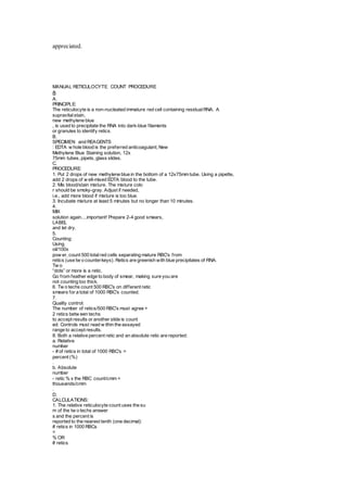
SlideShare a Scribd company logo
Reticulocyte Stains 
CHAPTER 1 I EXAMINATION OF BLOOD SAMPLES 13 
Reticulocyte stains are commercially available. Those wishing to prepare their 
own stain can do so by dissolving 0.5 g of new methylene blue and 1.6 g of 
potassium oxalate in 100 mL of distilled water. Following filtrations, equal 
volumes of blood and stain are mixed together in a test tube and incubated at 
room temperature for 10 to 20 minutes. After incubation, blood films are made 
and reticulocyte counts are performed by examining 1,000 erythrocytes and 
determining the percentage that are reticulocytes." The use of a Miller's disc in 
one of the microscope oculars saves time in performing the reticulocyte count. 
The blue-staining aggregates or "reticulum" seen in reticulocytes (Fig. 4D) 
does not occur as such in living cells but results from the precipitation of 
ribosomal ribonucleic acid (RNA; the same RNA that causes the bluish color 
seen in polychromatophilic erythrocytes) in immature erythrocytes during the 
staining process.I I As a reticulocyte matures, the number of ribosomes decreases 
until only small punctate (dotlike) inclusions are observed in erythrocytes 
(punctate reticulocytes) stained with the reticulocyte stain (Fig. 4E). To reduce 
the chance that a staining artifact would result in misclassifying a mature 
erythrocyte as a punctate reticulocyte using a reticulocyte stain. the cell in 
question should have two or more discrete blue granules that are visible without 
requiring fine-focus adjustment of the cell being evaluated to be classified 
as a punctate reticulocyte. 
In normal cats. as well as in cats with regenerative anemia. the number of 
punctate reticulocytes is much greater than that seen in other species." This 
apparently occurs because the maturation (loss of ribosomes) of reticulocytes in 
cats is slower than that in other species. Consequently, reticulocytes in cats are 
classified as aggregate (if coarse clumping is observed) or punctate (if small 
individual inclusions are present). Percentages of both types should be reported. 
Based on composite results from several authors, normal cats generally have 
from 0% to 0.5% aggregate and 1% to 10% punctate reticulocytes when determined 
by manual means. Higher punctate numbers of 2% to 17% have been 
reported using flow cytometry." 
The percentages of aggregate reticulocytes in cats correlate directly with 
the percentages of polychromatophilic erythrocytes observed in blood filmsIn contrast to those of 
the cat, most reticulocytes in other species are of 
the aggregate type. Consequently, no attempts are made to differentiate stages 
of reticulocytes in species other than the cat. The percentage of reticulocytes in 
most species correlates directly with the percentage of polychromatophilic erythrocytes 
observed on routinely stained blood films. 
Heinz bodies are composed of denatured, precipitated hemoglobin. They 
are spherical, stain pale blue with reticulocyte stains, and are usually found at 
the periphery of the erythrocyte. 
New Methylene Blue "Wet Mounts" 
A new methylene blue "wet mount" preparation can be used for rapid information 
concerning the number of reticulocytes, platelets, and Heinz bodies 
present. The stain consists of 0.5% new methylene blue dissolved in 0.85% 
NaCl. One mL of formalin is added per 100 mL of stain as a preservative. This 
stain is filtered after preparation and stored in dropper bottles. Alternately, the 
stain may be stored in a plastic syringe with a 0.2 IJ-m syringe filter attached so 
that the stain is filtered as it is used. Dry unfixed blood films are stained by 
placing a drop of stain between the coverslip and a glass slide. This preparation 
is not permanent and does not stain mature erythrocytes or eosinophil granules. 
Punctate reticulocytes are not demonstrated, but aggregate reticulocytes appear 
as erythrocyte ghosts containing blue to purple granular material (Fig. 4F). 
Platelets stain blue to purple, and Heinz bodies appear as refractile inclusions 
within erythrocyte ghosts. Although this staining method is not optimal for 
differential leukocyte counts, the number and type of leukocytes present can be
appreciated. 
MANUAL RETICULOCYTE COUNT PROCEDURE 
8 
A. 
PRINCIPLE: 
The reticulocyte is a non-nucleated immature red cell containing residual RNA. A 
supravital stain, 
new methylene blue 
, is used to precipitate the RNA into dark-blue f ilaments 
or granules to identify retics. 
B. 
SPECIMEN and REAGENTS 
: EDTA w hole blood is the preferred anticoagulant; New 
Methylene Blue Staining solution, 12x 
75mm tubes, pipets, glass slides. 
C. 
PROCEDURE: 
1. Put 2 drops of new methylene blue in the bottom of a 12x75mm tube. Using a pipette, 
add 2 drops of w ell-mixed EDTA blood to the tube. 
2. Mix blood/stain mixture. The mixture colo 
r should be smoky-gray. Adjust if needed, 
i.e., add more blood if mixture is too blue. 
3. Incubate mixture at least 5 minutes but no longer than 10 minutes. 
4. 
MIX 
solution again....important! Prepare 2-4 good smears, 
LABEL 
and let dry. 
5. 
Counting: 
Using 
oil/100x 
pow er, count 500 total red cells separating mature RBC's f rom 
retics (use tw o counter keys). Retics are greenish with blue precipitates of RNA. 
Tw o 
“dots” or more is a retic. 
Go f rom feather edge to body of smear, making sure you are 
not counting too thick. 
6. Tw o techs count 500 RBC's on dif ferent retic 
smears for a total of 1000 RBC's counted. 
7. 
Quality control: 
The number of retics/500 RBC's must agree + 
2 retics betw een techs 
to accept results or another slide is count 
ed. Controls must read w ithin the assayed 
range to accept results. 
8. Both a relative percent retic and an absolute retic are reported: 
a. Relative 
number 
- # of retics in total of 1000 RBC's = 
percent (%) 
. 
b. Absolute 
number 
- retic % x the RBC count/cmm = 
thousands/cmm 
. 
D. 
CALCULATIONS: 
1. The relative reticulocyte count uses the su 
m of the tw o techs answer 
s and the percent is 
reported to the nearest tenth (one decimal): 
# retics in 1000 RBCs 
= 
% OR 
# retics
x 100 = 
%. 
10 1000 RBCs 
2. The absolute reticulocyte count is repo 
rted to the nearest thousand/cmm using the 
follow ing calculation: 
# retics/cmm = # retics 
x RBC millions/cmm 
OR 
retic % 
x RBC/cmm 
1000 RBCs 100 
E. 
SOURCES OF ERROR: 
1. Inadequate mixing before making smears 
2. Counting artifact or other inclusions as retics......black/shiny inclusions are “junk”. 
3. Improper ratio of blood to stain. 
4. Not counting all of the retics.....two 
blue “dots” or more is a retic. 
5. Wrong calculati 
EXAMINATION OF STAINED BLOOD FILMS 
An overview and organized method of blood film examination are presented 
here. Descriptions and photographs of normal and abnormal blood cell morphology, 
inclusions, and infectious agents will be given in subsequent sections. 
Blood films are generally examined following staining with Romanowskytype 
stains such as Wright or Wright-Giemsa stains. These stains allow for 
examination of erythrocyte, leukocyte, and platelet morphology. Blood films 
should first be scanned using a low-power objective to estimate the total 
leukocyte count and to look for the presence of erythrocyte agglutination (Fig. 
SA), leukocyte aggregates (Fig. SB), platelet aggregates (Fig. SC), microfilaria 
(Fig. SD), and abnormal cells that might be missed during the differential 
leukocyte count. It is particularly important that the feathered end of blood 
films made on glass slides be examined because leukocytes (Fig. SE) and platelet 
aggregates (Fig. SF) may be concentrated in this area. Aggregates of cells tend 
to be in the center of coverslip blood films rather than at the feathered edge. 
When examining a glass-slide blood film, the blood film will be too thick 
to evaluate blood cell morphology at the back of the slide (Fig. 6A) and too 
thin at the feathered edge where cells are flattened (Fig. 6C). The optimal area 
for evaluation is generally in the front half of the smear behind the feathered 
edge (Fig. 6B). This area should appear as a well-stained monolayer (a field in 
which erythrocytes are dose together with approximately one half touching each 
other) of cells. 
Hemoglobinometry Hemoglobin Determination Decrease in 
hemoglobin concentration beyond established normal ranges for 
age and sex is called “ anemia”, whereas increase in hemoglobin 
concentration beyond established normal ranges for age , sex, and 
geographical distribution is called “polycythemia”. So that, for 
correct diagnosis it is important to determine accurately and 
precisely hemoglobin concentration. Many methods are available 
for the determination of hemoglobin, but among them the relevant, 
and the recommended one is the Modified Drabkin’s Method.
ICSH (International Committee for Standardization in Hematology) 
consider this method as the reference method for hemoglobin 
determination. Drabkin’s solution contains the following:- 
1- Potassium Ferricyanide 
2- Potassium Cyanide. 
3- Non- ionic Detergent 
4- Dihydrogen Potassium Phosphate. 
Well mixed EDTA anticoagulated blood is diluted in Drabkin’s 
solution; non-ionic detergent will lyse the red cells to (1) liberate 
hemoglobin, and to (2) decrease the turbidity caused by red cell 
membrane fragments by dissolving them. Then, hemoglobin is 
oxidized and converted to methemoglobin (Hi) by potassium 
ferricyanide, this step is accelerated by the dihydrogen potassium 
phosphate, and requires approximately 3 minutes for total 
conversion. Potassium cyanide will provide cyanide ions to form 
cyanomethemoglobin (HiCN), which have a broad spectrum of 
absorption at 540 nm. The absorption can then be compared with 
a hemoglobin standard with a known hemoglobin 
concentration, and by applying Beer’s law extract the hemoglobin 
concentration of the unknown (i.e. the patient). 
Hemoglobin + Potassium Ferricyanide Methemoglobin (Hi) 
Methemoglobin + Potassium Cyanide Cyanomethemoglobin

More Related Content

What's hot

Reticulocyte count
Reticulocyte countReticulocyte count
Reticulocyte countPrbn Shah
 
Updated - Blood film preparation and reporting
Updated - Blood film preparation and reportingUpdated - Blood film preparation and reporting
Updated - Blood film preparation and reporting
jadcaesar
 
Hematology technology
Hematology technologyHematology technology
Hematology technology
manueldsx
 
ABC of automated CBC
ABC of automated CBCABC of automated CBC
ABC of automated CBC
Dr. Parth Kaneria
 
Erythrocyte sedimentation rate
Erythrocyte sedimentation rateErythrocyte sedimentation rate
Erythrocyte sedimentation rate
Hajra Mehdi
 
Tlc dlc
Tlc dlcTlc dlc
Platelet count and hematocrit determination methods
Platelet count and hematocrit determination methodsPlatelet count and hematocrit determination methods
Platelet count and hematocrit determination methods
Negash Alamin
 
Automated cell counters
Automated cell countersAutomated cell counters
Automated cell counters
Kasturba Medical College
 
Histosch cw ieng
Histosch cw iengHistosch cw ieng
Histosch cw iengbabu3151
 
Total Leukocyte Count By Hemocytometer
Total Leukocyte Count By HemocytometerTotal Leukocyte Count By Hemocytometer
Total Leukocyte Count By Hemocytometer
Tshering Namgyal Wangdi
 
Automated cell counters: principle and types
Automated cell counters: principle and typesAutomated cell counters: principle and types
Automated cell counters: principle and types
Sivaranjini N
 
CBC Histogram DR NARMADA PRASAD TIWARI
 CBC Histogram DR NARMADA PRASAD TIWARI CBC Histogram DR NARMADA PRASAD TIWARI
CBC Histogram DR NARMADA PRASAD TIWARI
Narmada Tiwari
 
Cbc coulter principle
Cbc  coulter principleCbc  coulter principle
Cbc coulter principle
jadcaesar
 
Xtra online platelet_distribution_curves
Xtra online platelet_distribution_curvesXtra online platelet_distribution_curves
Xtra online platelet_distribution_curves
Alaa Fakhri
 
Automated cell counter & its quality control
Automated cell counter & its quality controlAutomated cell counter & its quality control
Automated cell counter & its quality control
Saikat Mandal
 
mbbs ims msu
mbbs ims msumbbs ims msu
mbbs ims msu
MBBS IMS MSU
 
Total Erythrocyte Count By Hemocytometer
Total Erythrocyte Count By HemocytometerTotal Erythrocyte Count By Hemocytometer
Total Erythrocyte Count By Hemocytometer
Tshering Namgyal Wangdi
 
Rbc indices
Rbc indicesRbc indices
Rbc indices
Manan Shah
 

What's hot (20)

Reticulocyte count
Reticulocyte countReticulocyte count
Reticulocyte count
 
Updated - Blood film preparation and reporting
Updated - Blood film preparation and reportingUpdated - Blood film preparation and reporting
Updated - Blood film preparation and reporting
 
Hematology technology
Hematology technologyHematology technology
Hematology technology
 
ABC of automated CBC
ABC of automated CBCABC of automated CBC
ABC of automated CBC
 
Erythrocyte sedimentation rate
Erythrocyte sedimentation rateErythrocyte sedimentation rate
Erythrocyte sedimentation rate
 
Tlc dlc
Tlc dlcTlc dlc
Tlc dlc
 
Platelet count and hematocrit determination methods
Platelet count and hematocrit determination methodsPlatelet count and hematocrit determination methods
Platelet count and hematocrit determination methods
 
Red blood cell count
Red blood cell countRed blood cell count
Red blood cell count
 
Automated cell counters
Automated cell countersAutomated cell counters
Automated cell counters
 
Histosch cw ieng
Histosch cw iengHistosch cw ieng
Histosch cw ieng
 
Total Leukocyte Count By Hemocytometer
Total Leukocyte Count By HemocytometerTotal Leukocyte Count By Hemocytometer
Total Leukocyte Count By Hemocytometer
 
Le cell
Le cellLe cell
Le cell
 
Automated cell counters: principle and types
Automated cell counters: principle and typesAutomated cell counters: principle and types
Automated cell counters: principle and types
 
CBC Histogram DR NARMADA PRASAD TIWARI
 CBC Histogram DR NARMADA PRASAD TIWARI CBC Histogram DR NARMADA PRASAD TIWARI
CBC Histogram DR NARMADA PRASAD TIWARI
 
Cbc coulter principle
Cbc  coulter principleCbc  coulter principle
Cbc coulter principle
 
Xtra online platelet_distribution_curves
Xtra online platelet_distribution_curvesXtra online platelet_distribution_curves
Xtra online platelet_distribution_curves
 
Automated cell counter & its quality control
Automated cell counter & its quality controlAutomated cell counter & its quality control
Automated cell counter & its quality control
 
mbbs ims msu
mbbs ims msumbbs ims msu
mbbs ims msu
 
Total Erythrocyte Count By Hemocytometer
Total Erythrocyte Count By HemocytometerTotal Erythrocyte Count By Hemocytometer
Total Erythrocyte Count By Hemocytometer
 
Rbc indices
Rbc indicesRbc indices
Rbc indices
 

Similar to دكتور عبد الامير عملي Reticulucyte stain

Artefacts in hemat part 2
Artefacts in hemat part 2Artefacts in hemat part 2
Artefacts in hemat part 2
DrShrikant Sonune
 
reticulocyte.pptx
reticulocyte.pptxreticulocyte.pptx
reticulocyte.pptx
DineshLaxmanRane
 
reticulocytecount-150706044130-lva1-app6892.pptx
reticulocytecount-150706044130-lva1-app6892.pptxreticulocytecount-150706044130-lva1-app6892.pptx
reticulocytecount-150706044130-lva1-app6892.pptx
MohanSinghDhakad1
 
Determination of Reticulocyte Count - Copy.pptx
Determination of Reticulocyte Count - Copy.pptxDetermination of Reticulocyte Count - Copy.pptx
Determination of Reticulocyte Count - Copy.pptx
islamkhan34
 
Total Leukocyte Count by Hemocytometer
Total Leukocyte Count by HemocytometerTotal Leukocyte Count by Hemocytometer
Total Leukocyte Count by Hemocytometer
Amjad Afridi
 
Dc
DcDc
PERIPHERAL SMEAR EXAMINATION
PERIPHERAL SMEAR EXAMINATIONPERIPHERAL SMEAR EXAMINATION
PERIPHERAL SMEAR EXAMINATION
Nithin Mathew
 
Hema I Chapter 13_Retics.ppt
Hema I Chapter 13_Retics.pptHema I Chapter 13_Retics.ppt
Hema I Chapter 13_Retics.ppt
deribew genetu
 
reticulocyte count.pptx
reticulocyte count.pptxreticulocyte count.pptx
reticulocyte count.pptx
abdiasis omar mohamed
 
Total leukocyte count by hemocytometer
Total leukocyte count by hemocytometerTotal leukocyte count by hemocytometer
Total leukocyte count by hemocytometer
Amjad Afridi
 
Differential leucocyte count & eosinophilia
Differential leucocyte count & eosinophiliaDifferential leucocyte count & eosinophilia
Differential leucocyte count & eosinophilia
sandeep singh
 
1 blood-smear
1 blood-smear1 blood-smear
1 blood-smear
UMC VICTORIA HOSPITAL
 
1-Blood-smear.ppt
1-Blood-smear.ppt1-Blood-smear.ppt
1-Blood-smear.ppt
chithankandeepan
 
Reticulocyte
ReticulocyteReticulocyte
Reticulocyte
Bhaikaka University
 
Hemocytometry
Hemocytometry Hemocytometry
Hemocytometry
Negash Alamin
 
Platelet
PlateletPlatelet
Laboratory diagnostic techniques
Laboratory diagnostic techniquesLaboratory diagnostic techniques
Laboratory diagnostic techniquesTarig Elfaki
 
DLC practical ppt
DLC  practical pptDLC  practical ppt
DLC practical ppt
Reena Gollapalli
 
lasercyte-dx-dot-plot-poster-en-2.pdf
lasercyte-dx-dot-plot-poster-en-2.pdflasercyte-dx-dot-plot-poster-en-2.pdf
lasercyte-dx-dot-plot-poster-en-2.pdf
FlorinPosastiuc
 
2nd, Blood Analysis
2nd, Blood Analysis2nd, Blood Analysis
2nd, Blood Analysisguestbc65a9
 

Similar to دكتور عبد الامير عملي Reticulucyte stain (20)

Artefacts in hemat part 2
Artefacts in hemat part 2Artefacts in hemat part 2
Artefacts in hemat part 2
 
reticulocyte.pptx
reticulocyte.pptxreticulocyte.pptx
reticulocyte.pptx
 
reticulocytecount-150706044130-lva1-app6892.pptx
reticulocytecount-150706044130-lva1-app6892.pptxreticulocytecount-150706044130-lva1-app6892.pptx
reticulocytecount-150706044130-lva1-app6892.pptx
 
Determination of Reticulocyte Count - Copy.pptx
Determination of Reticulocyte Count - Copy.pptxDetermination of Reticulocyte Count - Copy.pptx
Determination of Reticulocyte Count - Copy.pptx
 
Total Leukocyte Count by Hemocytometer
Total Leukocyte Count by HemocytometerTotal Leukocyte Count by Hemocytometer
Total Leukocyte Count by Hemocytometer
 
Dc
DcDc
Dc
 
PERIPHERAL SMEAR EXAMINATION
PERIPHERAL SMEAR EXAMINATIONPERIPHERAL SMEAR EXAMINATION
PERIPHERAL SMEAR EXAMINATION
 
Hema I Chapter 13_Retics.ppt
Hema I Chapter 13_Retics.pptHema I Chapter 13_Retics.ppt
Hema I Chapter 13_Retics.ppt
 
reticulocyte count.pptx
reticulocyte count.pptxreticulocyte count.pptx
reticulocyte count.pptx
 
Total leukocyte count by hemocytometer
Total leukocyte count by hemocytometerTotal leukocyte count by hemocytometer
Total leukocyte count by hemocytometer
 
Differential leucocyte count & eosinophilia
Differential leucocyte count & eosinophiliaDifferential leucocyte count & eosinophilia
Differential leucocyte count & eosinophilia
 
1 blood-smear
1 blood-smear1 blood-smear
1 blood-smear
 
1-Blood-smear.ppt
1-Blood-smear.ppt1-Blood-smear.ppt
1-Blood-smear.ppt
 
Reticulocyte
ReticulocyteReticulocyte
Reticulocyte
 
Hemocytometry
Hemocytometry Hemocytometry
Hemocytometry
 
Platelet
PlateletPlatelet
Platelet
 
Laboratory diagnostic techniques
Laboratory diagnostic techniquesLaboratory diagnostic techniques
Laboratory diagnostic techniques
 
DLC practical ppt
DLC  practical pptDLC  practical ppt
DLC practical ppt
 
lasercyte-dx-dot-plot-poster-en-2.pdf
lasercyte-dx-dot-plot-poster-en-2.pdflasercyte-dx-dot-plot-poster-en-2.pdf
lasercyte-dx-dot-plot-poster-en-2.pdf
 
2nd, Blood Analysis
2nd, Blood Analysis2nd, Blood Analysis
2nd, Blood Analysis
 

More from Ghadeer Albostany

Tests for fibrinogen/ عملي دكتور عبد الامير / تشخيصات
Tests for fibrinogen/  عملي  دكتور عبد الامير / تشخيصات Tests for fibrinogen/  عملي  دكتور عبد الامير / تشخيصات
Tests for fibrinogen/ عملي دكتور عبد الامير / تشخيصات
Ghadeer Albostany
 
المحاظرة الثالثة / معدية / دكتور اسعد /Shipping fever
المحاظرة الثالثة / معدية / دكتور اسعد /Shipping fever المحاظرة الثالثة / معدية / دكتور اسعد /Shipping fever
المحاظرة الثالثة / معدية / دكتور اسعد /Shipping fever
Ghadeer Albostany
 
المحاظرة الرابعة / معدية / دكتور اسعد /Glanders
المحاظرة الرابعة / معدية / دكتور اسعد /Glandersالمحاظرة الرابعة / معدية / دكتور اسعد /Glanders
المحاظرة الرابعة / معدية / دكتور اسعد /Glanders
Ghadeer Albostany
 
المحاظرة الخامسة / معدية / دكتور اسعد / Epizootic lymphangitis
المحاظرة الخامسة / معدية / دكتور اسعد / Epizootic lymphangitisالمحاظرة الخامسة / معدية / دكتور اسعد / Epizootic lymphangitis
المحاظرة الخامسة / معدية / دكتور اسعد / Epizootic lymphangitis
Ghadeer Albostany
 
دكتورة حلا المحاظرة الثانية الطباعة
دكتورة حلا المحاظرة الثانية الطباعةدكتورة حلا المحاظرة الثانية الطباعة
دكتورة حلا المحاظرة الثانية الطباعة
Ghadeer Albostany
 
Vs تشخيصات
Vs تشخيصات Vs تشخيصات
Vs تشخيصات
Ghadeer Albostany
 
Vesicular stomatitis تشخيصات
Vesicular stomatitis تشخيصات Vesicular stomatitis تشخيصات
Vesicular stomatitis تشخيصات
Ghadeer Albostany
 
Vesicular stomatitis (تشخيصات
Vesicular stomatitis (تشخيصات Vesicular stomatitis (تشخيصات
Vesicular stomatitis (تشخيصات
Ghadeer Albostany
 
Fmd1 تشخيصات
Fmd1 تشخيصاتFmd1 تشخيصات
Fmd1 تشخيصات
Ghadeer Albostany
 
Bovine ephemeral fever
Bovine ephemeral feverBovine ephemeral fever
Bovine ephemeral fever
Ghadeer Albostany
 
F.m.d تشخيصات
F.m.d تشخيصاتF.m.d تشخيصات
F.m.d تشخيصات
Ghadeer Albostany
 
دكتور عبد الامير Introduction to hematology
 دكتور عبد الامير Introduction to hematology دكتور عبد الامير Introduction to hematology
دكتور عبد الامير Introduction to hematology
Ghadeer Albostany
 
تشخيصات دكتور عبد الامير Haematology
 تشخيصات دكتور عبد الامير Haematology تشخيصات دكتور عبد الامير Haematology
تشخيصات دكتور عبد الامير Haematology
Ghadeer Albostany
 
Erythrocyte series عملي دكتور عبد الامير ّجامعة الكوفة تشخيصات
Erythrocyte series عملي دكتور عبد الامير ّجامعة الكوفة تشخيصاتErythrocyte series عملي دكتور عبد الامير ّجامعة الكوفة تشخيصات
Erythrocyte series عملي دكتور عبد الامير ّجامعة الكوفة تشخيصات
Ghadeer Albostany
 
Poikilocytes2 عبد الامير عملي
Poikilocytes2  عبد الامير عمليPoikilocytes2  عبد الامير عملي
Poikilocytes2 عبد الامير عمليGhadeer Albostany
 

More from Ghadeer Albostany (15)

Tests for fibrinogen/ عملي دكتور عبد الامير / تشخيصات
Tests for fibrinogen/  عملي  دكتور عبد الامير / تشخيصات Tests for fibrinogen/  عملي  دكتور عبد الامير / تشخيصات
Tests for fibrinogen/ عملي دكتور عبد الامير / تشخيصات
 
المحاظرة الثالثة / معدية / دكتور اسعد /Shipping fever
المحاظرة الثالثة / معدية / دكتور اسعد /Shipping fever المحاظرة الثالثة / معدية / دكتور اسعد /Shipping fever
المحاظرة الثالثة / معدية / دكتور اسعد /Shipping fever
 
المحاظرة الرابعة / معدية / دكتور اسعد /Glanders
المحاظرة الرابعة / معدية / دكتور اسعد /Glandersالمحاظرة الرابعة / معدية / دكتور اسعد /Glanders
المحاظرة الرابعة / معدية / دكتور اسعد /Glanders
 
المحاظرة الخامسة / معدية / دكتور اسعد / Epizootic lymphangitis
المحاظرة الخامسة / معدية / دكتور اسعد / Epizootic lymphangitisالمحاظرة الخامسة / معدية / دكتور اسعد / Epizootic lymphangitis
المحاظرة الخامسة / معدية / دكتور اسعد / Epizootic lymphangitis
 
دكتورة حلا المحاظرة الثانية الطباعة
دكتورة حلا المحاظرة الثانية الطباعةدكتورة حلا المحاظرة الثانية الطباعة
دكتورة حلا المحاظرة الثانية الطباعة
 
Vs تشخيصات
Vs تشخيصات Vs تشخيصات
Vs تشخيصات
 
Vesicular stomatitis تشخيصات
Vesicular stomatitis تشخيصات Vesicular stomatitis تشخيصات
Vesicular stomatitis تشخيصات
 
Vesicular stomatitis (تشخيصات
Vesicular stomatitis (تشخيصات Vesicular stomatitis (تشخيصات
Vesicular stomatitis (تشخيصات
 
Fmd1 تشخيصات
Fmd1 تشخيصاتFmd1 تشخيصات
Fmd1 تشخيصات
 
Bovine ephemeral fever
Bovine ephemeral feverBovine ephemeral fever
Bovine ephemeral fever
 
F.m.d تشخيصات
F.m.d تشخيصاتF.m.d تشخيصات
F.m.d تشخيصات
 
دكتور عبد الامير Introduction to hematology
 دكتور عبد الامير Introduction to hematology دكتور عبد الامير Introduction to hematology
دكتور عبد الامير Introduction to hematology
 
تشخيصات دكتور عبد الامير Haematology
 تشخيصات دكتور عبد الامير Haematology تشخيصات دكتور عبد الامير Haematology
تشخيصات دكتور عبد الامير Haematology
 
Erythrocyte series عملي دكتور عبد الامير ّجامعة الكوفة تشخيصات
Erythrocyte series عملي دكتور عبد الامير ّجامعة الكوفة تشخيصاتErythrocyte series عملي دكتور عبد الامير ّجامعة الكوفة تشخيصات
Erythrocyte series عملي دكتور عبد الامير ّجامعة الكوفة تشخيصات
 
Poikilocytes2 عبد الامير عملي
Poikilocytes2  عبد الامير عمليPoikilocytes2  عبد الامير عملي
Poikilocytes2 عبد الامير عملي
 

Recently uploaded

For Better Surat #ℂall #Girl Service ❤85270-49040❤ Surat #ℂall #Girls
For Better Surat #ℂall #Girl Service ❤85270-49040❤ Surat #ℂall #GirlsFor Better Surat #ℂall #Girl Service ❤85270-49040❤ Surat #ℂall #Girls
For Better Surat #ℂall #Girl Service ❤85270-49040❤ Surat #ℂall #Girls
Savita Shen $i11
 
Surat @ℂall @Girls ꧁❤8527049040❤꧂@ℂall @Girls Service Vip Top Model Safe
Surat @ℂall @Girls ꧁❤8527049040❤꧂@ℂall @Girls Service Vip Top Model SafeSurat @ℂall @Girls ꧁❤8527049040❤꧂@ℂall @Girls Service Vip Top Model Safe
Surat @ℂall @Girls ꧁❤8527049040❤꧂@ℂall @Girls Service Vip Top Model Safe
Savita Shen $i11
 
Novas diretrizes da OMS para os cuidados perinatais de mais qualidade
Novas diretrizes da OMS para os cuidados perinatais de mais qualidadeNovas diretrizes da OMS para os cuidados perinatais de mais qualidade
Novas diretrizes da OMS para os cuidados perinatais de mais qualidade
Prof. Marcus Renato de Carvalho
 
ACUTE SCROTUM.....pdf. ACUTE SCROTAL CONDITIOND
ACUTE SCROTUM.....pdf. ACUTE SCROTAL CONDITIONDACUTE SCROTUM.....pdf. ACUTE SCROTAL CONDITIOND
ACUTE SCROTUM.....pdf. ACUTE SCROTAL CONDITIOND
DR SETH JOTHAM
 
How STIs Influence the Development of Pelvic Inflammatory Disease.pptx
How STIs Influence the Development of Pelvic Inflammatory Disease.pptxHow STIs Influence the Development of Pelvic Inflammatory Disease.pptx
How STIs Influence the Development of Pelvic Inflammatory Disease.pptx
FFragrant
 
heat stroke and heat exhaustion in children
heat stroke and heat exhaustion in childrenheat stroke and heat exhaustion in children
heat stroke and heat exhaustion in children
SumeraAhmad5
 
Maxilla, Mandible & Hyoid Bone & Clinical Correlations by Dr. RIG.pptx
Maxilla, Mandible & Hyoid Bone & Clinical Correlations by Dr. RIG.pptxMaxilla, Mandible & Hyoid Bone & Clinical Correlations by Dr. RIG.pptx
Maxilla, Mandible & Hyoid Bone & Clinical Correlations by Dr. RIG.pptx
Dr. Rabia Inam Gandapore
 
NVBDCP.pptx Nation vector borne disease control program
NVBDCP.pptx Nation vector borne disease control programNVBDCP.pptx Nation vector borne disease control program
NVBDCP.pptx Nation vector borne disease control program
Sapna Thakur
 
Report Back from SGO 2024: What’s the Latest in Cervical Cancer?
Report Back from SGO 2024: What’s the Latest in Cervical Cancer?Report Back from SGO 2024: What’s the Latest in Cervical Cancer?
Report Back from SGO 2024: What’s the Latest in Cervical Cancer?
bkling
 
Triangles of Neck and Clinical Correlation by Dr. RIG.pptx
Triangles of Neck and Clinical Correlation by Dr. RIG.pptxTriangles of Neck and Clinical Correlation by Dr. RIG.pptx
Triangles of Neck and Clinical Correlation by Dr. RIG.pptx
Dr. Rabia Inam Gandapore
 
New Drug Discovery and Development .....
New Drug Discovery and Development .....New Drug Discovery and Development .....
New Drug Discovery and Development .....
NEHA GUPTA
 
Superficial & Deep Fascia of the NECK.pptx
Superficial & Deep Fascia of the NECK.pptxSuperficial & Deep Fascia of the NECK.pptx
Superficial & Deep Fascia of the NECK.pptx
Dr. Rabia Inam Gandapore
 
Are There Any Natural Remedies To Treat Syphilis.pdf
Are There Any Natural Remedies To Treat Syphilis.pdfAre There Any Natural Remedies To Treat Syphilis.pdf
Are There Any Natural Remedies To Treat Syphilis.pdf
Little Cross Family Clinic
 
Ocular injury ppt Upendra pal optometrist upums saifai etawah
Ocular injury  ppt  Upendra pal  optometrist upums saifai etawahOcular injury  ppt  Upendra pal  optometrist upums saifai etawah
Ocular injury ppt Upendra pal optometrist upums saifai etawah
pal078100
 
BRACHYTHERAPY OVERVIEW AND APPLICATORS
BRACHYTHERAPY OVERVIEW  AND  APPLICATORSBRACHYTHERAPY OVERVIEW  AND  APPLICATORS
BRACHYTHERAPY OVERVIEW AND APPLICATORS
Krishan Murari
 
Evaluation of antidepressant activity of clitoris ternatea in animals
Evaluation of antidepressant activity of clitoris ternatea in animalsEvaluation of antidepressant activity of clitoris ternatea in animals
Evaluation of antidepressant activity of clitoris ternatea in animals
Shweta
 
micro teaching on communication m.sc nursing.pdf
micro teaching on communication m.sc nursing.pdfmicro teaching on communication m.sc nursing.pdf
micro teaching on communication m.sc nursing.pdf
Anurag Sharma
 
Pharynx and Clinical Correlations BY Dr.Rabia Inam Gandapore.pptx
Pharynx and Clinical Correlations BY Dr.Rabia Inam Gandapore.pptxPharynx and Clinical Correlations BY Dr.Rabia Inam Gandapore.pptx
Pharynx and Clinical Correlations BY Dr.Rabia Inam Gandapore.pptx
Dr. Rabia Inam Gandapore
 
Physiology of Chemical Sensation of smell.pdf
Physiology of Chemical Sensation of smell.pdfPhysiology of Chemical Sensation of smell.pdf
Physiology of Chemical Sensation of smell.pdf
MedicoseAcademics
 
Couples presenting to the infertility clinic- Do they really have infertility...
Couples presenting to the infertility clinic- Do they really have infertility...Couples presenting to the infertility clinic- Do they really have infertility...
Couples presenting to the infertility clinic- Do they really have infertility...
Sujoy Dasgupta
 

Recently uploaded (20)

For Better Surat #ℂall #Girl Service ❤85270-49040❤ Surat #ℂall #Girls
For Better Surat #ℂall #Girl Service ❤85270-49040❤ Surat #ℂall #GirlsFor Better Surat #ℂall #Girl Service ❤85270-49040❤ Surat #ℂall #Girls
For Better Surat #ℂall #Girl Service ❤85270-49040❤ Surat #ℂall #Girls
 
Surat @ℂall @Girls ꧁❤8527049040❤꧂@ℂall @Girls Service Vip Top Model Safe
Surat @ℂall @Girls ꧁❤8527049040❤꧂@ℂall @Girls Service Vip Top Model SafeSurat @ℂall @Girls ꧁❤8527049040❤꧂@ℂall @Girls Service Vip Top Model Safe
Surat @ℂall @Girls ꧁❤8527049040❤꧂@ℂall @Girls Service Vip Top Model Safe
 
Novas diretrizes da OMS para os cuidados perinatais de mais qualidade
Novas diretrizes da OMS para os cuidados perinatais de mais qualidadeNovas diretrizes da OMS para os cuidados perinatais de mais qualidade
Novas diretrizes da OMS para os cuidados perinatais de mais qualidade
 
ACUTE SCROTUM.....pdf. ACUTE SCROTAL CONDITIOND
ACUTE SCROTUM.....pdf. ACUTE SCROTAL CONDITIONDACUTE SCROTUM.....pdf. ACUTE SCROTAL CONDITIOND
ACUTE SCROTUM.....pdf. ACUTE SCROTAL CONDITIOND
 
How STIs Influence the Development of Pelvic Inflammatory Disease.pptx
How STIs Influence the Development of Pelvic Inflammatory Disease.pptxHow STIs Influence the Development of Pelvic Inflammatory Disease.pptx
How STIs Influence the Development of Pelvic Inflammatory Disease.pptx
 
heat stroke and heat exhaustion in children
heat stroke and heat exhaustion in childrenheat stroke and heat exhaustion in children
heat stroke and heat exhaustion in children
 
Maxilla, Mandible & Hyoid Bone & Clinical Correlations by Dr. RIG.pptx
Maxilla, Mandible & Hyoid Bone & Clinical Correlations by Dr. RIG.pptxMaxilla, Mandible & Hyoid Bone & Clinical Correlations by Dr. RIG.pptx
Maxilla, Mandible & Hyoid Bone & Clinical Correlations by Dr. RIG.pptx
 
NVBDCP.pptx Nation vector borne disease control program
NVBDCP.pptx Nation vector borne disease control programNVBDCP.pptx Nation vector borne disease control program
NVBDCP.pptx Nation vector borne disease control program
 
Report Back from SGO 2024: What’s the Latest in Cervical Cancer?
Report Back from SGO 2024: What’s the Latest in Cervical Cancer?Report Back from SGO 2024: What’s the Latest in Cervical Cancer?
Report Back from SGO 2024: What’s the Latest in Cervical Cancer?
 
Triangles of Neck and Clinical Correlation by Dr. RIG.pptx
Triangles of Neck and Clinical Correlation by Dr. RIG.pptxTriangles of Neck and Clinical Correlation by Dr. RIG.pptx
Triangles of Neck and Clinical Correlation by Dr. RIG.pptx
 
New Drug Discovery and Development .....
New Drug Discovery and Development .....New Drug Discovery and Development .....
New Drug Discovery and Development .....
 
Superficial & Deep Fascia of the NECK.pptx
Superficial & Deep Fascia of the NECK.pptxSuperficial & Deep Fascia of the NECK.pptx
Superficial & Deep Fascia of the NECK.pptx
 
Are There Any Natural Remedies To Treat Syphilis.pdf
Are There Any Natural Remedies To Treat Syphilis.pdfAre There Any Natural Remedies To Treat Syphilis.pdf
Are There Any Natural Remedies To Treat Syphilis.pdf
 
Ocular injury ppt Upendra pal optometrist upums saifai etawah
Ocular injury  ppt  Upendra pal  optometrist upums saifai etawahOcular injury  ppt  Upendra pal  optometrist upums saifai etawah
Ocular injury ppt Upendra pal optometrist upums saifai etawah
 
BRACHYTHERAPY OVERVIEW AND APPLICATORS
BRACHYTHERAPY OVERVIEW  AND  APPLICATORSBRACHYTHERAPY OVERVIEW  AND  APPLICATORS
BRACHYTHERAPY OVERVIEW AND APPLICATORS
 
Evaluation of antidepressant activity of clitoris ternatea in animals
Evaluation of antidepressant activity of clitoris ternatea in animalsEvaluation of antidepressant activity of clitoris ternatea in animals
Evaluation of antidepressant activity of clitoris ternatea in animals
 
micro teaching on communication m.sc nursing.pdf
micro teaching on communication m.sc nursing.pdfmicro teaching on communication m.sc nursing.pdf
micro teaching on communication m.sc nursing.pdf
 
Pharynx and Clinical Correlations BY Dr.Rabia Inam Gandapore.pptx
Pharynx and Clinical Correlations BY Dr.Rabia Inam Gandapore.pptxPharynx and Clinical Correlations BY Dr.Rabia Inam Gandapore.pptx
Pharynx and Clinical Correlations BY Dr.Rabia Inam Gandapore.pptx
 
Physiology of Chemical Sensation of smell.pdf
Physiology of Chemical Sensation of smell.pdfPhysiology of Chemical Sensation of smell.pdf
Physiology of Chemical Sensation of smell.pdf
 
Couples presenting to the infertility clinic- Do they really have infertility...
Couples presenting to the infertility clinic- Do they really have infertility...Couples presenting to the infertility clinic- Do they really have infertility...
Couples presenting to the infertility clinic- Do they really have infertility...
 

دكتور عبد الامير عملي Reticulucyte stain

  • 1. Reticulocyte Stains CHAPTER 1 I EXAMINATION OF BLOOD SAMPLES 13 Reticulocyte stains are commercially available. Those wishing to prepare their own stain can do so by dissolving 0.5 g of new methylene blue and 1.6 g of potassium oxalate in 100 mL of distilled water. Following filtrations, equal volumes of blood and stain are mixed together in a test tube and incubated at room temperature for 10 to 20 minutes. After incubation, blood films are made and reticulocyte counts are performed by examining 1,000 erythrocytes and determining the percentage that are reticulocytes." The use of a Miller's disc in one of the microscope oculars saves time in performing the reticulocyte count. The blue-staining aggregates or "reticulum" seen in reticulocytes (Fig. 4D) does not occur as such in living cells but results from the precipitation of ribosomal ribonucleic acid (RNA; the same RNA that causes the bluish color seen in polychromatophilic erythrocytes) in immature erythrocytes during the staining process.I I As a reticulocyte matures, the number of ribosomes decreases until only small punctate (dotlike) inclusions are observed in erythrocytes (punctate reticulocytes) stained with the reticulocyte stain (Fig. 4E). To reduce the chance that a staining artifact would result in misclassifying a mature erythrocyte as a punctate reticulocyte using a reticulocyte stain. the cell in question should have two or more discrete blue granules that are visible without requiring fine-focus adjustment of the cell being evaluated to be classified as a punctate reticulocyte. In normal cats. as well as in cats with regenerative anemia. the number of punctate reticulocytes is much greater than that seen in other species." This apparently occurs because the maturation (loss of ribosomes) of reticulocytes in cats is slower than that in other species. Consequently, reticulocytes in cats are classified as aggregate (if coarse clumping is observed) or punctate (if small individual inclusions are present). Percentages of both types should be reported. Based on composite results from several authors, normal cats generally have from 0% to 0.5% aggregate and 1% to 10% punctate reticulocytes when determined by manual means. Higher punctate numbers of 2% to 17% have been reported using flow cytometry." The percentages of aggregate reticulocytes in cats correlate directly with the percentages of polychromatophilic erythrocytes observed in blood filmsIn contrast to those of the cat, most reticulocytes in other species are of the aggregate type. Consequently, no attempts are made to differentiate stages of reticulocytes in species other than the cat. The percentage of reticulocytes in most species correlates directly with the percentage of polychromatophilic erythrocytes observed on routinely stained blood films. Heinz bodies are composed of denatured, precipitated hemoglobin. They are spherical, stain pale blue with reticulocyte stains, and are usually found at the periphery of the erythrocyte. New Methylene Blue "Wet Mounts" A new methylene blue "wet mount" preparation can be used for rapid information concerning the number of reticulocytes, platelets, and Heinz bodies present. The stain consists of 0.5% new methylene blue dissolved in 0.85% NaCl. One mL of formalin is added per 100 mL of stain as a preservative. This stain is filtered after preparation and stored in dropper bottles. Alternately, the stain may be stored in a plastic syringe with a 0.2 IJ-m syringe filter attached so that the stain is filtered as it is used. Dry unfixed blood films are stained by placing a drop of stain between the coverslip and a glass slide. This preparation is not permanent and does not stain mature erythrocytes or eosinophil granules. Punctate reticulocytes are not demonstrated, but aggregate reticulocytes appear as erythrocyte ghosts containing blue to purple granular material (Fig. 4F). Platelets stain blue to purple, and Heinz bodies appear as refractile inclusions within erythrocyte ghosts. Although this staining method is not optimal for differential leukocyte counts, the number and type of leukocytes present can be
  • 2. appreciated. MANUAL RETICULOCYTE COUNT PROCEDURE 8 A. PRINCIPLE: The reticulocyte is a non-nucleated immature red cell containing residual RNA. A supravital stain, new methylene blue , is used to precipitate the RNA into dark-blue f ilaments or granules to identify retics. B. SPECIMEN and REAGENTS : EDTA w hole blood is the preferred anticoagulant; New Methylene Blue Staining solution, 12x 75mm tubes, pipets, glass slides. C. PROCEDURE: 1. Put 2 drops of new methylene blue in the bottom of a 12x75mm tube. Using a pipette, add 2 drops of w ell-mixed EDTA blood to the tube. 2. Mix blood/stain mixture. The mixture colo r should be smoky-gray. Adjust if needed, i.e., add more blood if mixture is too blue. 3. Incubate mixture at least 5 minutes but no longer than 10 minutes. 4. MIX solution again....important! Prepare 2-4 good smears, LABEL and let dry. 5. Counting: Using oil/100x pow er, count 500 total red cells separating mature RBC's f rom retics (use tw o counter keys). Retics are greenish with blue precipitates of RNA. Tw o “dots” or more is a retic. Go f rom feather edge to body of smear, making sure you are not counting too thick. 6. Tw o techs count 500 RBC's on dif ferent retic smears for a total of 1000 RBC's counted. 7. Quality control: The number of retics/500 RBC's must agree + 2 retics betw een techs to accept results or another slide is count ed. Controls must read w ithin the assayed range to accept results. 8. Both a relative percent retic and an absolute retic are reported: a. Relative number - # of retics in total of 1000 RBC's = percent (%) . b. Absolute number - retic % x the RBC count/cmm = thousands/cmm . D. CALCULATIONS: 1. The relative reticulocyte count uses the su m of the tw o techs answer s and the percent is reported to the nearest tenth (one decimal): # retics in 1000 RBCs = % OR # retics
  • 3. x 100 = %. 10 1000 RBCs 2. The absolute reticulocyte count is repo rted to the nearest thousand/cmm using the follow ing calculation: # retics/cmm = # retics x RBC millions/cmm OR retic % x RBC/cmm 1000 RBCs 100 E. SOURCES OF ERROR: 1. Inadequate mixing before making smears 2. Counting artifact or other inclusions as retics......black/shiny inclusions are “junk”. 3. Improper ratio of blood to stain. 4. Not counting all of the retics.....two blue “dots” or more is a retic. 5. Wrong calculati EXAMINATION OF STAINED BLOOD FILMS An overview and organized method of blood film examination are presented here. Descriptions and photographs of normal and abnormal blood cell morphology, inclusions, and infectious agents will be given in subsequent sections. Blood films are generally examined following staining with Romanowskytype stains such as Wright or Wright-Giemsa stains. These stains allow for examination of erythrocyte, leukocyte, and platelet morphology. Blood films should first be scanned using a low-power objective to estimate the total leukocyte count and to look for the presence of erythrocyte agglutination (Fig. SA), leukocyte aggregates (Fig. SB), platelet aggregates (Fig. SC), microfilaria (Fig. SD), and abnormal cells that might be missed during the differential leukocyte count. It is particularly important that the feathered end of blood films made on glass slides be examined because leukocytes (Fig. SE) and platelet aggregates (Fig. SF) may be concentrated in this area. Aggregates of cells tend to be in the center of coverslip blood films rather than at the feathered edge. When examining a glass-slide blood film, the blood film will be too thick to evaluate blood cell morphology at the back of the slide (Fig. 6A) and too thin at the feathered edge where cells are flattened (Fig. 6C). The optimal area for evaluation is generally in the front half of the smear behind the feathered edge (Fig. 6B). This area should appear as a well-stained monolayer (a field in which erythrocytes are dose together with approximately one half touching each other) of cells. Hemoglobinometry Hemoglobin Determination Decrease in hemoglobin concentration beyond established normal ranges for age and sex is called “ anemia”, whereas increase in hemoglobin concentration beyond established normal ranges for age , sex, and geographical distribution is called “polycythemia”. So that, for correct diagnosis it is important to determine accurately and precisely hemoglobin concentration. Many methods are available for the determination of hemoglobin, but among them the relevant, and the recommended one is the Modified Drabkin’s Method.
  • 4. ICSH (International Committee for Standardization in Hematology) consider this method as the reference method for hemoglobin determination. Drabkin’s solution contains the following:- 1- Potassium Ferricyanide 2- Potassium Cyanide. 3- Non- ionic Detergent 4- Dihydrogen Potassium Phosphate. Well mixed EDTA anticoagulated blood is diluted in Drabkin’s solution; non-ionic detergent will lyse the red cells to (1) liberate hemoglobin, and to (2) decrease the turbidity caused by red cell membrane fragments by dissolving them. Then, hemoglobin is oxidized and converted to methemoglobin (Hi) by potassium ferricyanide, this step is accelerated by the dihydrogen potassium phosphate, and requires approximately 3 minutes for total conversion. Potassium cyanide will provide cyanide ions to form cyanomethemoglobin (HiCN), which have a broad spectrum of absorption at 540 nm. The absorption can then be compared with a hemoglobin standard with a known hemoglobin concentration, and by applying Beer’s law extract the hemoglobin concentration of the unknown (i.e. the patient). Hemoglobin + Potassium Ferricyanide Methemoglobin (Hi) Methemoglobin + Potassium Cyanide Cyanomethemoglobin