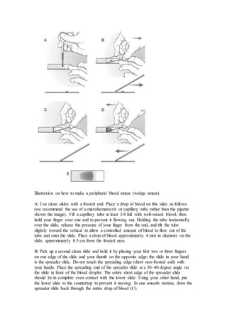
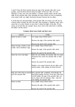
This document provides an introduction to hematology and performing a complete blood count. It defines hematology as the study of blood and its components. The objectives include being able to define hematology, list the components and functions of blood, describe blood values, and perform tests to determine packed cell volume and plasma protein values. The document describes the cellular components of blood, functions of blood, normal blood values, organs of the circulatory system, and procedures for a complete blood count including packed cell volume and plasma protein determination.
2008 my lecture introduction to hematologyBruno Mmassy
Manage all clotting disorders by providing supportive care, controlling bleeding if present, and transporting promptly for definitive care. Monitor closely for signs of worsening condition.
Hematology is the study of blood and blood forming organs. It involves the formation and development of blood cells through hematopoiesis. Hematopoiesis begins in the yolk sac and later the liver, spleen, and bone marrow take over production of blood cells. The bone marrow contains stem cells that can differentiate into various blood cell types through committed progenitor cells. It maintains homeostasis by replacing blood cells that have a limited lifespan.
The document provides an introduction to the topic of hematology, covering definitions of hematology, the composition and functions of blood, different cell types found in blood and their roles, basic hematological tests, and branches of hematology including immunohematology and transfusion medicine. Key concepts covered include the components and volume of blood, functions of red blood cells, white blood cells, platelets, and the processes of erythropoiesis and hemostasis.
This document provides an overview of hematopoiesis, the process by which blood cells are produced. It begins with a discussion of hematopoietic stem cells and their ability to differentiate into various blood cell types. The stages of red blood cell development from pronormoblast to reticulocyte to mature erythrocyte are described. Granulocyte development from myeloblast to mature neutrophil takes 5-12 days. Various hematopoietic growth factors that regulate blood cell production are also listed, including erythropoietin, GM-CSF, G-CSF, M-CSF, and others.
This document provides an introduction to the topic of hematology. It discusses that hematology is the study of blood and blood diseases, including their diagnosis, treatment and prevention. It also covers some key areas in hematology, such as clotting time (how blood clots), bleeding time (how long it takes for bleeding to stop), total red blood cell count and white blood cell count procedures. The document also briefly discusses blood types and the basic procedure for making a blood smear.
Introduction to veterinary hematology / Assist.Prof.Sr. Salah Al - KubaisiSalah Mahmoud
- Hematology is the study of blood cells, which include red blood cells (RBCs), platelets, and white blood cells (WBCs). RBCs are responsible for oxygen transport.
- RBCs are produced through erythropoiesis in the red bone marrow. This process takes 4-7 days to complete. When demand for RBCs increases, the bone marrow increases production by allowing younger cells to enter circulation or by speeding up maturation.
- RBCs survive for 2-5 months before being broken down. Their hemoglobin is broken into parts, with iron recycled and other parts excreted, giving feces their color.
- Erythro
The summary provides an overview of the key components of the hematologic system including:
- Bone marrow is the primary site of blood cell production and contains red and yellow marrow.
- The three main cellular components of blood are erythrocytes, leukocytes, and thrombocytes which are involved in oxygen transport, immune function, and hemostasis respectively.
- Important blood groups are ABO and Rh which determine transfusion compatibility.
- Common lab tests include complete blood count, coagulation studies, and blood typing to evaluate the hematologic system.
This document provides an overview of hematology and the basic functions and components of blood. It discusses [1] the properties and cellular elements of blood including red blood cells, white blood cells, and platelets, [2] the coagulation and fibrinolysis processes, and [3] defense mechanisms such as inflammation. Various diagrams and tables are included to illustrate hematopoiesis, blood cell types, the coagulation pathway, and the inflammatory response.
Blood transports oxygen, nutrients, hormones, and removes waste throughout the body. It consists of plasma and formed elements including red blood cells, white blood cells, and platelets. Centrifuging blood separates its components based on density. Red blood cells carry oxygen and make up about 45% of blood volume. Blood groups are determined by antigens on red blood cells. Hemostasis stops bleeding through vessel constriction, platelet plug formation, and a coagulation cascade involving clotting factors.
This document summarizes key topics in hematology, including cellular components of blood, components of blood such as red blood cells, white blood cells and platelets, diseases that affect these components, blood typing, transfusion, and coagulation disorders. Some of the major areas covered are red blood cell production and diseases like sickle cell disease and polycythemia, white blood cell disorders including leukemia and lymphoma, platelet disorders, and bleeding disorders like hemophilia. Treatment approaches focus on following general guidelines, supportive care, and preventing further injury or infection.
This document provides an overview of blood physiology for midwifery students. It discusses the major functions of blood including distribution of oxygen, nutrients, waste removal, and temperature regulation. The key components of blood - plasma, red blood cells, white blood cells, and platelets - are described along with their production and roles in clotting and immunity. Hematopoiesis, the production of blood cells in the bone marrow, is regulated by various hormones and growth factors.
physiology of Blood for nursery students dina merzeban
1. Red blood cells are biconcave disks that carry oxygen and carbon dioxide via hemoglobin. They require iron, vitamin B12, and folic acid for synthesis and maturation.
2. White blood cells include granulocytes like neutrophils, eosinophils, and basophils, and agranulocytes like lymphocytes and monocytes. They protect the body from pathogens and destroy abnormal cells.
3. Blood typing involves the presence or absence of antigens A, B, and Rh factor. Type O blood is the universal donor while type AB is the universal recipient. Sensitization can occur in Rh-negative mothers carrying Rh-positive fetuses.
The document provides an overview of hematology and the components of blood. It describes how blood circulates from the heart through arteries and veins, and how gases are exchanged in capillaries. The main components of blood are plasma, red blood cells, white blood cells, and platelets. It details the formation of blood cells through hematopoiesis in the bone marrow, and the different types of white blood cells. Common blood tests like complete blood count and disorders of the blood components are also summarized.
This document discusses diseases of the haemopoietic system. It begins by describing blood cells and bone marrow haemopoiesis. The main types of diseases are then outlined as anaemias, haematological malignancies, and bleeding disorders. Iron deficiency anaemia is discussed in detail as the most common type of anaemia. It is caused by blood loss, increased demands, malabsorption or dietary deficiencies. Clinical features include pallor, spoon-shaped nails, and cognitive effects in children. Laboratory tests can identify microcytic hypochromic blood cells characteristic of iron deficiency.
Blood is composed of plasma and blood cells suspended in plasma. Plasma is mostly water but also contains proteins, electrolytes, nutrients, waste products, hormones and gases. The three main types of blood cells are red blood cells, white blood cells, and platelets. Red blood cells contain hemoglobin and transport oxygen and carbon dioxide. White blood cells help fight infection. Platelets help with blood clotting to stop bleeding. Transfusions require matching blood types to avoid rejection reactions from antibodies in the plasma.
It is a brief review on blood and its cellular components. The ppt contains knowledge about types of blood, blood coagulation pathway and disorders of blood.
This document summarizes information on haemostasis and bleeding disorders. It discusses the mechanisms of haemostasis, screening tests for bleeding disorders, platelet disorders like ITP and TTP, coagulation factor deficiencies like hemophilia A and B, and vessel wall abnormalities. Key points covered are the stages of haemostasis, common causes of thrombocytopenia and thrombocytosis, management of ITP, and clinical features and treatment of disorders like hemophilia, von Willebrand disease, and Henoch-Schonlein purpura.
This document discusses blood formation and types of anemia. It explains that blood is composed of red cells, white cells, platelets, and plasma. Blood formation begins in the yolk sac in the 2nd week and later occurs chiefly in the liver and spleen, then the bone marrow. The bone marrow contains stem cells that can differentiate into mature blood cells. Various growth factors are involved in blood formation. The document also classifies and describes different types of anemia, including iron deficiency anemia, and lists causes and treatment for anemia.
This document summarizes the components and functions of blood. Blood is composed of plasma and formed elements including red blood cells, white blood cells, and platelets. Red blood cells contain hemoglobin and transport oxygen and carbon dioxide. White blood cells help fight infection and antibodies. Platelets help with clotting to stop bleeding. The document also discusses blood typing and the antigens and antibodies involved in the main blood types A, B, AB, and O.
This document summarizes the components and functions of human blood. It discusses the three main cellular components - red blood cells, white blood cells, and platelets. Red blood cells are described as biconcave discs that contain hemoglobin and lack a nucleus. Their main function is to transport oxygen from the lungs to tissues and assist with carbon dioxide removal. The production of new red blood cells occurs through erythropoiesis in the red bone marrow. Possible deficiencies or abnormalities in red blood cell levels, such as anemia or polycythemia, are also mentioned.
This document provides an introduction to hematology and summarizes key topics including:
1. The components of blood and cellular elements such as red blood cells, white blood cells, and platelets.
2. Principles of hematologic diagnosis including medical history, physical examination, and laboratory evaluations like complete blood count and peripheral blood smear.
3. Causes of anemia including hypoproliferative anemias like anemia of chronic disease and anemia of renal disease.
4. Aplastic anemia, its definition, epidemiology, etiology including acquired, inherited, and secondary causes.
The document discusses several key aspects of blood:
1) Blood transports oxygen, nutrients, waste, and more throughout the body using red blood cells, white blood cells, platelets, and plasma.
2) Red blood cells contain hemoglobin which gives blood its red color and allows it to carry oxygen.
3) The coagulation process forms blood clots to stop bleeding through vessel constriction, platelet plugs, and fibrin formation.
Blood is composed of plasma and formed elements such as red blood cells, white blood cells, and platelets. Red blood cells are biconcave discs that contain hemoglobin and transport oxygen and carbon dioxide. White blood cells provide defense against pathogens through mechanisms like phagocytosis. Platelets help in blood clotting when blood vessels are damaged. All blood cells are produced through hematopoiesis, which occurs primarily in the bone marrow.
RBC
WBC
1. Granulocytes
Neutrophils
Eosinophil’s
Basophils
2. Agranulocytes
Lymphocytes
Monocyte
PLATELETS
Blood is a bright red, viscous, slightly alkaline fluid that accounts for approximately 7 % of total body weightThe average human has 5 litres of blood (Average Blood Volume is 4 to 6 liters).
It is a transporting fluid.
Red colour is due to the presence of oxyhaemoglobin.
Ph - 7.4 slightly alkaline.
Specific gravity - 1.060
Viscosity is 5 times greater then the water i.e thicker than water.
Blood is the only fluid tissue.
Blood is a complex connective tissue in which living cells, the formed elements, are suspended in fluid componenet called plasma.
Functions of Blood
Transport of:
Gases, nutrients, waste products
Processed molecules
Regulatory molecules.
Regulation of pH and osmosis.
Maintenance of body temperature.
Protection against foreign substances.
Clot formation.
Blood composition
55% Plasma (fluid matrix of water, salts, proteins, etc.)
45% Cellular elements:
Red Blood Cells (RBCs) (Erythrocytes) : 5-6 million RBCs/ml of blood.
Contain hemoglobin which transport oxygen and CO2.
White Blood Cells (WBCs) (Leukocytes) : 5,000-10,000 WBCs/ml of blood.
Play an essential role in immunity and defense.
Include:
Granulocytes
Neutrophils 40-70%
Eosinophil's 0-1%
Basophils 1-5%
Agranulocytes
Lymphocytes 25-40% T cells and B cells
Monocyte 2-8% (phagocytes)
Platelets (Thrombocytes) : Cellular fragments, 250,000- 400,000/ml of blood.
Important in blood clotting.
Blood functions to transport oxygen, carbon dioxide, nutrients, hormones and waste throughout the body. It also defends the body against pathogens. Blood is composed of plasma, which carries red blood cells, white blood cells, and platelets in suspension. Red blood cells transport oxygen, white blood cells fight infection, and platelets help the blood clot to stop bleeding. In summary, blood is a circulating connective tissue that sustains life through these critical functions and cellular components.
This chapter will discuss blood composition, the physical and chemical properties of blood, blood cells including erythrocytes, leukocytes and platelets, hemostasis, blood groups, and principles of blood transfusion. Specifically, it will cover the components and characteristics of plasma, hematopoiesis and the life cycle of red blood cells, the roles and regulation of white blood cells and platelets, and the clinical significance of blood groups and cross-matching for transfusion.
The document provides an overview of haematology and blood components. It discusses the composition of blood including plasma and cellular components such as red blood cells, white blood cells, and platelets. It describes the structure and functions of red blood cells, including haemoglobin synthesis and the erythrocyte life cycle. The document also covers leukopoiesis, immunity basics, blood groups, and the coagulation process. Learning outcomes focus on describing blood components and discussing the formation and functions of red and white blood cells.
This document summarizes the composition, properties, functions and volume of blood. It discusses that blood consists of cells suspended in plasma. The main cells are red blood cells, white blood cells and platelets. Plasma is mostly water with dissolved proteins and electrolytes. Bone marrow produces blood cells. Key functions of blood include transport, defense, regulation and hemostasis. The normal blood volume in adults is 5-6 liters. The hematocrit level indicates the proportion of red blood cells and can be used to screen for anemia. The blood and plasma volumes are regulated to maintain homeostasis.
This document discusses hematology and the components and functions of blood. It describes how hematology is the study of blood and the circulatory system. The key components of blood are plasma, red blood cells, white blood cells, and platelets. Plasma acts as a transport medium and contains proteins, electrolytes, nutrients, wastes, gases, and hormones. Red blood cells transport oxygen and carbon dioxide through their hemoglobin content. White blood cells provide protection through immune functions. Platelets contribute to hemostasis and blood clotting. Tests like complete blood count, hematocrit, and erythrocyte sedimentation rate are used to analyze blood and diagnose conditions.
The complete blood count (CBC) test measures the quantity of the main types of blood cells - white blood cells, red blood cells, and platelets. It provides information on cell counts and other parameters to help identify issues like infections, anemia, bleeding disorders, and certain cancers. The CBC is one of the most commonly ordered blood tests and involves drawing a small blood sample, usually from a vein in the arm. The results are interpreted based on normal reference ranges to evaluate any abnormalities.
Blood carries oxygen, nutrients, hormones and waste products throughout the body. It is composed of plasma and formed elements including red blood cells, white blood cells and platelets. Red blood cells contain hemoglobin and transport oxygen, while white blood cells help fight infection. Platelets help form clots to stop bleeding. Blood type is determined by antigens on red blood cells and the presence of antibodies in plasma. A blood transfusion matches the donor's blood type to the recipient to avoid an immune reaction.
This document provides an overview of the cardiovascular and lymphatic systems topics to be covered, including the structure and function of the heart, blood vessels, blood pressure regulation, properties of blood, formation of blood cells, blood clotting, and the lymphatic system. The key topics are the components and functions of blood, blood cell formation in the bone marrow, the roles of red blood cells, white blood cells, and platelets, and the process of hemostasis to stop bleeding through vascular spasms, platelet plugs, and blood clotting.
Physicians working in the field of hematology are called hematologists. Initially, hematologists complete a four-year medical degree and this is followed by three or four years in an internship or residency program. Thereafter, they spend two or three more years learning how to diagnose and treat blood disorders.
Blood has three main functions: transport, protection, and regulation. It transports gases, nutrients, hormones, heat, and waste throughout the body. Blood also protects the body through white blood cells that destroy pathogens and platelets that initiate clotting. Additionally, blood helps regulate pH, water balance, and other processes. Blood is composed of plasma, red blood cells, white blood cells, and platelets, each with distinct roles in these important functions.
Blood is a connective tissue composed of plasma, red blood cells, white blood cells, and platelets. Plasma is the liquid portion that transports nutrients, waste, hormones, and other critical components. Red blood cells contain hemoglobin and carry oxygen from the lungs to tissues. White blood cells defend the body against infection and disease. Platelets help the body form blood clots to stop bleeding. Together, these components perform vital functions for circulation and transport throughout the body.
Blood is a specialized fluid that transports necessary substances like oxygen and nutrients to cells and removes waste products from cells. It is composed of plasma, red blood cells, white blood cells, and platelets suspended in plasma. Red blood cells contain hemoglobin which carries oxygen. White blood cells help fight infections. Platelets help with clotting to stop bleeding. Blood is circulated by the heart through arteries and veins to oxygenate tissues and remove carbon dioxide.
The cellular components of blood are erythrocytes (red blood cells, or RBCs), leukocytes (white blood cells, or WBCs), and thrombocytes (platelets). By volume, the RBCs constitute about 45% of whole blood, the plasma about 54.3%, and white blood cells about 0.7%. Platelets make up less than 1%.
This document summarizes the composition and functions of blood. It describes that blood consists of plasma and formed elements suspended in plasma. The formed elements are red blood cells, white blood cells, and platelets. The document then discusses the composition of plasma and the different types of white blood cells and platelets. It concludes by outlining the key functions of blood, which include transport of respiratory gases, nutrients, waste removal, regulation of pH, temperature, blood pressure, and playing a defensive role through coagulation and the immune response.
This document provides information about the composition and functions of blood. It discusses the different components of blood including plasma, red blood cells, white blood cells and platelets. It describes the functions of blood such as transport, defense, regulation and more. It also covers topics like blood viscosity, coagulation, and the lifespan and development of red blood cells.
Body Fluids And Circulation Class 11thNehaRohtagi1
Created By: NehaRohtagi1
Class 11th CBSE [NCERT]
Biology Chapter 18
Notes on the topic: Body Fluids and Circulation
For Class - 11th
I hope that you will found this presentation useful and it will help you out for your concept understanding.
Thank You!
Please give feedbacks and suggestions to get presentations on more interesting topics.
Do not repost or copy.
The circulatory system transports blood throughout the body using the heart as a pumping organ and a network of arteries, veins, and capillaries. It carries oxygen, nutrients, hormones, and cellular waste products. The document defines the key components of circulation including blood composition and the roles of red blood cells, white blood cells, and platelets. It also provides details on the production and functions of these blood components.
Hematology tests measure components of blood to diagnose diseases. A complete blood count measures red blood cells, white blood cells, and platelets to assess health and detect issues like anemia or infection. Additional tests examine hemoglobin levels, hematocrit, and other factors to further evaluate red blood cell counts and identify potential deficiencies or disorders. Blood types are also classified based on antigen and antibody presence to ensure safe transfusions.
Blood performs several essential functions:
- It transports oxygen, nutrients, wastes, hormones and defenses throughout the body.
- Blood maintains pH, fluid volume and temperature homeostasis.
- It prevents blood loss through clot formation and prevents infection using antibodies and white blood cells.
Blood is composed of fluid plasma and formed elements including red blood cells, white blood cells and platelets. Plasma proteins generate oncotic pressure and buffer pH while transporting substances. Red blood cells transport oxygen using hemoglobin and carbon dioxide via bicarbonate ions.
Blood is a transport fluid that carries nutrients, waste products, gases, and blood cells throughout the body. It is composed of plasma and formed elements. Plasma is 90% water and contains proteins, salts, and other dissolved substances. Formed elements include red blood cells, white blood cells, and platelets. Red blood cells contain hemoglobin and transport oxygen and carbon dioxide. Blood volume varies based on factors like age, sex, and body composition. Blood types are determined by antigens on red blood cells and the presence of corresponding antibodies.
Blood is a connective tissue composed of plasma and cells. It transports oxygen, nutrients, hormones, and waste products throughout the body. Blood contains red blood cells, white blood cells, and platelets suspended in plasma. Red blood cells contain hemoglobin and transport oxygen and carbon dioxide. White blood cells help fight infection. Platelets help form blood clots to stop bleeding. The document discusses blood components, functions, disorders, and blood grouping in detail.
The document discusses the circulatory system, including its components and functions. It describes the closed circulatory system in humans, which involves the heart pumping blood through arteries, arterioles, capillaries, venules and veins. The circulatory system transports nutrients, gases, wastes and more throughout the body. Blood consists of plasma and formed elements like red blood cells, white blood cells and platelets. Red blood cells contain hemoglobin and transport oxygen, while white blood cells help fight infection. Platelets help the blood clot to stop bleeding.
Blood contains plasma, red blood cells, white blood cells, and platelets. Plasma is mostly water containing proteins, ions, nutrients, and waste. Red blood cells carry oxygen to tissues and carbon dioxide back to the lungs using hemoglobin. Platelets form blood clots to prevent blood loss from damaged vessels. White blood cells include neutrophils, lymphocytes, monocytes, eosinophils, and basophils which fight infection and disease. Each blood component has specialized functions in transporting substances and protecting the body.
Similar to دكتور عبد الامير Introduction to hematology (20)
Tests for fibrinogen/ عملي دكتور عبد الامير / تشخيصات Ghadeer Albostany
There are three main ways to measure fibrinogen concentration: 1) heat precipitation of EDTA samples to measure elevated levels in inflammation/renal disease, 2) thrombin clot time to measure functional fibrinogen levels and detect deficiencies, 3) clottable fibrinogen concentration determined using thrombin clot time and compared to a standard curve. Normal fibrinogen levels vary between species but are generally 100-500 mg/dl in horses, sheep, and dogs and 300-700 mg/dl in cows. Elevated levels indicate inflammation.
Equine influenza is a highly contagious viral respiratory disease of horses characterized by fever and persistent cough. It is caused by two influenza A viruses, H7N7 and H3N8. Major outbreaks have occurred in Europe and North America in the 1950s-1980s. The virus can be transmitted via inhalation of droplets from infected horses, ingestion of contaminated food/water, or breeding. Clinical signs include fever, dry cough that becomes moist, depression, and stiffness. Complications can include bronchitis, pneumonia, and death in young foals. Diagnosis is based on symptoms, virus isolation, serology, and necropsy findings showing bronchopneumonia. There is no specific treatment but
Glanders is a highly contagious and often fatal disease that affects horses, mules and donkeys. It is caused by the bacterium Burkholderia mallei. The disease causes nodules and ulcers in the respiratory tract and on the skin. It occurs sporadically in parts of Europe, Asia, Africa and the Middle East. Horses with poor nutrition and living conditions are most at risk. Humans can also be infected through contact with infected animals. Diagnosis involves tests like the mallein test and isolating the bacteria from nodules. There is no vaccine and infected animals must be quarantined or destroyed to control the spread of the disease.
EPIZOOTIC LYMPHANGITIS is a chronic, contagious fungal disease that affects horses, donkeys, and mules. It is characterized by suppurative lymphangitis, lymphadenitis, and skin ulcers. The causative agent is the fungus Histoplasma farciminosum. The disease spreads through direct contact with infected animals or contact with contaminated soil or flies. Clinical signs include spreading skin lesions and lymphangitis along the limbs. Diagnosis involves identifying the fungus microscopically or through culture. Treatment has limited success. Control relies on identifying and destroying infected animals, quarantining other animals, and disinfecting equipment.
This document summarizes ovulation and corpus luteum lutelysis. It describes the process of oogenesis where oogonia develop into primary and secondary oocytes. It explains that ovulation occurs when the preovulatory follicle ruptures and releases the secondary oocyte. After ovulation, the collapsed follicle forms the corpus hemorrhagicum and later the corpus luteum, which secretes progesterone. The LH surge stimulates ovulation and luteinization. Ovulation can be spontaneous or induced. The corpus luteum will lutelize due to secretion of PGF2α by the uterus, except in cases where the uterus is removed or contains a fetus, inflammation, or foreign body.
Vesicular stomatitis is a viral disease that affects cattle, horses, and swine. It causes the development of blisters or vesicles in the mouth and on the feet. The most common symptoms are excessive salivation and lameness. While the clinical signs are similar to foot-and-mouth disease, vesicular stomatitis can be differentiated based on the species affected, presence of systemic illness, and location of lesions. Definitive diagnosis requires antibody testing or virus isolation. There is no treatment for infected animals. To control spread, infected animals should be isolated, insect vectors controlled, and movement of animals from infected premises restricted for at least 21 days after lesions have healed.
Vesicular stomatitis is caused by a virus of the family Rhabdoviridae that primarily affects the mouths and feet of horses and cattle, causing blisters and sores. It is transmitted by black flies and biting midges and has an incubation period of 2-8 days. The lesions are initially small blisters that rupture, leaving raw erosions and making eating painful. The disease cannot be distinguished from foot-and-mouth disease without laboratory tests.
Vesicular stomatitis is caused by a vesiculovirus that primarily affects the oral mucosa of horses and cattle, causing blisters and sores that are painful and prevent eating. While its reservoir is unknown, it is transmitted by black flies and biting midges. The incubation period is 2-8 days, after which time blisters develop on the tongue, lips, and feet. It cannot be clinically distinguished from foot-and-mouth disease. Definitive diagnosis requires antibody testing or virus isolation.
Foot-and-mouth disease is caused by an aphthovirus that occurs in seven major serotypes. It affects cloven-hooved animals and is endemic in parts of Africa, Asia, South America, and Europe. The virus infects the pharynx and causes a brief viremia before appearing in excretions and causing vesicles in the mouth and feet. Young animals are more severely affected and can develop myocarditis.
Bovine ephemeral fever is a viral disease of cattle and water buffalo caused by the bovine ephemeral fever virus. It occurs in parts of Africa, Asia, Australia, and the Middle East, often in widespread outbreaks. Clinical signs include fever, lameness, edema, and recumbency. While mortality is usually low, it can reach 30% in very fat cattle. The disease is transmitted by arthropod vectors like mosquitoes and biting midges.
Foot-and-mouth disease is caused by an aphthovirus that has seven major serotypes. It affects cloven-hooved animals and is endemic in parts of Africa, Asia, South America, and Europe. The virus is transmitted through inhalation, ingestion, or contact between animals and infects the pharynx before spreading to other areas. This causes fever, vesicles in the mouth and on the feet, and can occasionally lead to sudden death in young animals.
Reticulocyte stains are used to identify immature red blood cells (reticulocytes) by precipitating their residual RNA. A stain is prepared using new methylene blue and potassium oxalate or commercial stains can be purchased. Equal volumes of blood and stain are mixed and incubated before making blood films. Reticulocytes are identified and counted under a microscope by their blue aggregates or granules of precipitated RNA. Counts of reticulocytes are an indicator of bone marrow response to anemia. The percentage of reticulocytes correlates with the level of polychromatophilic erythrocytes seen on blood films.
This document provides an introduction to hematology and performing a complete blood count. It defines hematology as the study of blood and its components. The objectives include being able to define hematology, list the components and functions of blood, describe how to perform a complete blood count, and interpret normal hematology values in various species. The document describes the cellular components of blood, functions of blood, normal hematology ranges, and procedures for performing a packed cell volume test and plasma protein determination.
Erythrocyte series عملي دكتور عبد الامير ّجامعة الكوفة تشخيصاتGhadeer Albostany
This document describes the maturation process of red blood cells from stem cells to reticulocytes to mature red blood cells. It also lists common abnormalities in red blood cell shape and membrane, including acanthocytes, target cells, spherocytes, and stomatocytes. Various staining patterns are explained, such as basophilic stippling from residual RNA, Howell-Jolly bodies from splenic dysfunction, and Heinz bodies from oxidative damage. Causes of different anemias are provided, like microcytic hypochromic cells in iron deficiency anemia.
This document discusses various abnormal red blood cell (RBC) shapes, called poikilocytes, that can occur in dogs and cats. It describes several specific poikilocyte types including spherocytes, acanthocytes, schistocytes, and leptocytes. Spherocytes are spherical RBCs that have lost their normal biconcave shape. Acanthocytes have fingerlike surface projections related to abnormal lipid accumulation. The document also discusses RBC inclusions such as Heinz bodies, basophilic stippling, and Howell-Jolly bodies, which can indicate various underlying conditions.
Rasamanikya is a excellent preparation in the field of Rasashastra, it is used in various Kushtha Roga, Shwasa, Vicharchika, Bhagandara, Vatarakta, and Phiranga Roga. In this article Preparation& Comparative analytical profile for both Formulationon i.e Rasamanikya prepared by Kushmanda swarasa & Churnodhaka Shodita Haratala. The study aims to provide insights into the comparative efficacy and analytical aspects of these formulations for enhanced therapeutic outcomes.
Integrating Ayurveda into Parkinson’s Management: A Holistic ApproachAyurveda ForAll
Explore the benefits of combining Ayurveda with conventional Parkinson's treatments. Learn how a holistic approach can manage symptoms, enhance well-being, and balance body energies. Discover the steps to safely integrate Ayurvedic practices into your Parkinson’s care plan, including expert guidance on diet, herbal remedies, and lifestyle modifications.
- Video recording of this lecture in English language: https://youtu.be/kqbnxVAZs-0
- Video recording of this lecture in Arabic language: https://youtu.be/SINlygW1Mpc
- Link to download the book free: https://nephrotube.blogspot.com/p/nephrotube-nephrology-books.html
- Link to NephroTube website: www.NephroTube.com
- Link to NephroTube social media accounts: https://nephrotube.blogspot.com/p/join-nephrotube-on-social-media.html
Adhd Medication Shortage Uk - trinexpharmacy.comreignlana06
The UK is currently facing a Adhd Medication Shortage Uk, which has left many patients and their families grappling with uncertainty and frustration. ADHD, or Attention Deficit Hyperactivity Disorder, is a chronic condition that requires consistent medication to manage effectively. This shortage has highlighted the critical role these medications play in the daily lives of those affected by ADHD. Contact : +1 (747) 209 – 3649 E-mail : sales@trinexpharmacy.com
Histololgy of Female Reproductive System.pptxAyeshaZaid1
Dive into an in-depth exploration of the histological structure of female reproductive system with this comprehensive lecture. Presented by Dr. Ayesha Irfan, Assistant Professor of Anatomy, this presentation covers the Gross anatomy and functional histology of the female reproductive organs. Ideal for students, educators, and anyone interested in medical science, this lecture provides clear explanations, detailed diagrams, and valuable insights into female reproductive system. Enhance your knowledge and understanding of this essential aspect of human biology.
TEST BANK For Basic and Clinical Pharmacology, 14th Edition by Bertram G. Kat...rightmanforbloodline
TEST BANK For Basic and Clinical Pharmacology, 14th Edition by Bertram G. Katzung, Verified Chapters 1 - 66, Complete Newest Version.
Here is the updated list of Top Best Ayurvedic medicine for Gas and Indigestion and those are Gas-O-Go Syp for Dyspepsia | Lavizyme Syrup for Acidity | Yumzyme Hepatoprotective Capsules etc
Vestibulocochlear Nerve by Dr. Rabia Inam Gandapore.pptx
دكتور عبد الامير Introduction to hematology
1. Introduction to Hematology
Objectives
After completing this unit you should be able to:
1. Define hematology
2. List the components and functions of blood
3. List the units of measurement for RBC counts, WBC counts, platelet
counts, plasma proteins
4. Describe the functions of organs associated with the circulatory system
5. List the components of a complete blood count (CBC)
6. Given a sample of blood, perform the tests to determine the PCV and
plasma protein values
7. Estimate erythrocyte and hemoglobin values given the PCV
8. List the PCV and plasma protein values for normal farm animals ,
canine and feline blood
Introduction
Hematology is the study of blood and an important part of clinical pathology and the
diagnostic process. It includes not only the examination of the cellular and fluid
potions of blood, but also includes a study of the tissues that form, store and circulate
blood cells. A veterinarian uses the results of hematology tests to help determine the
health of an animal. These results are used in conjunction with the history, physical
exam and other laboratory findings. In this unit you will be introduced to the
components of blood and the procedures involved in a complete blood count.
Components of blood
Blood is a tissue consisting of cells within a fluid matrix. Blood creates an internal
environment which directly or indirectly baths all cells of the body and protects it
from the external environment.
Blood contains red blood cells (erythrocytes), white blood cells (leukocytes) and
platelets (thrombocytes). Figure 2.3 on page 10 of Voigt illustrates the cellular
components of blood. Red blood cells contain hemoglobin and are responsible for
carrying oxygen from the lungs to the cells throughout the body, as well as carbon
dioxide from the tissues to the lungs for excretion. White blood cells are either
“granulocytes” (contain granules in the cytoplasm) or “agranulocytes” (do not contain
granules in the cytoplasm.) Granular leukoytes include neutrophils, eosinophils and
basophils. Agranulocytes include lymphocytes and monocytes. WBCs are a critical
component of the immune system. Platelets are cell fragments from large
multnucleated cells (megakaryocytes). Platelets are important in blood clotting or
hemostasis.
2. The clear to pale-yellow fluid portion of blood is called plasma. Five to ten percent
of the plasma consists of proteins. The majority of the proteins are albumin, globulins
and fibrinogen. Albumins transport numerous substances in the blood and are the
main determinant of the osmotic pressure. Globulins (alpha, beta and gamma) are
important in transport and immunity. Fibrinogen is important in blood clotting and
the inflammatory cascade. If blood is allowed to clot, the clotting factors are removed
from the plasma and the remaining fluid portion of the blood is called serum.
Functions of blood
The three main functions of blood are transportation, regulation and defense. Many
of these functions will be covered in detail in other units.
Transports Oxygen and nutrients to cells in the body
Carbon dioxide and waste materials from cells in body
Hormones from glands to target organs
Regulates Body temperature
Water balance
pH
Electrolytes
Defense Phagocytosis of foreign invaders
Involved with immunity
Blood clotting
Blood values
The amount of blood present in an animal averages 7 % of body weight (20 – 50
ml/lb). A 100 pound animal would have about 3150 ml of blood. As a general rule
0.2 ml of blood/ lb of body weight can be safely removed from a healthy animal
3. without detrimental effects. The blood volume, then, of a 100 lb dog would be: 45 kg
X 0.7 = 31.5 kg of blood, and since 1 kg = 1 liter, this is equivalent to 3,150 mL of
blood.
Red blood cells are the most abundant cells in the blood. The average red blood cell
count in dogs is 6,800,000 red blood cells per micro liter of blood. This value is
routinely written as 6.8 x 10⁶ cells/᪽l. Feline RBC values normally averages 7.5 x
106 cells/᪽l. The life span of a canine red blood cell is 120 days, thus in a healthy
animal red blood cells are constantly being produced and destroyed. It is estimated
this replacement occurs at the rate of 35 million cells per second. RBCs are produced
in the bone marrow in response to the hormone erythropoietin. They are destroyed in
the spleen when they are too old, or damaged.
White blood cell values in an animal are constantly changing depending on the
degree of disease and stress present. The average white blood cell count in a healthy
dog is 11.5 x 10³ ᪽l and the average value for a healthy cat is 12.5 x 10³ ᪽l. (The
difference between 103 and 106 is a magnitude of 1000. Therefore, there is normally
1 white blood cell for every 1000 red blood cells in healthy animals.) The life span of
white blood cells vary from a few hours to years, depending on the type of the cell,
the physiology of the animal, and other factors. WBCs are produced in the bone
marrow, mature in lymphatic tissues, bone marrow and spleen, and circulate through
the blood and tissues where they are destroyed.
Platelet values in normal animals can vary a great deal with referenced “normal
ranges” from 350,000 to 500,000 platelets /ul. The values can also be written as 3.5 -
5 x 10⁵/ ᪽l. Platelets are produced in the bone marrow, and either used up in the
clotting cascade, or destroyed and filtered out of the blood in the spleen.
Plasma protein values range between 6 to 8 gm/dl for adult domestic animals and 4
to 6 gm/dl for younger animals. Plasma protein is an important indicator of the
patient's hydration status, and over all health. It is one of the most important
diagnostic values you can obtain.
Tables 2-4, 2-5 and 2-6 on pages 71 and 72 of the Hendrix text contains “normal”
reference hematologic values for domestic animals.
Organs associated with circulatory system
Many organs are associated with the circulatory system. They include:
Heart: Pumps blood throughout the body
Lungs: Gas exchange: Oxygen and carbon dioxide
Liver: Produces clotting factors and albumin
Removes waste material from blood
4. Spleen: Blood storage
Removal of dead and damaged RBC’s
WBC production in the lymphoid tissues of the
spleen
Bone marrow: RBC and WBC production
Complete blood count
A complete blood count (CBC) provides information to the veterinarian that can be
used to determine the health of an animal. A CBC should contain, at a minimum, the
following information
PVC – Packed Cell Volume
Plasma protein, also referred to as Total Protein, or
Total Solids
Total WBC count
Blood smear examination
Differential
RBC morphology
Reticulocyte count when patient is anemic
Platelet estimation
Hemoglobin concentration
Estimated RBC count
RBC indices
Complete blood counts are done manually and with automated equipment. Some
determinations, such as the differential, are better made manually. Other
determinations such as hemoglobin concentration are better made using
instrumentation. This course will emphasize manual procedures for performing a
CBC. Automated procedures will be briefly covered. In this unit you will learn to
perform a PVC and a plasma protein determination.
Packed cell volume (Hematocrit)
The PCV, also known as the hematocrit, is one of the most common blood test
performed in a veterinary clinic. PCV is the percentage of RBC in the blood. It is
easy, inexpensive, reliable, and provides valuable information. The term “packed cell
volume” describes the principle behind the test. Blood is drawn into a capillary tube,
being careful to not overfill the tube. The tube is centrifuged and the cellular
components are”packed” into the bottom of the capillary tube. The red blood cells
make up the red layer at the bottom of the tube, the buffy coat contains leukocytes and
platelets and the clear to yellowish top layer is plasma.
5. Procedures for PCV
1. Fill two hematocrit tubed ᪽ full with blood containing
anticoagulant. If blood is obtained directly from the patient, use a
hematocrit tube containing an anticoagulant.
2. Wipe the tube with a kimwipe
3. Place finger over the “non-blood” end of the tube and push the
opposite end into a clay sealant 3-4 times
4. Place the hematocrit tube in a centrifuge with the clay end toward
the periphery
5. Centrifuge for 2 – 5 minutes. (3 minutes at 15,000; 5 minutes at
10,000)
6. Place centrifuged hematocrit tube on a reader with the top of the
clay sealant at the 0% mark and the top of the plasma layer at 100%
7. Read the % of RBC which is read at the top of the RBC layer, do
not include the buffy coat
8. Note and record the color of the plasma (ex. Clear and transparent,
white and cloudy, etc)
9. Note an increase or decrease in the size of the buffy coat. The buffy
coat is usually less than 1 mm wide. Also, note the color of the
buffy coat which is normally white
6. Filling capillary tube Centrifuge with
Hematocrit reader
clay sealant in background hematocrit tubes
After the hematocrit is read, the plasma in the capillary tube is used to measure the
concentration of plasma proteins in the blood. A refractometer or TS meter is used
for the measurement. The refractometer (TS meter) measures the total solids present
in a solution. The principle behind the test is that light rays bend when they travel
through a solution. The amount of bending is proportional to the concentration of
solids in the solution.
Procedure for plasma protein determination
1. Break the hematocrit tube above the buffy coat
2. Place a drop of the plasma on the glass plate of the refractometer and
close the plastic cover. The drop should be obtained from the
non-broken end of the tube so that fragments of glass do not get
on the refractometer.
3. While holding the plastic cover in place, point the refractometer
toward a strong light source.
4. The horizontal line between the bright and dark area is used to read
the scale for TS. Be sure this line is sharp and in focus. If it is fuzzy
or blurred, you may need to add more sample to the glass surface, or
be sure there are no air bubbles between the cover and the glass
surface.
5. Record the value for plasma proteins (TS) -- don't forget the units of
measure!
6. Clean the refractometer using distilled water and lens paper or chem
wipes -- DO NOT use running tap water to clean your
refractometer. Be sure it is thoroughly dry before using again.
7. Opening plastic cover Loading
refractometer
of refractometer with plasma
Estimation of values
PCV values can be used to estimate the RBC count and hemoglobin value of the
blood.
To estimate RBC count divide the PCV by 6 and record as value x 106 cells/᪽l.
For example if the PCV is 42, the RBC count would be 7 x 10⁶ cells/᪽l
To estimate hemoglobin value divide the PCV by 3 and record as g/dl
For example if the PCV is 42, the hemoglobin value would be 14 g/dl
SUMMARY OF CANINE AND EQUINE VALUES (MEANS)
Canine (Mean) Feline (Mean)
PCV (%) 37 - 55 (45) 24 - 45 (37)
RBC (x 106 cells/᪽l) 5.5 - 8.5 (6.8) 5 - 10 (7.5)
8. WBC (x 103 cells/᪽l) 6 - 17 (11.5 ) 5.5 - 19.5 (12.5)
Total protein (g/dl) 6 - 7.5 6 - 7.5
Platelets (x 105/ ᪽l) 2 - 9 3 - 7 (4.5)
Hemoglobin (g/dl) 12 - 18 (15) 8 - 15 (12)
Assignment:
1. Practice doing PCV's under the guidance of your mentor. Estimate the
RBC count and hemoglobin values.
2. Practice doing plasma protein determinations under the guidance of your
mentor.
3. Once your are confident with your technique in doing a PCV and plasma
protein determination, perform the tests on two samples and report your
findings on Lab Report #1. You will find the lab report form for lab # 1 in
the What's New Section of the website. Don't forget to answer the
questions included in the lab report.
4. Email Dr. Durham a copy of your lab report #1 -- Be sure to put "Lab
Report # 1" in the subject line of your email so it is easily
recognizable.
Skip to main content
Cornell University Veterinary Medicine
SEARCH:
AHDC Cornell
Sign in | Register
Test and Fee Search
My Account
9. o Register
Tests & Submissions
o Test & Fee Information
o Sample Guidelines
o Forms & Submission Information
o Shipping
o Supplies
o Vet Support Services
Results
o Standard Results
o NYSCHAP Results
Lab Sections
o Anatomic Pathology
o Avian Disease
o Bacteriology/Mycology
o Clinical Pathology
o Comparative Coagulation
o Endocrinology
o Molecular Diagnostics
o NYSCHAP
o Parasitology
o Referrals
o Quality Milk (QMPS)
o Serology & Immunology
o Toxicology
o Virology
Technical Resources
Research
People
About
Giving
Clinical
Pathology
10. Clinical Pathology Testing
Test Info & Protocols
Research/Development
Educational Resources
ClinPath Residency
About the Clinical Pathology Section
Personnel
Contact Clinical Pathology
Dept of Population Medicine & Diagnostic Sciences
College of Veterinary Medicine
News & Announcements
Contact AHDC
CVM Home > AHDC > Sects > ClinPath > Sample > Test
Samples for Hematology
General information
Making a blood smear
Clotted samples
Non-mammalian hemograms
General information
In general, hematology testing is performed on EDTA- (purple top tube)
anticoagulated blood. This is the only type of anticoagulant that can be assayed with
our hematology analyzer, therefore all hematology tests performed with this analyzer
(routine hemograms, red and white cell counts, etc) will only be done from EDTA
tubes. Heparin (green top tube) is not recommended as an anticoagulant for cell
counts, because the cells clump in heparin, invalidating counts. Citrate (blue top tube)
is not recommended due to the dilution of the blood by the liquid citrate. These
guidelines should be followed for collecting blood for hematology tests:
11. A full EDTA tube should be submitted. Partially filled EDTA tubes affect the
cells because EDTA is hypertonic (e.g. echinocytes will form in underfilled
EDTA tubes and red cells shrink, decreasing the mean cell volume and
increasing the mean cell corpuscular hemoglobin concentration). EDTA tubes
should ideally be more than half full.
Ensure that the blood is mixed promptly with the EDTA to avoid sample
clotting. This is especially pertinent with microtainers. This should be done by
rolling the tube between your palms or gentle inversion several times. Do not
shake the tube!!
Microtainers should be avoided. If only a small amount of blood can be
collected, e.g. from a young dog or cat, or very sick animals in which multiple,
sequential samples are going to be collected, the blood should be collected
into a microtainer. The microtainer should be full. Full microtainers are
required, because if we have too little blood, we may not be able to perform
other tests that may be required, e.g. diluting the sample, checking counts etc.
The tubes should be labeled with the patient identification and owner name at
the minimum. A request form with pertinent history details should be
submitted concurrently with the sample, e.g. dog administered oxyglobin.
If there is going to be a delay between sample collection and submission,
always make 2-3 blood smears from the sample and submit with the EDTA
tube (see making a blood smear below).
o Smears should be submitted unfixed, unstained, with the EDTA blood
for any hemogram or test involving counts or blood smear
examination. We do not charge any extra for these blood smears, and
we always (provided smear quality is sufficient) do our blood smear
examination from the submitted smears.
o We request these smears is because changes occur in cells when they
are stored for more than a few hours. Platelets begin to clump, white
cells become pyknotic and undergo nuclear swelling so that many
neutrophils look like bands when they actually are not. The red cells
may lyse. Red cells also consistently swell in vitro, such that old
samples (usually > 24 hours) have macrocytic hypochromic red blood
cells.
Some hematology samples, e.g. packed cell volume and total protein by
refractometer, can be performed on heparin or citrate anticoagulants. We can
also perform cell counts on these anticoagulants, however this will only be
done on specific research samples or on individual patients, after consultation
with the Clinical pathologist on duty. In these cases, our automated
hematology analyzer will not be used for counts; instead we will use bench
methods, including an impedance-based cell counter for white and red cell
counts and a hemocytometer for manual leukocyte or platelet counts. Note that
for a fecal occult blood, we need feces, not blood!
EDTA blood should be kept refrigerated until submission or mailing and
should be mailed on a cold pack, but should be kept out of direct contact with
the pack (insert paper towels between the blood and the icepack). Direct
contact may cause freezing of red cells, with subsequent hemolysis.
Furthermore, blood smears should not be refrigerated (causes cell lysis) or
exposed to formalin (alters staining characteristics).
Making a blood smear
12. When there is going to be a delay between sample collection and submission, e.g.
samples shipped to the laboratory or collected after hours, always make 2-3 peripheral
blood smears. We have provided tips and an illustration for making a good blood
smear below.
Tips for making a good blood smear
Clean slides: It is imperative to use clean high-quality glass slides with clean
edges. Touching the edges of the spreader slide will affect the quality of the
smear.
The size of the drop: If the drop is too large, the smear will be too long and
thick. A small drop may not be fully representative of the blood.
Speed of spreading action: The speed at which the spreader slide is moved is
very important. If you move it too fast, the smear is too short and all the cells
are at the feathered edge. If you go too slow, the smear is too long (lacks a
feathered edge).
Angle of the spreader slide: The angle determines the length of the smear.
An angle of approximately 30-40° is optimal. If you use a larger angle (45°),
the smear is very short. If you use a lower angle, the smear will be too long.
Maintain this angle through the duration of the spreading action.
Even contact: Even contact between the two slides is essential throughout the
smear preparation process – do not add much downward pressure onto the
spreader (top) slide.
13. Illustration on how to make a peripheral blood smear (wedge smear).
A: Use clean slides with a frosted end. Place a drop of blood on this slide as follows
(we recommend the use of a microhematocrit or capillary tube rather than the pipette
shown the image). Fill a capillary tube at least 3/4 full with well-mixed blood; then
hold your finger over one end to prevent it flowing out. Holding the tube horizontally
over the slide, release the pressure of your finger from the end, and tilt the tube
slightly toward the vertical to allow a controlled amount of blood to flow out of the
tube and onto the slide. Place a drop of blood approximately 4 mm in diameter on the
slide, approximately 0.5 cm from the frosted area.
B: Pick up a second clean slide and hold it by placing your first two or three fingers
on one edge of the slide and your thumb on the opposite edge; the slide in your hand
is the spreader slide. Do not touch the spreading edge (short non-frosted end) with
your hands. Place the spreading end of the spreader slide at a 30–40 degree angle on
the slide in front of the blood droplet. The entire short edge of the spreader slide
should be in complete even contact with the lower slide. Using your other hand, pin
the lower slide to the countertop to prevent it moving. In one smooth motion, draw the
spreader slide back through the entire drop of blood (C).
14. C and D: Once the blood spreads along the edge of the spreader slide (this occurs
quickly), push the blood forward along the length of the lower slide. It is very
important to relax your wrist and maintain a constant smooth motion and the same
angle for the spreader slide when spreading the drop of blood as well as consistently
even contact (with very slight downward pressure) between the two slides.
E: If the drop size and speed/angle of the spreader slide are correct, you will run out
of blood before reaching the end of the slide, thus producing a “feathered edge” and a
smear that is no more extends no more than ¾ along the length of the slide. If your
smears do not look like the example shown above, look at the table below to identify
the fault(s) and the cure(s).
Common blood smear faults and their cures
FAULT CURE
Smear too short or small Use a larger drop of blood and/or
Decrease the angle of the spreader slide and/or
Decrease the speed of the spreader slide.
Smear too long, extends to end
of slide with no feathered edge
Use a smaller drop of blood and/or
Increase the angle of the spreader slide and/or
Increase the speed of the spreader slide.
Smear has waves and ridges Relax the wrist holding the spreader slide (too much
downward force causes the spreader slide to skip)
and/or
Increase the speed of the spreader slide.
Maintain even contact between the two slides and a
smooth motion while pushing the blood forward
Only part of the drop was picked
up by the spreader slide
Draw spreader slide completely back through the
drop before pushing forward. If one side of the drop
was left behind, the edge of the spreader slide was
not in contact with the stationary slide - relax the
wrist holding the spreader slide.
Smear too thick Use a smaller drop of blood and/or
Decrease the angle of the spreader slide and/or
Increase the speed of the spreader slide.
15. Smear too thin Use a larger drop of blood and/or
Increase the angle of the spreader slide and/or
Decrease the speed of the spreader slide.
Clotted samples
If blood has clotted in the EDTA tube, the sample will not be analyzed. Clotting
affects our automated hematology analyzer adversely and also invalidates cell counts
in an unpredictable fashion. For CUHA, we make every effort to notify the
clinician/technician/student that a sample has clotted so that a new sample can be
drawn from that patient. Furthermore, as soon as we know the sample is clotted, the
test is cancelled in the computer. For samples submitted through the Animal Health
Diagnostic Center, we cancel hemograms or tests involving counts if the sample is
clotted. However, if a blood smear is provided with the sample, we will add on a
blood smear examination, which can provide valuable information.
Non-mammalian samples
Only small amounts of blood can be collected from these species, necessitating the
use of microtainer tubes. Similar to mammals, EDTA is the preferred anticoagulant
for non-mammalian hematology. However, there are certain species of birds, e.g.
cranes, and reptiles, e.g. turtles, whose blood hemolyzes on contact with EDTA. This
hemolysis invalidates the PCV and affects assessment of red blood cell morphology
during blood smear examination. For these species, blood can be collected directly
from the needle into citrate anticioagulant. However, the correct citrate to blood ratio
must be maintained, i.e. 1 part citrate to 9 parts blood. Ideally, the citrate should be
placed into the syringe and the appropriate volume of blood withdrawn directly into
anticoagulant. For example, to collect 1 ml blood, 0.1 ml citrate is placed into a
syringe and 0.9 ml of blood is taken from the patient (collect blood up to the 1 ml
mark). If less blood is collected, you will have to resample, hence make sure you can
obtain the correct amount of blood. We require at least 500 μL of blood for
performing a hemogram, hence you can collect only this amount of blood, which is
achievable in most non-mammalian patients. The correct amount of citrate to blood
must be maintained because citrate dilutes the blood; this dilution must be corrected
for when evaluating the hemogram (i.e. each value should be multiplied by 1.1 for a
1:9 citrate:blood ratio). We do not make this correction in our reports. Heparin is not
recommended as an anticoagulant because leukocytes and thrombocytes clump,
invalidating WBC counts and differential cell counts.
Samples for Coagulation Tests
As of September 1, 2001, the Clinical Pathology Lab will no longer be performing
coagulation tests; these tests will instead be offered exclusively by the Comparative
Coagulation Laboratory in the Diagnostic Lab at Cornell University. The Clinical
Pathology lab will continue to offer the Fibrinogen by Heat Precipitation test.