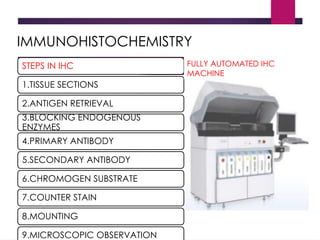
PATHOLOGY POSTING
DATE:19.04.20 TO 24.04.20
 OBJECTIVES:
 Preparation of thick and thin smear.
 Staining and interpretation of peripheral blood smear.
 Interpretation of chromatograms for
hemoglobinopathies.
 Preparation and staining of bone marrow aspiration
and biopsy.
 Staining of FNAC aspirate.
 Observation of IHC & PAP smear.
Preparation of thick and thin smear: slide method
THIN SMEAR PREPARATION
 Place a small drop of blood on the pre-
cleaned, labeled slide, near its frosted
end.
 Bring another slide at a 30-45° angle up
to the drop, allowing the drop to spread
along the contact line of the 2 slides.
 Quickly push the upper (spreader) slide
toward the unfrosted end of the lower
slide to make a tongue shape.
 Air dry.
 Place a small drop of blood in the
center of the pre-cleaned, labeled
slide.
 Using the corner of another slide
or an applicator stick, spread the
drop in a circular pattern until it
is the size of a dime (1.5 cm2).
 A thick smear of proper density is
one which, if placed (wet) over
newsprint, allows you to barely
read the words.
 Air dried 30 minutes.
THICK SMEAR PREPARATION
 Good smear:
 Tongue shaped
with a smooth
tail.
 Doesn’t cover
the entire area of
the slide.
 Has both thin
and thick areas.
THIN SMEAR THICK SMEAR
 A thin blood smear is a drop
of blood that is spread across
a large area of the slide.
 WBC, platelets and parasites
are visualized.
 Thin blood smears helps
discover what species of
malaria is causing the
infection.
 It is fixed in methanol.
 A thick blood smear is a drop
of blood on a glass slide.
 parasites are visualized.
 Thick blood smears are most
useful for detecting the
presence of parasites, because
they examine a larger sample
of blood.
 It is not fixed in methanol.
Three basic steps to make blood film:
1. Preparation of blood smear.
2. Fixation of blood smear.
3. Staining of blood smear.
 Purpose of fixation:
To preserve the morphology of the cells.
 Fixator:
Methyl alcohol(methanol)is the choice ,although
ethyl alcohol can be used.
Stains for peripheral blood
smear:
 Romanowsky stains are universally employed for staining.
 All Romanowsky stains have
methylene blue acidic dye eosin/azure basic dye
has affinity for basic has affinity for acidic
components of cell component of cell
i.e. cytoplasm i.e. nucleus
 Most Romanowsky stains are prepared in methyl alcohol
so that they combine fixation and staining.
Various stains included under Romanowsky stain:
 Leishman stain(used for peripheral smear)
 May Grunwald -Giemsa stain(used for bone marrow
staining)
 Wright stain
 Field stain
 Jenner stain
 JSB stain
Procedure for staining:
 Freshly prepared, air dried blood film is taken.
 It is then covered with Leishman Stain and allowed to
act for 1 minute.
 Methanol in the stain fixes the preparation.
 Double the volume of distilled water is added and
mixed.
 The diluted stain is allowed to act for 10-12 minutes.
 The film is then washed with distilled
water,drained,dried and examined.
Interpretation of peripheral
smear:
 Macroscopic view:Quality of smear present,
any abnormal particles present.
 Microscopic analysis:Begins on lower power 10x
to assess quality of the preparation,to assess
whether red cell agglutination excessive rouleaux
formation or platelet aggregation.
 On high power(40x):to obtain a WBC estimate.
 Oil immersion100x:detailed analysis of cellular
elements.
RBC: PERIPHERAL SMEAR
Normal human red blood cells
are:
• biconcave discs
• mean diameter of about 7.5 μm.
• They are slightly smaller than small
lymphocytes.
• The hemoglobin of red cells is
located peripherally, leaving an area
of central pallor
• equal to approximately 30 to 45% of
the diameter of the cell..
WBC TYPE NUCLEUS CYTOPLASM
GRANULOCYTES
NEUTROPHIL SEGMENTED
LOBES:2 -5
MOSTLY TRILOBED.
PINK /ORANGE,
FINE GRANULATION.
EOSINOPHIL BILOBED. SHERICAL
GOLD,ORANGE
COARSE GRANULATION.
BASOPHIL IRREGULAR,DENSE. DARK
BLUE,PURPLE,LARGE
GRANULATION.
AGRANULOCYTES
MONOCYTE LARGE,CURVED,HORSE
SHOE SHAPED.
GREY BLUE CYTOPLASM.
LYMPHOCYTE SINGLE,SHARPLY
DEFINED
GREY BLUE.
Platelet: peripheral
smear
• Size:1-3 µm
• Non nucleated cells
derived from
cytoplasmic
fragments of
megakaryocytes.
• Has purple red
granules.
• Lilac colour.
 A RBC
 B REACTIVE
LYMPHOCYTE
 C ,E ,I
NEUTROPHIL
 D EOSINOPHIL
 F MONOCYTE
 H
LYMPHOCYTE
 J BASOPHIL
HYPOCHROMIA:
 ↓ Hemoglobin content of RBC
 ↑ Central pallor >1/3rd
.
 ↓ in MCH and MCHC
 Seen in
IRON DEFICIENCY
ANAEMIA,
THALASSAEMIA.
SICKLE CELL ANAEMIA:
• Cells are sickle (boat) or
crescent shaped.
• Present in film of patient
with homozygous HbS
• Absent in neonates and rare
in high HbF percentage.
Hemoglobinopathy
THALASSAEMIA SYNDROMES:
 Beta thalassemia
Thalassemia major
Thalassemia intermedia
Thalassemia trait
 Alpha thalassemia
Hydrops fetalis
HbH disease
Alpha thalassemia trait.
Misc. thalassemia syndromes:HbS, HbE,
HbD,Delta-beta, Gamma, delta.
Sickle cell disease
HbS
 Heterozygous : Sickle cell trait : Hb AS
 Homozygous: Sickle cell anemia : Hb SS
 Combined : Sickle cell β thalassemia :
Sickle cell Hemoglobin C disease
 Hb S provides protection against falciparum malaria
Interpretation of Hemoglobinopathies by
HPLC
 The HPLC Machine
(BIORAD D10 Hb Testing System)
Chromatography:
• Chromatography is the separation of a mixture into
its components
Principle & Instrumentation of HPLC
Pump
Sample
Injector
Cartridge
Photometer
Mobile Phases
Gradient
Controller
•
Chromatogram
Stationary Phase: Cation Exchange Cartridge
Carboxyl groups attached to a resin base
Direction of flow Detector
Principle: Cation Exchange Chromatography
Hemoglobin Introduction
Positively charged hemoglobin fragments attach to the
carboxyl groups at varying strengths.
Starting Gradient:
Low Ionic Strength Buffer
The gradient starts with a low % of Buffer B and high %
Buffer A
At this gradient, hemoglobin fragments with an ionic
strength lower than the buffer gradient, such as F, are
displaced from the cartridge and pass into the detector
Ending Gradient:
High Ionic Strength Buffer
• As the % of the High Ionic Strength Buffer B increases, more
hemoglobin fragments will be displaced.
• Once the gradient is 100% Buffer B all remaining hemoglobin
fragments, including any variant hemoglobin such as S, D and C,
will be removed.
Chromatogram & Retention Time
•

Voltage signal from the detector
is plotted against the retention
time (0- 6.5min).

The area counts of the peak for
each component are calculated as
a percentage of total area.

Check the chromatogram for:
Base line - straight.
Peak shape- Sharp and
symmetrical.
Area under curve= 1-4 million
Normal
chromatogram
Peak Description
Peak Name Window
(mins)
Description
A1a 0.16-0.26 normal upto 2.5%
A1b 0.24-0.36 normal upto 2.5%
F 0.42-0.62 Raised in homozygous beta Thalassemia, HPFH, delta beta Thalassemia
LA1c/CHb-1 0.62-0.93 normal upto 4%
LA1c/CHb-2 0.66-0.93 normal upto 4%
A1c 0.70-1.04 changes with the glycemic status
P3 1.23-1.63 upto 8 % acceptable; 8 to 12 % may indicate sample deterioration; 15 to 25 %
indicate Hb J
A0 1.55-1.85 non glycated fraction of Adult hemoglobin
A2 2.80-3.50 Normal <3.6%; 3.6-3.9% ??Silent Beta Thalassemia.
S 4.02-4.30 22 to 40% (heterozygous ); 70 to 90 % (homozygous ); (upto 10 % not
significant)
C 4.70-4.90
Unknown ↕ may appear anywhere; Upto 8% is not significant
Unknown
HbD Punjab elutes as Unknown peak (3.8 +/- 0.1 min).
HbQ India elutes at 4.45 +/- 0.02 min
First evaluate age and transfusion
history .
If transfusion is involved
report with parental screening.
If no transfusion is involved
Look for the following :
Increased NRBC count
Marked variation in shape and size
Hb F elevated upto 90%
Reduced Hb A
Normal or elevated Hb A2
Beta Thalassemia
Major
(HbF peak present)
HbS Trait
HbSS
Beta Thalassemia trait
with HbQ India
Additional points :
 P3 – upto 6 % acceptable ,
– 6 to 12 % may indicate sample
detioration.
– 15 to 25 % indicate Hb J
 Iron deficiency – Hb A2 found to be
slightly lower.
 Megaloblastic anemia – Hb A2 found to be
higher.
Preparation and staining
bone marrow aspiration
and biopsy
BONE MARROW
ASPIRATION
Preparation:
Make films of aspirated marrow 3-5
cm in length using:
Smooth edge glass spreader 2cm in
width.
Put a drop of marrow on slide.
Drag the drop by spreader leaving a
trail of cells behind it.
Fix the film and stain them.
Staining:
Most commonly done using May
Grunwald -Giemsa stain or Wright
stain.
FNAC
STAINING :
 AIR DRYING: Followed by staining with a hematological stain such as MG-Giemsa stain or
Jenner Giemsa stain .
 ALCOHOL FIXATION: Followed by staining according to PAP or with H&E.
Pap smear and IHC
 PAP SMEAR
 PAPANICOLAOU STAIN:
Contents: Harris Hematoxylin
orange G6
EA 50
CLUE CELLS
Squamous cell in PAP
IMMUNOHISTOCHEMISTRY
STEPS IN IHC
1.TISSUE SECTIONS
2.ANTIGEN RETRIEVAL
3.BLOCKING ENDOGENOUS
ENZYMES
4.PRIMARY ANTIBODY
5.SECONDARY ANTIBODY
6.CHROMOGEN SUBSTRATE
7.COUNTER STAIN
8.MOUNTING
9.MICROSCOPIC OBSERVATION
FULLY AUTOMATED IHC
MACHINE
THANK YOU

path presentation of laboratory pre.pptx

  • 1.
    PATHOLOGY POSTING DATE:19.04.20 TO24.04.20  OBJECTIVES:  Preparation of thick and thin smear.  Staining and interpretation of peripheral blood smear.  Interpretation of chromatograms for hemoglobinopathies.  Preparation and staining of bone marrow aspiration and biopsy.  Staining of FNAC aspirate.  Observation of IHC & PAP smear.
  • 2.
    Preparation of thickand thin smear: slide method THIN SMEAR PREPARATION  Place a small drop of blood on the pre- cleaned, labeled slide, near its frosted end.  Bring another slide at a 30-45° angle up to the drop, allowing the drop to spread along the contact line of the 2 slides.  Quickly push the upper (spreader) slide toward the unfrosted end of the lower slide to make a tongue shape.  Air dry.
  • 3.
     Place asmall drop of blood in the center of the pre-cleaned, labeled slide.  Using the corner of another slide or an applicator stick, spread the drop in a circular pattern until it is the size of a dime (1.5 cm2).  A thick smear of proper density is one which, if placed (wet) over newsprint, allows you to barely read the words.  Air dried 30 minutes. THICK SMEAR PREPARATION
  • 4.
     Good smear: Tongue shaped with a smooth tail.  Doesn’t cover the entire area of the slide.  Has both thin and thick areas.
  • 5.
    THIN SMEAR THICKSMEAR  A thin blood smear is a drop of blood that is spread across a large area of the slide.  WBC, platelets and parasites are visualized.  Thin blood smears helps discover what species of malaria is causing the infection.  It is fixed in methanol.  A thick blood smear is a drop of blood on a glass slide.  parasites are visualized.  Thick blood smears are most useful for detecting the presence of parasites, because they examine a larger sample of blood.  It is not fixed in methanol.
  • 6.
    Three basic stepsto make blood film: 1. Preparation of blood smear. 2. Fixation of blood smear. 3. Staining of blood smear.  Purpose of fixation: To preserve the morphology of the cells.  Fixator: Methyl alcohol(methanol)is the choice ,although ethyl alcohol can be used.
  • 7.
    Stains for peripheralblood smear:  Romanowsky stains are universally employed for staining.  All Romanowsky stains have methylene blue acidic dye eosin/azure basic dye has affinity for basic has affinity for acidic components of cell component of cell i.e. cytoplasm i.e. nucleus  Most Romanowsky stains are prepared in methyl alcohol so that they combine fixation and staining.
  • 8.
    Various stains includedunder Romanowsky stain:  Leishman stain(used for peripheral smear)  May Grunwald -Giemsa stain(used for bone marrow staining)  Wright stain  Field stain  Jenner stain  JSB stain
  • 9.
    Procedure for staining: Freshly prepared, air dried blood film is taken.  It is then covered with Leishman Stain and allowed to act for 1 minute.  Methanol in the stain fixes the preparation.  Double the volume of distilled water is added and mixed.  The diluted stain is allowed to act for 10-12 minutes.  The film is then washed with distilled water,drained,dried and examined.
  • 10.
    Interpretation of peripheral smear: Macroscopic view:Quality of smear present, any abnormal particles present.  Microscopic analysis:Begins on lower power 10x to assess quality of the preparation,to assess whether red cell agglutination excessive rouleaux formation or platelet aggregation.  On high power(40x):to obtain a WBC estimate.  Oil immersion100x:detailed analysis of cellular elements.
  • 11.
    RBC: PERIPHERAL SMEAR Normalhuman red blood cells are: • biconcave discs • mean diameter of about 7.5 μm. • They are slightly smaller than small lymphocytes. • The hemoglobin of red cells is located peripherally, leaving an area of central pallor • equal to approximately 30 to 45% of the diameter of the cell..
  • 12.
    WBC TYPE NUCLEUSCYTOPLASM GRANULOCYTES NEUTROPHIL SEGMENTED LOBES:2 -5 MOSTLY TRILOBED. PINK /ORANGE, FINE GRANULATION. EOSINOPHIL BILOBED. SHERICAL GOLD,ORANGE COARSE GRANULATION. BASOPHIL IRREGULAR,DENSE. DARK BLUE,PURPLE,LARGE GRANULATION. AGRANULOCYTES MONOCYTE LARGE,CURVED,HORSE SHOE SHAPED. GREY BLUE CYTOPLASM. LYMPHOCYTE SINGLE,SHARPLY DEFINED GREY BLUE.
  • 13.
    Platelet: peripheral smear • Size:1-3µm • Non nucleated cells derived from cytoplasmic fragments of megakaryocytes. • Has purple red granules. • Lilac colour.
  • 14.
     A RBC B REACTIVE LYMPHOCYTE  C ,E ,I NEUTROPHIL  D EOSINOPHIL  F MONOCYTE  H LYMPHOCYTE  J BASOPHIL
  • 15.
    HYPOCHROMIA:  ↓ Hemoglobincontent of RBC  ↑ Central pallor >1/3rd .  ↓ in MCH and MCHC  Seen in IRON DEFICIENCY ANAEMIA, THALASSAEMIA.
  • 16.
    SICKLE CELL ANAEMIA: •Cells are sickle (boat) or crescent shaped. • Present in film of patient with homozygous HbS • Absent in neonates and rare in high HbF percentage.
  • 17.
    Hemoglobinopathy THALASSAEMIA SYNDROMES:  Betathalassemia Thalassemia major Thalassemia intermedia Thalassemia trait  Alpha thalassemia Hydrops fetalis HbH disease Alpha thalassemia trait. Misc. thalassemia syndromes:HbS, HbE, HbD,Delta-beta, Gamma, delta.
  • 18.
    Sickle cell disease HbS Heterozygous : Sickle cell trait : Hb AS  Homozygous: Sickle cell anemia : Hb SS  Combined : Sickle cell β thalassemia : Sickle cell Hemoglobin C disease  Hb S provides protection against falciparum malaria
  • 19.
    Interpretation of Hemoglobinopathiesby HPLC  The HPLC Machine (BIORAD D10 Hb Testing System)
  • 20.
    Chromatography: • Chromatography isthe separation of a mixture into its components
  • 21.
    Principle & Instrumentationof HPLC Pump Sample Injector Cartridge Photometer Mobile Phases Gradient Controller • Chromatogram
  • 22.
    Stationary Phase: CationExchange Cartridge Carboxyl groups attached to a resin base Direction of flow Detector Principle: Cation Exchange Chromatography
  • 23.
    Hemoglobin Introduction Positively chargedhemoglobin fragments attach to the carboxyl groups at varying strengths.
  • 24.
    Starting Gradient: Low IonicStrength Buffer The gradient starts with a low % of Buffer B and high % Buffer A At this gradient, hemoglobin fragments with an ionic strength lower than the buffer gradient, such as F, are displaced from the cartridge and pass into the detector
  • 25.
    Ending Gradient: High IonicStrength Buffer • As the % of the High Ionic Strength Buffer B increases, more hemoglobin fragments will be displaced. • Once the gradient is 100% Buffer B all remaining hemoglobin fragments, including any variant hemoglobin such as S, D and C, will be removed.
  • 26.
    Chromatogram & RetentionTime •  Voltage signal from the detector is plotted against the retention time (0- 6.5min).  The area counts of the peak for each component are calculated as a percentage of total area.  Check the chromatogram for: Base line - straight. Peak shape- Sharp and symmetrical. Area under curve= 1-4 million
  • 27.
  • 28.
    Peak Description Peak NameWindow (mins) Description A1a 0.16-0.26 normal upto 2.5% A1b 0.24-0.36 normal upto 2.5% F 0.42-0.62 Raised in homozygous beta Thalassemia, HPFH, delta beta Thalassemia LA1c/CHb-1 0.62-0.93 normal upto 4% LA1c/CHb-2 0.66-0.93 normal upto 4% A1c 0.70-1.04 changes with the glycemic status P3 1.23-1.63 upto 8 % acceptable; 8 to 12 % may indicate sample deterioration; 15 to 25 % indicate Hb J A0 1.55-1.85 non glycated fraction of Adult hemoglobin A2 2.80-3.50 Normal <3.6%; 3.6-3.9% ??Silent Beta Thalassemia. S 4.02-4.30 22 to 40% (heterozygous ); 70 to 90 % (homozygous ); (upto 10 % not significant) C 4.70-4.90 Unknown ↕ may appear anywhere; Upto 8% is not significant Unknown HbD Punjab elutes as Unknown peak (3.8 +/- 0.1 min). HbQ India elutes at 4.45 +/- 0.02 min
  • 29.
    First evaluate ageand transfusion history . If transfusion is involved report with parental screening. If no transfusion is involved Look for the following : Increased NRBC count Marked variation in shape and size Hb F elevated upto 90% Reduced Hb A Normal or elevated Hb A2 Beta Thalassemia Major (HbF peak present)
  • 30.
  • 31.
  • 32.
  • 33.
    Additional points : P3 – upto 6 % acceptable , – 6 to 12 % may indicate sample detioration. – 15 to 25 % indicate Hb J  Iron deficiency – Hb A2 found to be slightly lower.  Megaloblastic anemia – Hb A2 found to be higher.
  • 34.
    Preparation and staining bonemarrow aspiration and biopsy BONE MARROW ASPIRATION Preparation: Make films of aspirated marrow 3-5 cm in length using: Smooth edge glass spreader 2cm in width. Put a drop of marrow on slide. Drag the drop by spreader leaving a trail of cells behind it. Fix the film and stain them. Staining: Most commonly done using May Grunwald -Giemsa stain or Wright stain.
  • 35.
    FNAC STAINING :  AIRDRYING: Followed by staining with a hematological stain such as MG-Giemsa stain or Jenner Giemsa stain .  ALCOHOL FIXATION: Followed by staining according to PAP or with H&E.
  • 36.
    Pap smear andIHC  PAP SMEAR  PAPANICOLAOU STAIN: Contents: Harris Hematoxylin orange G6 EA 50 CLUE CELLS Squamous cell in PAP
  • 37.
    IMMUNOHISTOCHEMISTRY STEPS IN IHC 1.TISSUESECTIONS 2.ANTIGEN RETRIEVAL 3.BLOCKING ENDOGENOUS ENZYMES 4.PRIMARY ANTIBODY 5.SECONDARY ANTIBODY 6.CHROMOGEN SUBSTRATE 7.COUNTER STAIN 8.MOUNTING 9.MICROSCOPIC OBSERVATION FULLY AUTOMATED IHC MACHINE
  • 38.

Editor's Notes

  • #21 Mobile Phases - Component solvents/mobile phases to make up gradient Gradient Controller - Sets up gradient - linearity, steps, ramps, number of solvents/mobile phases (binary, ternary, quaternary). Pump - Dual piston, Pulse free, Able to deliver 4000PSI, Precision flow rates of 0.001mL/min, Flow range 0.001-10.001 mL/min.