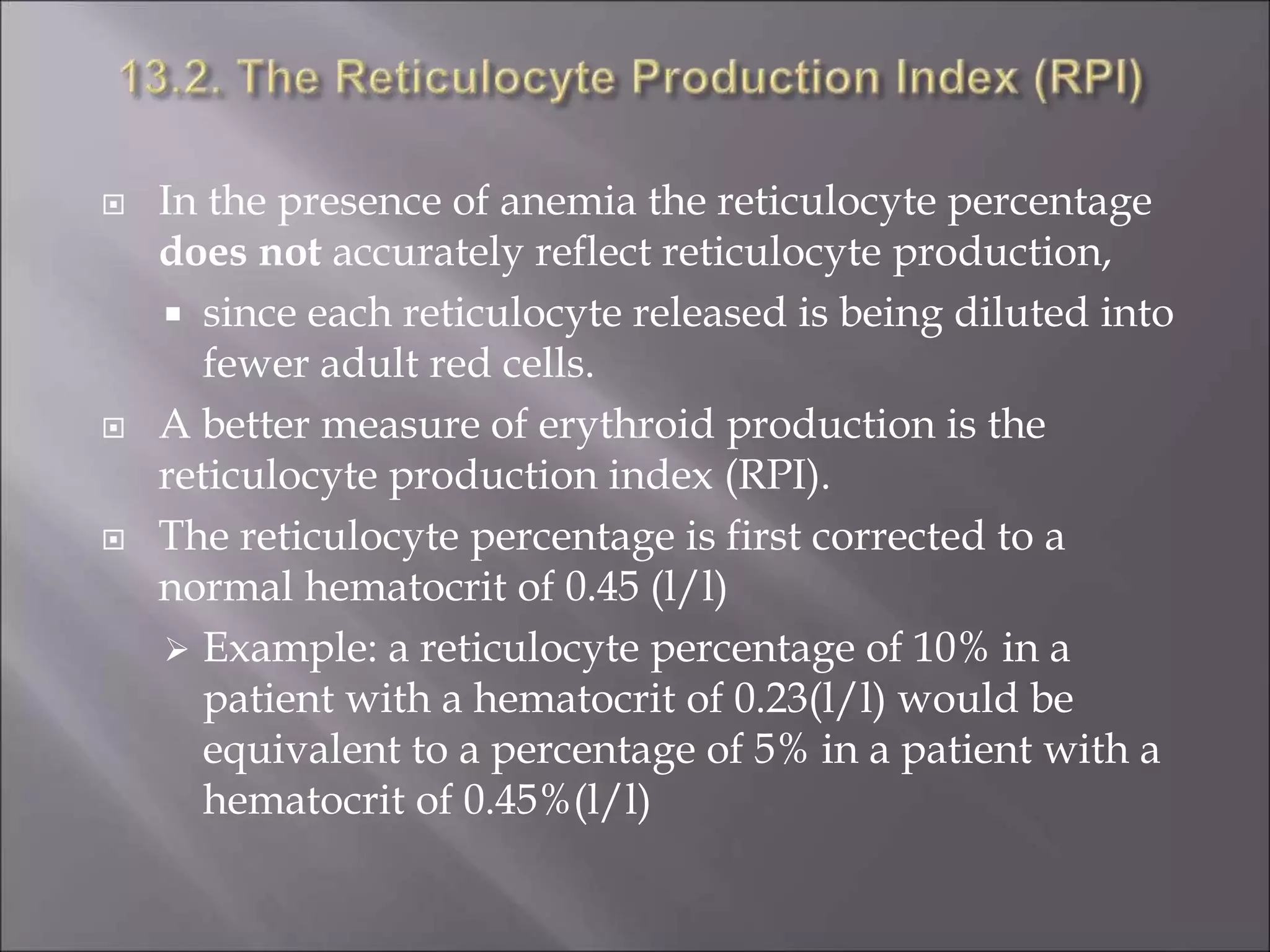
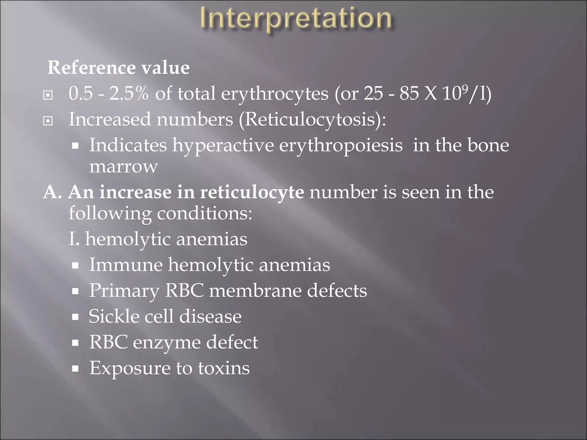
Nathan Kikku Mubiru
Medical Laboratory
Scientific Officer (MLSO)
At the end of this chapter, the student will be able to:
 Define reticulocytes
 Explain the relationship between the number of
reticulocytes in the peripheral blood and erythropoietic
activity in the bone marrow
 Discuss the reticulocyte production index
 Define supravital dyes
 Prepare supravital dyes in the right proportion and stain
a sample of blood
 Perform reticulocyte count on a sample of blood
 Indicate the normal range for reticulocyte count
 Discuss the clinical implications of the reticulocyte count
 Perform QC measures in reticulocyte counting
 List possible sources of error and their remedies
 Introduction
 RPI
 Supravital staining
 Counting of reticulocytes
 Clinical implication
 QC in reticulocyte count
 Reticulocytes (Retics) are juvenile red cells
 They contain remnants of the ribosomal RNA
 The most immature reticulocytes are those with the
largest amount of precipitable material
 In the least immature Retics, only a few dots or strands
are seen.
Reticulocytes after counter staining with
Romanowsky dyes (left panel) and supravital
dyes (right)
 Supravital staining is staining of cells while they are
viable
 The number of reticulocytes in the peripheral blood is a
fairly accurate reflection of erythropoietic activity
 Complete loss of basophilic material probably occurs as
a rule in the blood stream after the cells have left the
bone marrow.
 The ripening process is thought to take 2-3 days, of
which about 24 hours are spent in the circulation.
 When there is an increased erythropoietic stimulus as
in hemolytic anemia there will be premature release of
reticulocytes into the circulation as their transit time in
the bone marrow is reduced, the so-called 'stress' or
'shift' reticulocytosis.
 To assist physicians in the diagnosis, treatment or
monitoring of patients with various anemias
 The reticulocyte count is an index of bone marrow red
cell production
 It measures erythropoietic activity
 The count is based on the property of ribosomal RNA
to react with basic dyes such as new methylene blue or
brilliant cresyl blue to form a blue precipitate of
granules or filaments. Retics are larger than mature red
cells and show diffuse basophilic staining
(polychromasia) in Romanowsky stained films.
Note: polychromasia on Romanowsky stains are
indicative of Reticulocytosis
 Only supravital staining techniques enable their
number to be determined with sufficient accuracy.
 Better and more reliable results are obtained with
new methylene blue than brilliant cresyl blue, the
former stains the reticulo-filamentous material in the
reticulocytes more deeply and more uniformly than
does the latter
Staining Solution:
 1% New methylene blue (NMB)
 1% Brilliant cresyl blue.
 EDTA whole blood:
 Specimen can be stored at room temperature for 8
hours or refrigerated at 2-8oC for 24 hours
1. Deliver 2-3 drops of the dye solution into 75 X 10mm
glass or plastic tube using a Pasteur pipette.
2. Add 2-4 drops of the patient’s EDTA anticoagulated
blood to the dye solution and mix.
3. Plug the tube and incubate at 370C for 10-15 minutes.
 The exact volume of blood to be added to the dye
solution for optimal staining depends upon the red
cell count.
 A larger volume of blood in anemic condition
 a smaller proportion in polycythemic blood should be
added than normal blood.
4. After incubation, resuspend the cells by gentle mixing and make
films on glass slides in the usual way
5. Allow to air dry
6. Examine the films without fixing or counter staining.
 In a successful preparation, the reticulofilamentous material
should be stained deep blue and the non-reticulated cells
stained diffuse shades of pale greenish blue.
Pt
name-
Almaz
Card
no-
2453/09
Date
–
12/09/09
1% NMB stained
smear
Counting
 An area of the film should be chosen for the count
where the cells are undistorted and where the staining
is good.
 To count the cells:
 eye pieces provided with an adjustable diaphragm
could be used
 or cardboard diaphragm with a small cut in the
center can be inserted into an eyepiece and used as a
substitute
 When the reticulocyte count is expected to be 10%, a
total of 500 red cells should be counted noting the
number of reticulocytes.
 If less than 10% reticulocytes are expected, at least 1000
red cells should be counted.
 Reticulocyte count (%) = Reticulocyte number X 100
RBC number
 Absolute reticulocyte count = Reticulocyte count (%) X RBC count
 An alternative method is based on the
principle of ‘balanced sampling’ using a
Miller ocular.
 An eyepiece giving a square field in the
corner of which is a second ruled square
one-ninth of the area of the total square.
 Reticulocytes are counted in the large
square and red cells in the small square in
successive fields until at least 300 red cells
are counted
Reticulocyte count (%) = Reticulocyte number X 100
RBC number X 9
 In the presence of anemia the reticulocyte percentage
does not accurately reflect reticulocyte production,
 since each reticulocyte released is being diluted into
fewer adult red cells.
 A better measure of erythroid production is the
reticulocyte production index (RPI).
 The reticulocyte percentage is first corrected to a
normal hematocrit of 0.45 (l/l)
 Example: a reticulocyte percentage of 10% in a
patient with a hematocrit of 0.23(l/l) would be
equivalent to a percentage of 5% in a patient with a
hematocrit of 0.45%(l/l)
 Another correction is made because erythropoietin
production in response to anemia leads to premature release
of newly formed reticulocytes and these stress reticulocytes
take up to two days to mature into adult erythrocytes
 If many polychromatophils are seen on the stained blood
film, then a correction factor of 2 is divided into the
corrected reticulocyte percentage
 Example:
RPI = 10 x 23/45 (hematocrit correction) = 2.5
2.0 (maturation time correction)
 Maturation factors from 1.0-2.0 are used
 the higher numbers if there is a great deal of
polychromatophilia in the peripheral blood film, and
 the lower numbers if there is little
polychromatophilia
 The RPI is an approximate measure of effective red cell
production in the marrow
 A normal marrow has an index of 1.0.
 In hemolytic anemia with excessive destruction of
red cells in the peripheral blood and a functionally
normal marrow, this index may be 3-7 times higher
than normal.
 the index is less than expected for the degree of
anemia, i.e., 2 or less
 When there is marrow damage
 erythropoietin suppression
 a deficiency of iron, vitamin B12 or folic acid
 If the bone marrow contains many normoblasts but
the RPI is low, this may indicate ineffective
erythropoiesis, with intramedullary (marrow)
destruction of erythroid precursors
1. Insufficient number of cells counted.
2. Confusion of reticulocytes with red cell inclusions like
Basophilic stippling, Howell-Jolly bodies,
Pappenheimer bodies and
3. Recent blood transfusion
4. Mishandling, contamination, or inadequate
refrigeration of the sample
Pappenheimer bodies Basophilic stippling
Reference value
 0.5 - 2.5% of total erythrocytes (or 25 - 85 X 109/l)
 Increased numbers (Reticulocytosis):
 Indicates hyperactive erythropoiesis in the bone
marrow
A. An increase in reticulocyte number is seen in the
following conditions:
I. hemolytic anemias
 Immune hemolytic anemias
 Primary RBC membrane defects
 Sickle cell disease
 RBC enzyme defect
 Exposure to toxins
II. Following hemorrhage
III. Following treatment of anemias where an increase in
the reticulocyte number may be used as an index of
the effectiveness of treatment.
e.g.
 After doses of iron in iron deficiency anemia, the
reticulocyte count may exceed 20%
 Proportional increase of Retic count when pernicious
anemia is treated by transfusion or vitamin B12
therapy.
IV. Physiologic increase in pregnancy and in infants
B. Decreased levels: means that the bone marrow is not
producing enough erythrocytes.
Conditions associated with low Retic count:
 Iron deficiency anemia
 Aplastic anemia
 Radiation therapy
 Untreated pernicious anemia
 Tumour in marrow
 Use of commercially available controls
 Strict adherence to SOP (preparation, counting, etc)
1. What are reticulocytes?
2. How could the number of reticulocytes in the peripheral
blood be a fairly accurate reflection of erythropoietic activity
in the bone marrow?
3. Define supravital staining.
4. What is the difference between a Romanowsky and
supravital stains
5. How do you manage to count the number of reticulocytes in
each field of the microscope after you stain the cells with
supravital dyes?
6. How do you calculate the relative number of reticulocytes in
the patient sample?
7. Briefly discuss RPI.
7) What is the clinical interpretation of an increase in the
number of reticulocytes in the peripheral blood in
general terms?
8) list sources of errors in reticulocyte counting and their
remedies

Retics

  • 1.
    Nathan Kikku Mubiru MedicalLaboratory Scientific Officer (MLSO)
  • 2.
    At the endof this chapter, the student will be able to:  Define reticulocytes  Explain the relationship between the number of reticulocytes in the peripheral blood and erythropoietic activity in the bone marrow  Discuss the reticulocyte production index  Define supravital dyes  Prepare supravital dyes in the right proportion and stain a sample of blood  Perform reticulocyte count on a sample of blood
  • 3.
     Indicate thenormal range for reticulocyte count  Discuss the clinical implications of the reticulocyte count  Perform QC measures in reticulocyte counting  List possible sources of error and their remedies
  • 4.
     Introduction  RPI Supravital staining  Counting of reticulocytes  Clinical implication  QC in reticulocyte count
  • 5.
     Reticulocytes (Retics)are juvenile red cells  They contain remnants of the ribosomal RNA  The most immature reticulocytes are those with the largest amount of precipitable material  In the least immature Retics, only a few dots or strands are seen.
  • 6.
    Reticulocytes after counterstaining with Romanowsky dyes (left panel) and supravital dyes (right)
  • 7.
     Supravital stainingis staining of cells while they are viable  The number of reticulocytes in the peripheral blood is a fairly accurate reflection of erythropoietic activity  Complete loss of basophilic material probably occurs as a rule in the blood stream after the cells have left the bone marrow.  The ripening process is thought to take 2-3 days, of which about 24 hours are spent in the circulation.  When there is an increased erythropoietic stimulus as in hemolytic anemia there will be premature release of reticulocytes into the circulation as their transit time in the bone marrow is reduced, the so-called 'stress' or 'shift' reticulocytosis.
  • 8.
     To assistphysicians in the diagnosis, treatment or monitoring of patients with various anemias  The reticulocyte count is an index of bone marrow red cell production  It measures erythropoietic activity
  • 9.
     The countis based on the property of ribosomal RNA to react with basic dyes such as new methylene blue or brilliant cresyl blue to form a blue precipitate of granules or filaments. Retics are larger than mature red cells and show diffuse basophilic staining (polychromasia) in Romanowsky stained films. Note: polychromasia on Romanowsky stains are indicative of Reticulocytosis  Only supravital staining techniques enable their number to be determined with sufficient accuracy.
  • 10.
     Better andmore reliable results are obtained with new methylene blue than brilliant cresyl blue, the former stains the reticulo-filamentous material in the reticulocytes more deeply and more uniformly than does the latter Staining Solution:  1% New methylene blue (NMB)  1% Brilliant cresyl blue.
  • 11.
     EDTA wholeblood:  Specimen can be stored at room temperature for 8 hours or refrigerated at 2-8oC for 24 hours
  • 12.
    1. Deliver 2-3drops of the dye solution into 75 X 10mm glass or plastic tube using a Pasteur pipette. 2. Add 2-4 drops of the patient’s EDTA anticoagulated blood to the dye solution and mix. 3. Plug the tube and incubate at 370C for 10-15 minutes.  The exact volume of blood to be added to the dye solution for optimal staining depends upon the red cell count.  A larger volume of blood in anemic condition  a smaller proportion in polycythemic blood should be added than normal blood.
  • 13.
    4. After incubation,resuspend the cells by gentle mixing and make films on glass slides in the usual way 5. Allow to air dry 6. Examine the films without fixing or counter staining.  In a successful preparation, the reticulofilamentous material should be stained deep blue and the non-reticulated cells stained diffuse shades of pale greenish blue. Pt name- Almaz Card no- 2453/09 Date – 12/09/09 1% NMB stained smear
  • 14.
    Counting  An areaof the film should be chosen for the count where the cells are undistorted and where the staining is good.  To count the cells:  eye pieces provided with an adjustable diaphragm could be used  or cardboard diaphragm with a small cut in the center can be inserted into an eyepiece and used as a substitute
  • 15.
     When thereticulocyte count is expected to be 10%, a total of 500 red cells should be counted noting the number of reticulocytes.  If less than 10% reticulocytes are expected, at least 1000 red cells should be counted.  Reticulocyte count (%) = Reticulocyte number X 100 RBC number  Absolute reticulocyte count = Reticulocyte count (%) X RBC count
  • 16.
     An alternativemethod is based on the principle of ‘balanced sampling’ using a Miller ocular.  An eyepiece giving a square field in the corner of which is a second ruled square one-ninth of the area of the total square.  Reticulocytes are counted in the large square and red cells in the small square in successive fields until at least 300 red cells are counted Reticulocyte count (%) = Reticulocyte number X 100 RBC number X 9
  • 17.
     In thepresence of anemia the reticulocyte percentage does not accurately reflect reticulocyte production,  since each reticulocyte released is being diluted into fewer adult red cells.  A better measure of erythroid production is the reticulocyte production index (RPI).  The reticulocyte percentage is first corrected to a normal hematocrit of 0.45 (l/l)  Example: a reticulocyte percentage of 10% in a patient with a hematocrit of 0.23(l/l) would be equivalent to a percentage of 5% in a patient with a hematocrit of 0.45%(l/l)
  • 18.
     Another correctionis made because erythropoietin production in response to anemia leads to premature release of newly formed reticulocytes and these stress reticulocytes take up to two days to mature into adult erythrocytes  If many polychromatophils are seen on the stained blood film, then a correction factor of 2 is divided into the corrected reticulocyte percentage  Example: RPI = 10 x 23/45 (hematocrit correction) = 2.5 2.0 (maturation time correction)
  • 19.
     Maturation factorsfrom 1.0-2.0 are used  the higher numbers if there is a great deal of polychromatophilia in the peripheral blood film, and  the lower numbers if there is little polychromatophilia  The RPI is an approximate measure of effective red cell production in the marrow  A normal marrow has an index of 1.0.  In hemolytic anemia with excessive destruction of red cells in the peripheral blood and a functionally normal marrow, this index may be 3-7 times higher than normal.
  • 20.
     the indexis less than expected for the degree of anemia, i.e., 2 or less  When there is marrow damage  erythropoietin suppression  a deficiency of iron, vitamin B12 or folic acid  If the bone marrow contains many normoblasts but the RPI is low, this may indicate ineffective erythropoiesis, with intramedullary (marrow) destruction of erythroid precursors
  • 21.
    1. Insufficient numberof cells counted. 2. Confusion of reticulocytes with red cell inclusions like Basophilic stippling, Howell-Jolly bodies, Pappenheimer bodies and 3. Recent blood transfusion 4. Mishandling, contamination, or inadequate refrigeration of the sample Pappenheimer bodies Basophilic stippling
  • 22.
    Reference value  0.5- 2.5% of total erythrocytes (or 25 - 85 X 109/l)  Increased numbers (Reticulocytosis):  Indicates hyperactive erythropoiesis in the bone marrow A. An increase in reticulocyte number is seen in the following conditions: I. hemolytic anemias  Immune hemolytic anemias  Primary RBC membrane defects  Sickle cell disease  RBC enzyme defect  Exposure to toxins
  • 23.
    II. Following hemorrhage III.Following treatment of anemias where an increase in the reticulocyte number may be used as an index of the effectiveness of treatment. e.g.  After doses of iron in iron deficiency anemia, the reticulocyte count may exceed 20%  Proportional increase of Retic count when pernicious anemia is treated by transfusion or vitamin B12 therapy.
  • 24.
    IV. Physiologic increasein pregnancy and in infants B. Decreased levels: means that the bone marrow is not producing enough erythrocytes. Conditions associated with low Retic count:  Iron deficiency anemia  Aplastic anemia  Radiation therapy  Untreated pernicious anemia  Tumour in marrow
  • 25.
     Use ofcommercially available controls  Strict adherence to SOP (preparation, counting, etc)
  • 26.
    1. What arereticulocytes? 2. How could the number of reticulocytes in the peripheral blood be a fairly accurate reflection of erythropoietic activity in the bone marrow? 3. Define supravital staining. 4. What is the difference between a Romanowsky and supravital stains 5. How do you manage to count the number of reticulocytes in each field of the microscope after you stain the cells with supravital dyes? 6. How do you calculate the relative number of reticulocytes in the patient sample? 7. Briefly discuss RPI.
  • 27.
    7) What isthe clinical interpretation of an increase in the number of reticulocytes in the peripheral blood in general terms? 8) list sources of errors in reticulocyte counting and their remedies

Editor's Notes

  • #2 This material for Hematology I is a draft document which needs further revision. Please forward your comments to School of Medical Laboratory Sciences, Addis Ababa University. Thanks!!
  • #3 Instructor notes : please make Reference concerning pictures and figure
  • #4 Instructor notes : please make Reference concerning pictures and figure
  • #6 Reference:
  • #7 Reference:
  • #10 Note to the instructor: The instructor should define medical and hematological terminologies where necessary
  • #14 Reference:
  • #17 Fig. 2.5 The appearance of a Miller ocular micrometer for use in counting reticulocytes. it consists of two squares whose areas have a ratio 1:9
  • #20 Note to the instructor: The instructor should define medical and hematological terminologies where necessary
  • #21 Note to the instructor: The instructor should define medical and hematological terminologies where necessary
  • #22 Recent Reference:
  • #23 Note to the instructor: The instructor should define medical and hematological terminologies where necessary
  • #25 Note to the instructor: The instructor should define medical and hematological terminologies where necessary