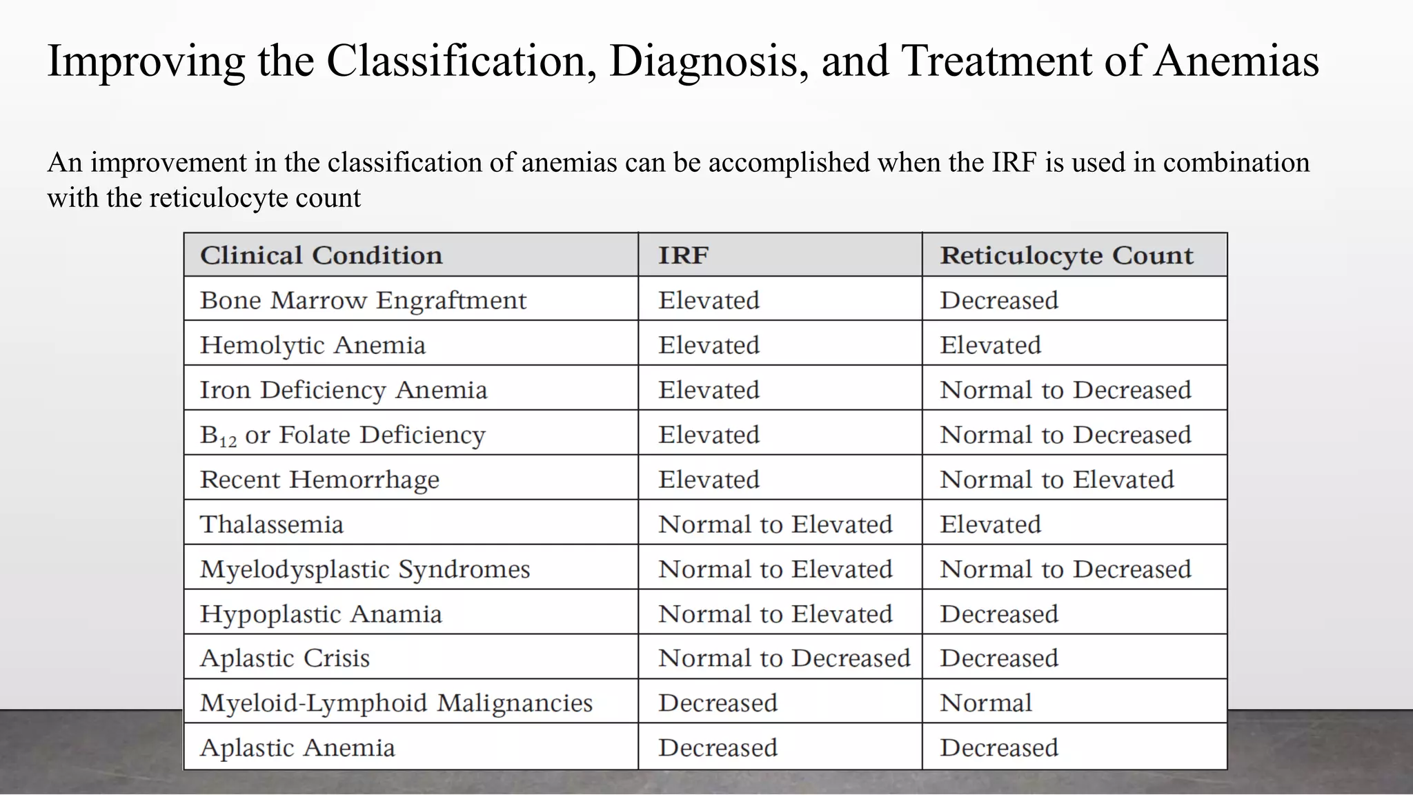
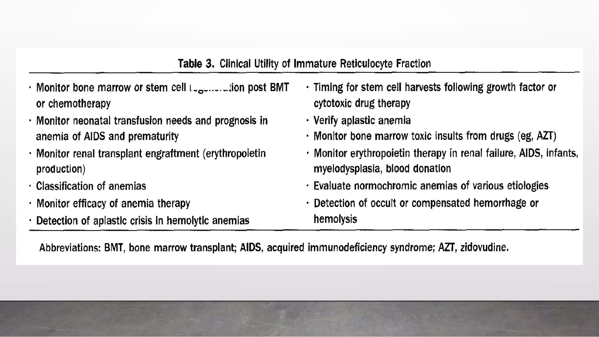
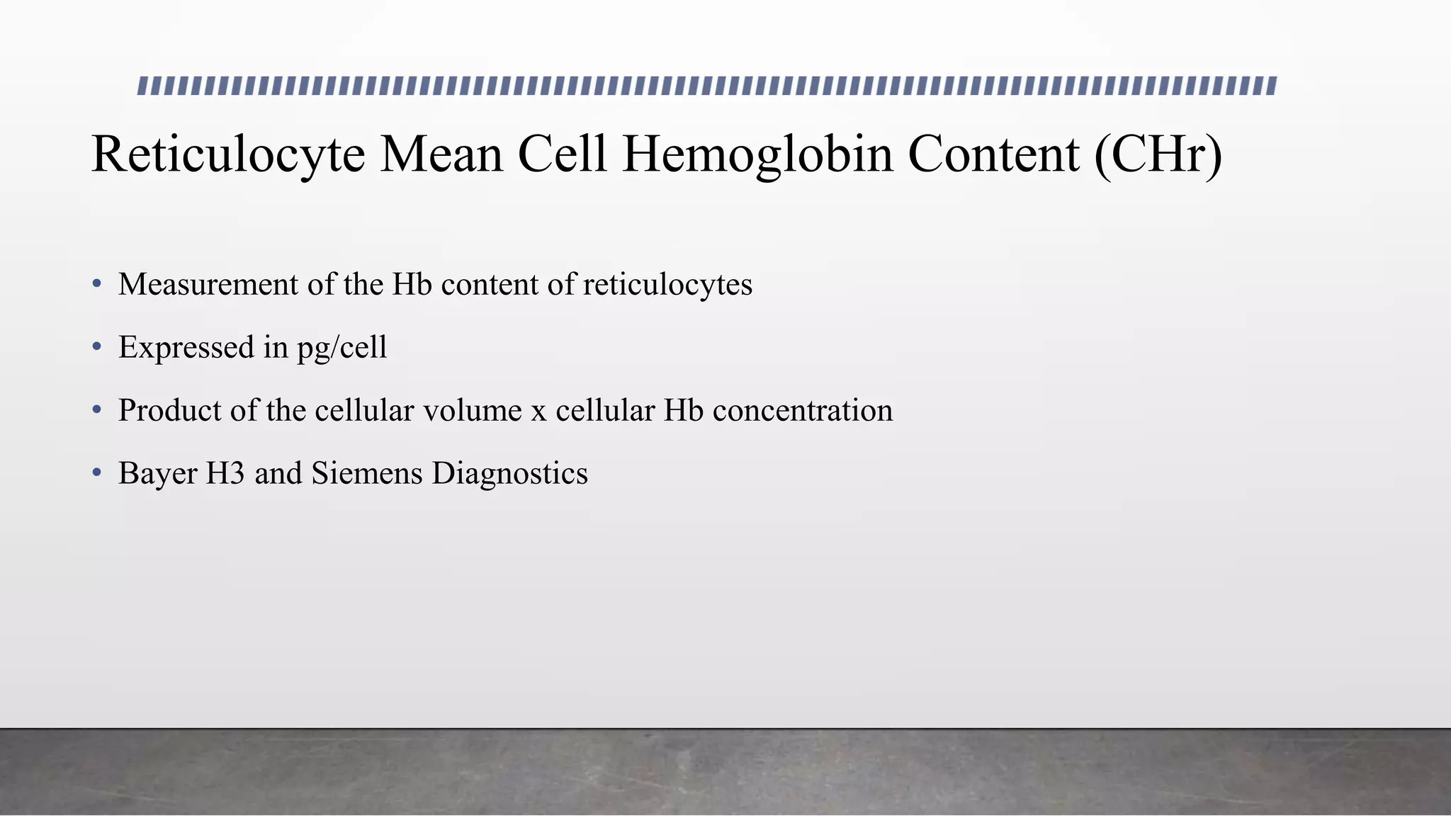
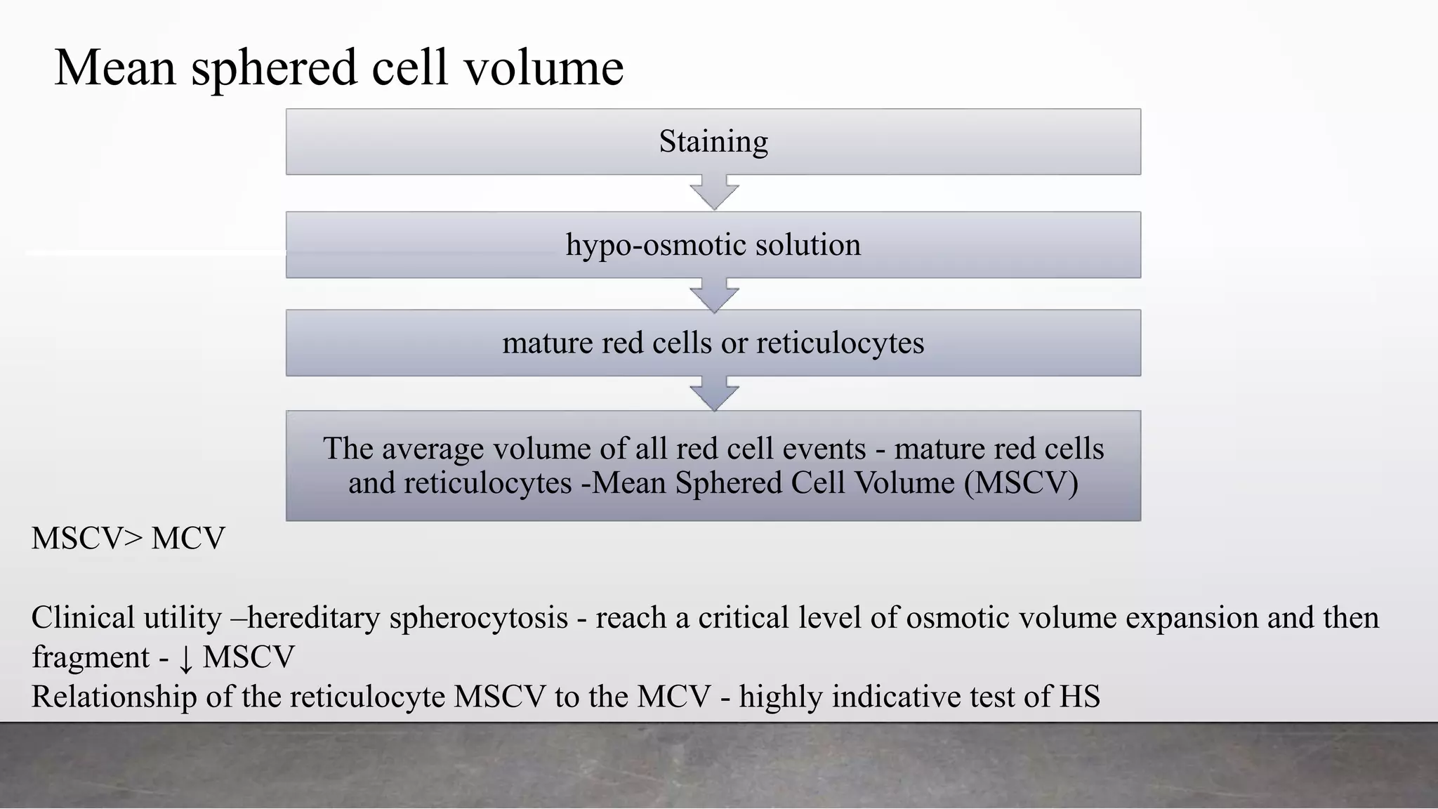
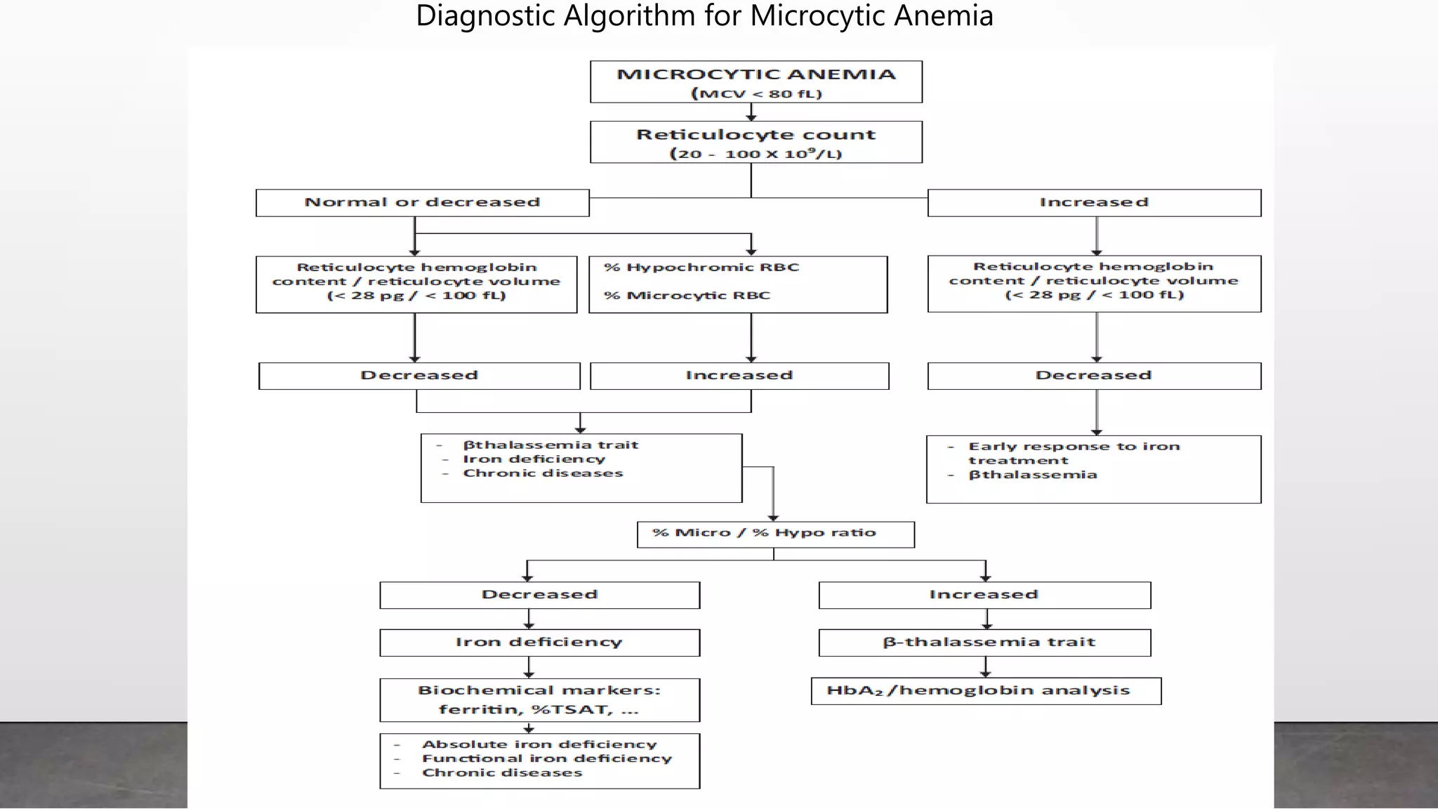
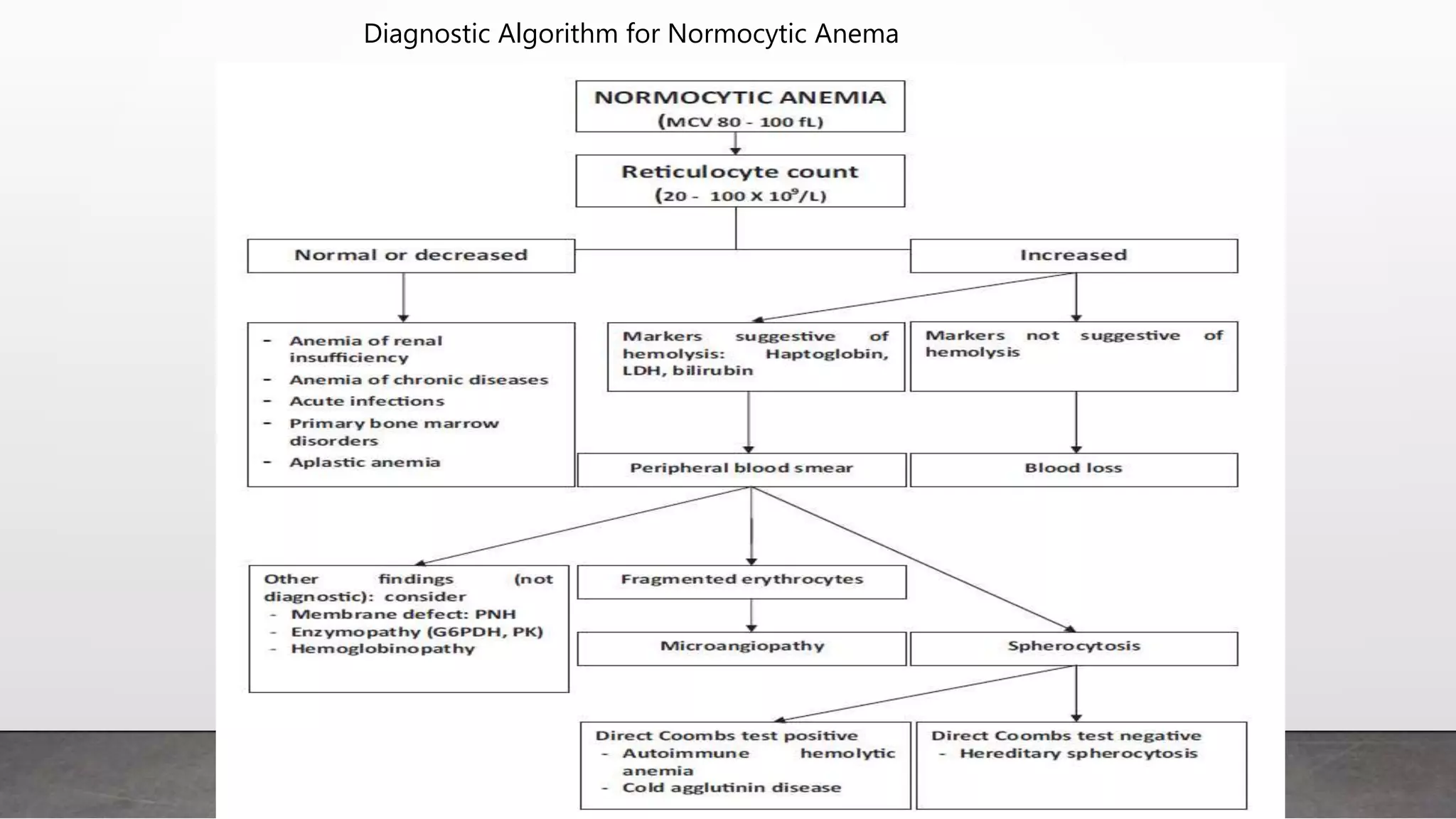
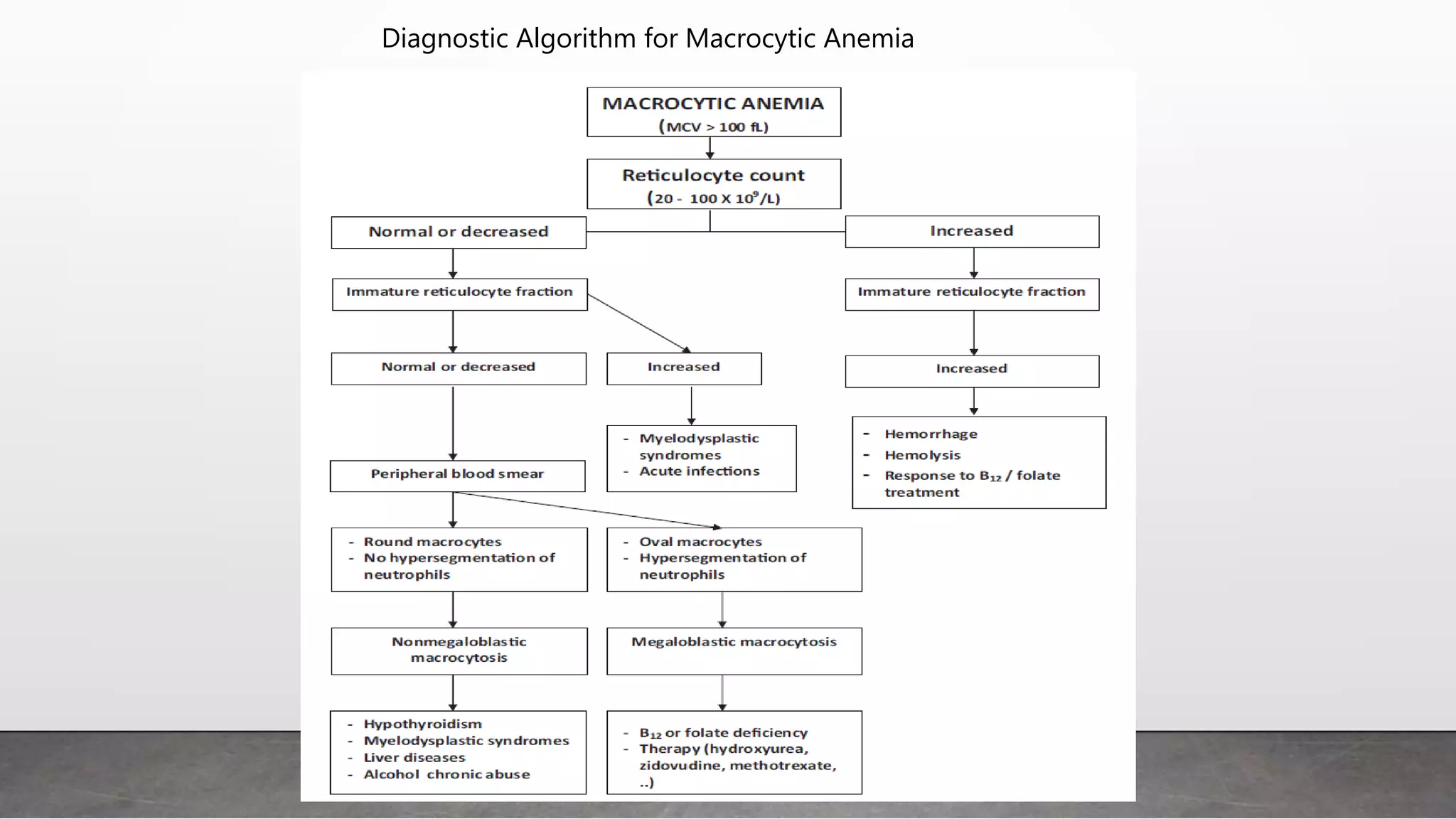
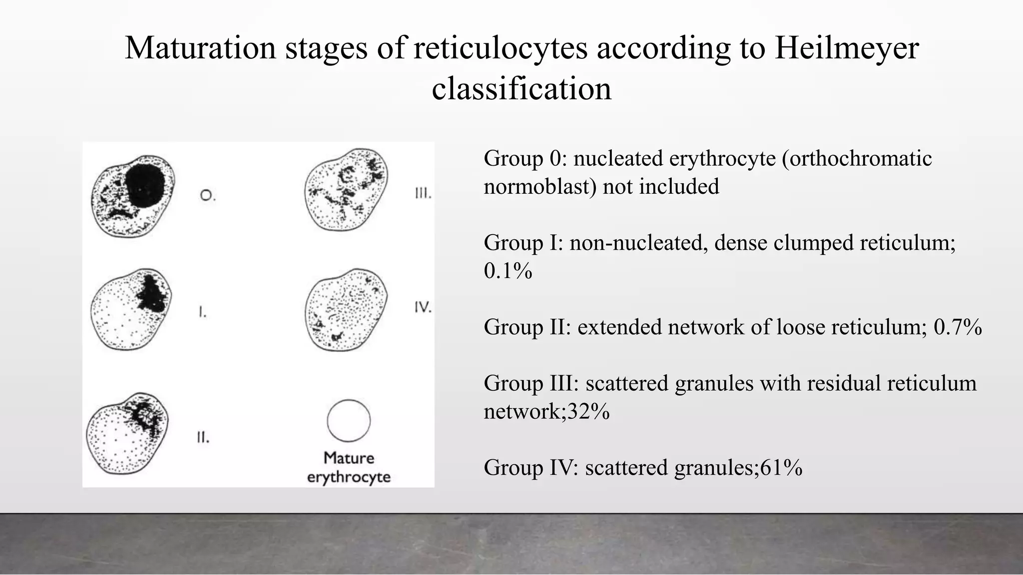
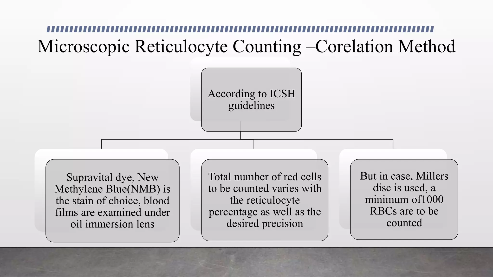
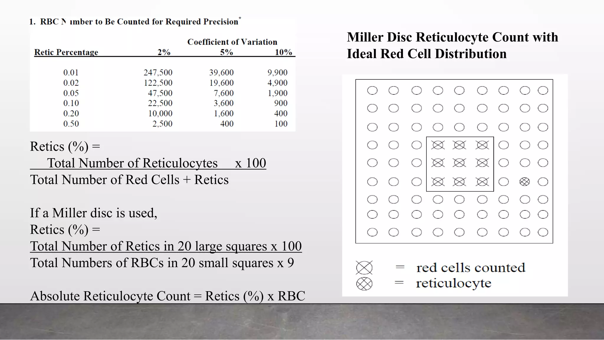
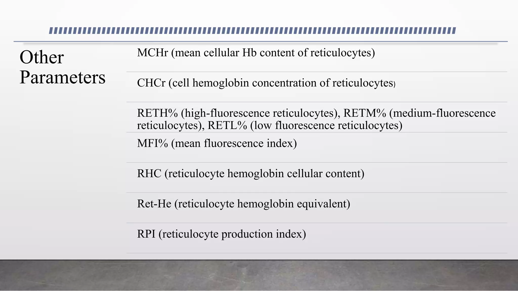
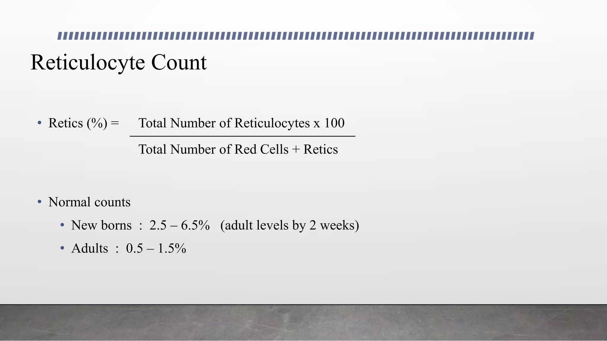
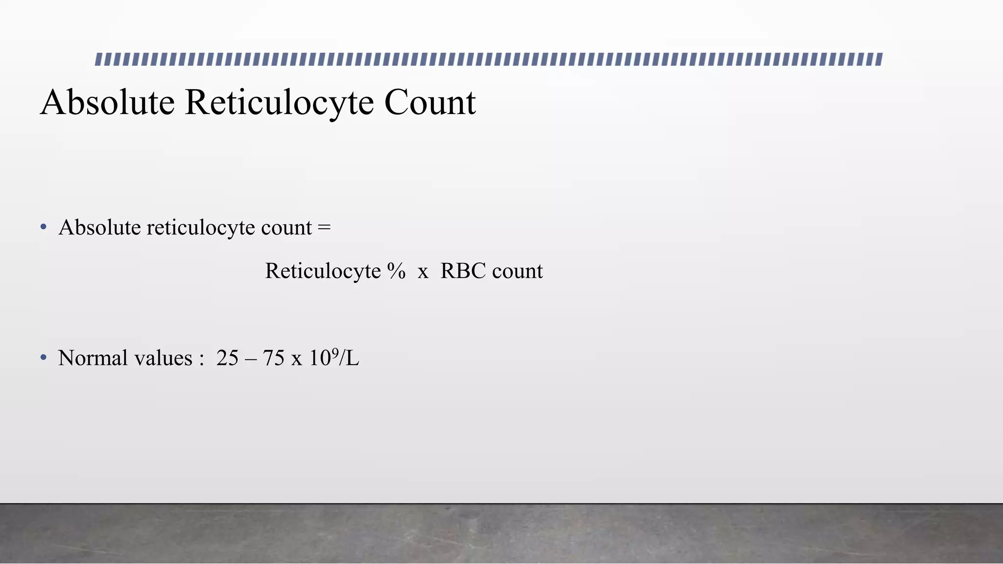
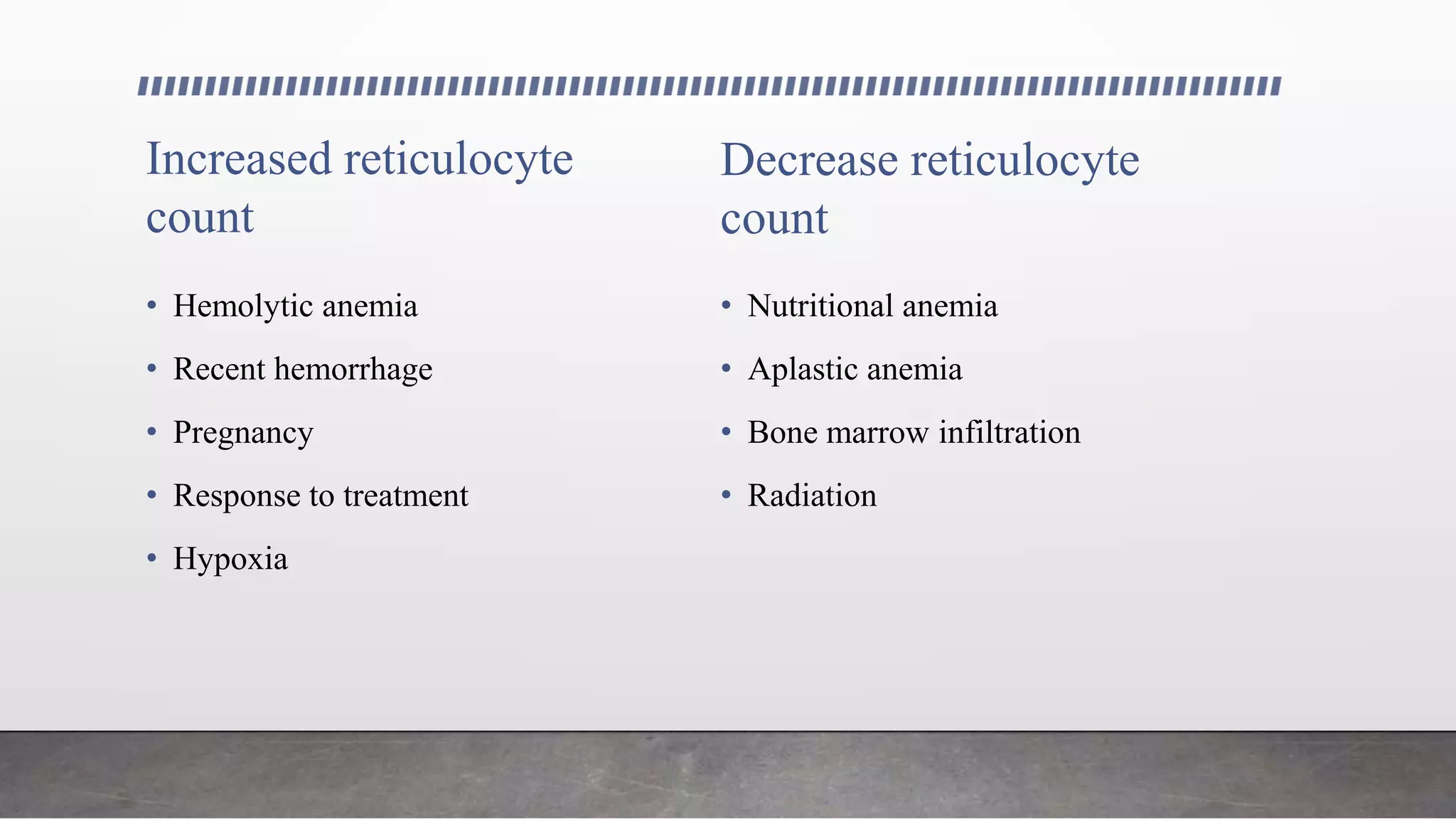
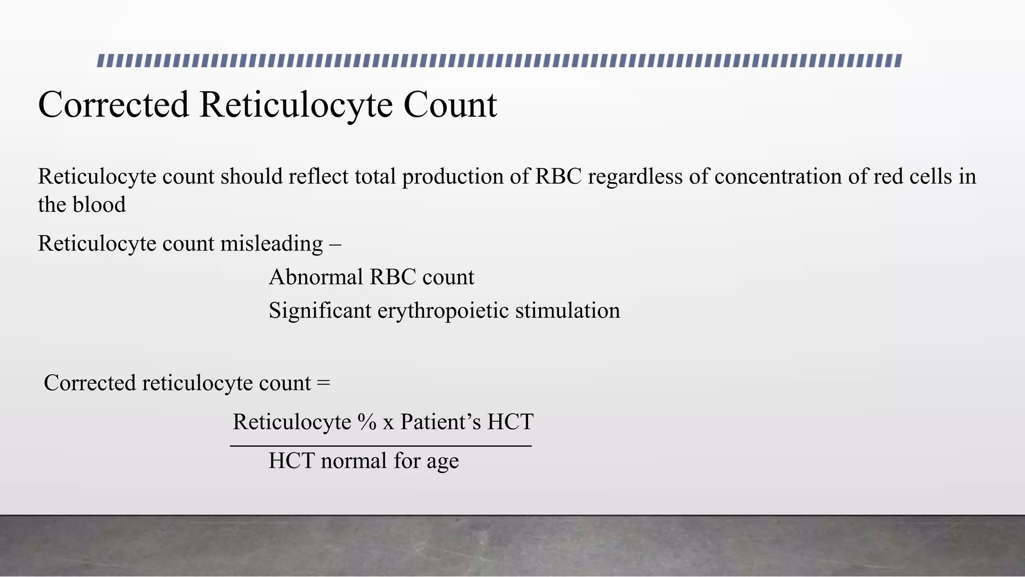
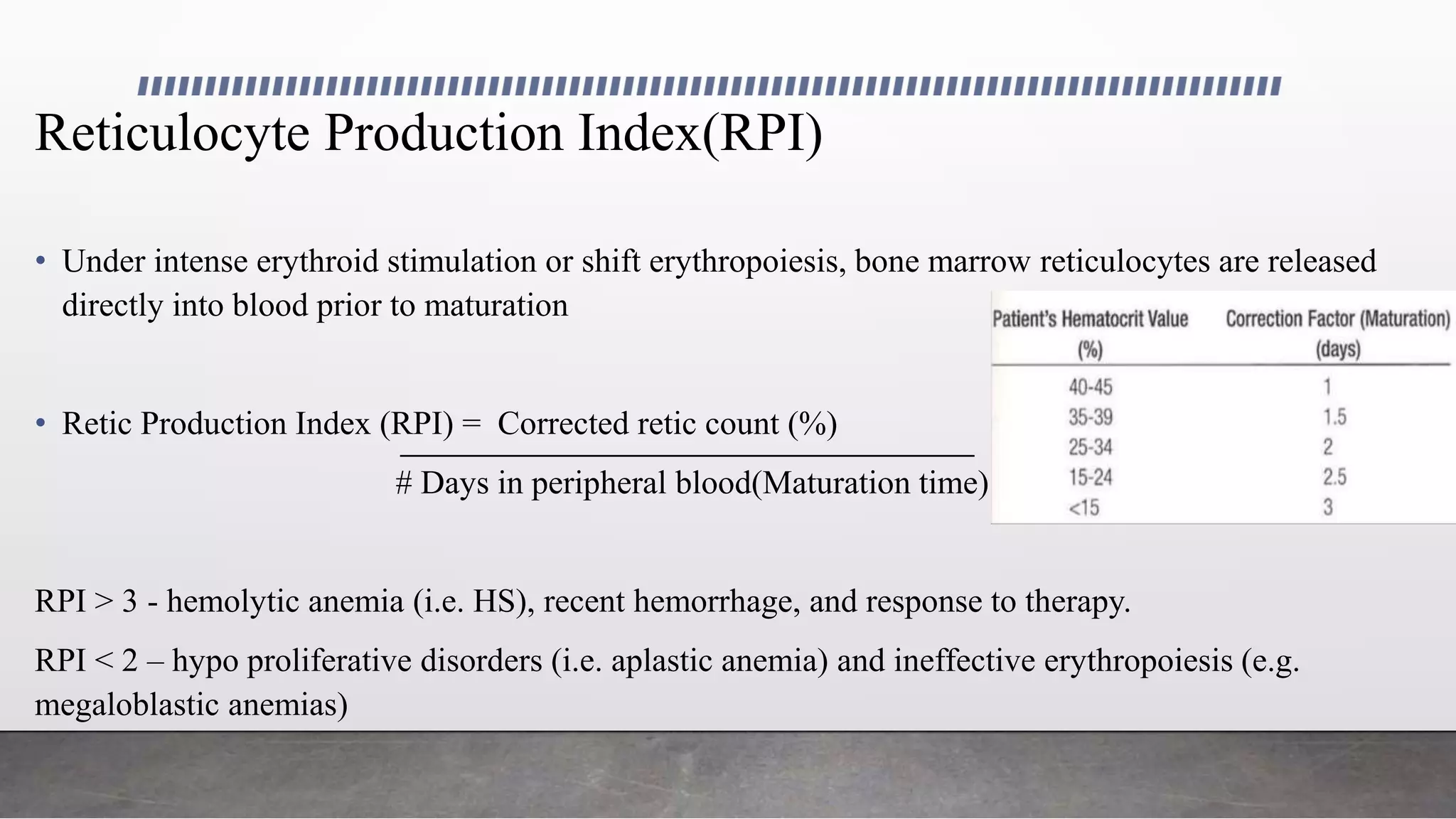
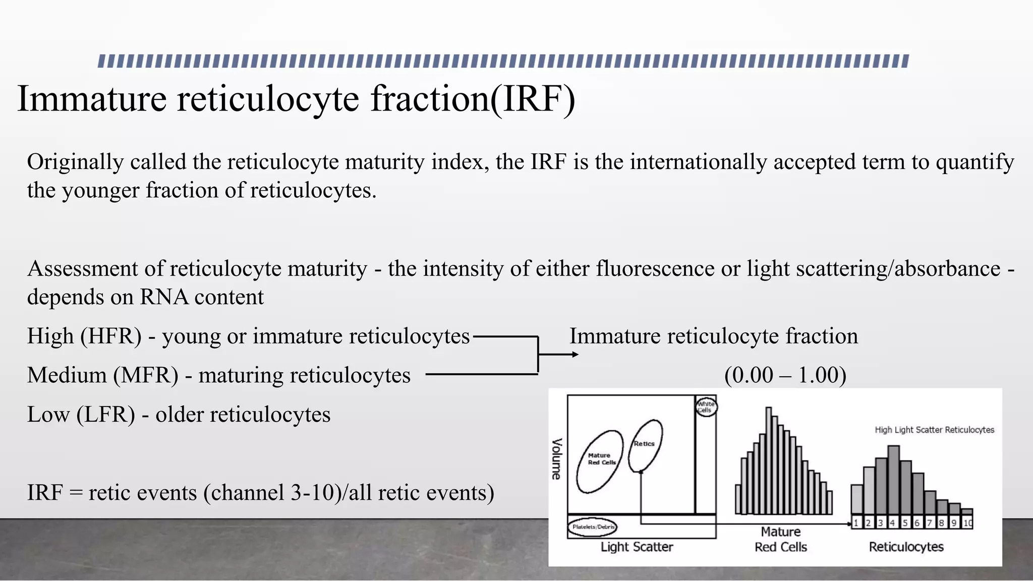
Utility of Reticulocyte Parameters
Dr. Muneerah Saeed
History of Reticulocytes
• 1865 -Heinrich Erb -“granulated” erythrocytes - Transition of WBC to RBC
• Late 1800 - Paul Ehrlich –Properties of Reticulum - senescent RBCs
• Early 1900 - Theobald Smith - young red cells
• 1922 - Edward Bell - term “reticulocyte”
• 1932 - Ludwig Heilmeyer - classification of reticulocytes maturity
Definition of Reticulocyte
• Term derived- reticulum of RNA and protein - supravital dyes
• ICSH definition - “Reticulocytes must have at least 2 blue staining granules, visible without
fine microscope adjustment and located away from the cell margin”
• Benefit - standardization of the reticulocyte morphology, disadvantage- does not take into
account fluorescent dyes
Properties
Reticulocytes, in comparison with mature erythrocytes, show:
• Greater volume: +24%
• Slightly higher Hb content: +3%
• Lower Hb concentration: -6.7%
• Constant volume ratio between reticulocytes and erythrocytes: 1.24
Reticulocyte Maturation
• Late-stage erythroblast loses its nucleus- reticulocyte
• Bone marrow for 3 days - released into circulation - maturation completed in 1 day
• Maturation – continuous process - membrane remodeling, volume changes, and elimination of
organelles and ribosomes
• Maturation is complete when the basophilic filamentous substance disappear
Group 0: nucleated erythrocyte (orthochromatic
normoblast) not included
Group I: non-nucleated, dense clumped reticulum;
0.1%
Group II: extended network of loose reticulum; 0.7%
Group III: scattered granules with residual reticulum
network;32%
Group IV: scattered granules;61%
Maturation stages of reticulocytes according to Heilmeyer
classification
Microscopic Reticulocyte Counting –Corelation Method
According to ICSH
guidelines
Supravital dye, New
Methylene Blue(NMB) is
the stain of choice, blood
films are examined under
oil immersion lens
Total number of red cells
to be counted varies with
the reticulocyte
percentage as well as the
desired precision
But in case, Millers
disc is used, a
minimum of1000
RBCs are to be
counted
Miller Disc Reticulocyte Count with
Ideal Red Cell Distribution
Retics (%) =
Total Number of Reticulocytes x 100
Total Number of Red Cells + Retics
If a Miller disc is used,
Retics (%) =
Total Number of Retics in 20 large squares x 100
Total Numbers of RBCs in 20 small squares x 9
Absolute Reticulocyte Count = Retics (%) x RBC
Automated Reticulocyte Analysis
• Precision
• Statistical error
• Interobserver variability and subjectivity
• Turn around time
• Variability resulting from staining, dilution, and incubation
Beckman Coulter- DxH 800
Addition of NMB
dye in stain
chamber and
incubation(precipi
tates and stains
RNA)
Hypo osmotic agent
added in retic
chambers (cell
swelling, hb clearing
and fixation in a
spherical form)
Submitted to a
multi transducer
module -diode laser
module, flow cell
and two optical
sensor assemblies
Flow cytometric
digital analysis using
different angles of
light-scattering
detection,
impedance,
radiofrequency
(VCSn)
The new features and parameter measurements include:
• New flow cell with new optical sensors
• Temperature controlled sample preparation and
analysis
• Seven distinct parameter measurements for each cell
passing through the flow cell
- Volume
- Conductivity
- Axial light loss (AL2)
- Low angle light scatter (LALS)
- Median angle light scatter (MALS)
- Lower median angle light scatter (LMALS)
- Upper median angle light scatter (UMALS)
Reticulocyte Parameters
Utilized parameters Research- use- only parameters
Coulter DxH800
Utilized parameters
• Reticulocyte count
• Absolute reticulocyte count
• IRF( immature reticulocyte
fraction)
• MRV (mean reticulocyte
volume)
Research- use- only parameters
• HLR (high light scatter reticulocytes)
• RDWR-CV (reticulocyte distribution
width, coefficient of variation)
• RDWR-SD (reticulocyte distribution
width, standard deviation)
• RSF (red cell size factor)
DxH-
Other
Parameters
MCHr (mean cellular Hb content of reticulocytes)
CHCr (cell hemoglobin concentration of reticulocytes)
RETH% (high-fluorescence reticulocytes), RETM% (medium-fluorescence
reticulocytes), RETL% (low fluorescence reticulocytes)
MFI% (mean fluorescence index)
RHC (reticulocyte hemoglobin cellular content)
Ret-He (reticulocyte hemoglobin equivalent)
RPI (reticulocyte production index)
Reticulocyte Count
• Retics (%) = Total Number of Reticulocytes x 100
Total Number of Red Cells + Retics
• Normal counts
• New borns : 2.5 – 6.5% (adult levels by 2 weeks)
• Adults : 0.5 – 1.5%
Inaccurate Reticulocyte Count
• Erythrocyte inclusions- Howell-Jolly bodies
Heinz bodies
Pappenheimer bodies
Parasites- Malaria & Babesia
Basophilic stippling bodies
Hb H inclusion bodies
• Cellular interferences - High number of lymphocytes
Cellular fragments
Giant platelets
Platelet/red cell coincidence
nRBC
Platelet clumps
• Auto fluorescence – drugs( anthracyclines)
Porphyria
• Paraproteins
• Cold agglutinins
• Hemolysis
Absolute Reticulocyte Count
• Absolute reticulocyte count =
Reticulocyte % x RBC count
• Normal values : 25 – 75 x 109/L
Increased reticulocyte
count
• Hemolytic anemia
• Recent hemorrhage
• Pregnancy
• Response to treatment
• Hypoxia
Decrease reticulocyte
count
• Nutritional anemia
• Aplastic anemia
• Bone marrow infiltration
• Radiation
Corrected Reticulocyte Count
Reticulocyte count should reflect total production of RBC regardless of concentration of red cells in
the blood
Reticulocyte count misleading –
Abnormal RBC count
Significant erythropoietic stimulation
Corrected reticulocyte count =
Reticulocyte % x Patient’s HCT
HCT normal for age
Reticulocyte Production Index(RPI)
• Under intense erythroid stimulation or shift erythropoiesis, bone marrow reticulocytes are released
directly into blood prior to maturation
• Retic Production Index (RPI) = Corrected retic count (%)
# Days in peripheral blood(Maturation time)
RPI > 3 - hemolytic anemia (i.e. HS), recent hemorrhage, and response to therapy.
RPI < 2 – hypo proliferative disorders (i.e. aplastic anemia) and ineffective erythropoiesis (e.g.
megaloblastic anemias)
Immature reticulocyte fraction(IRF)
Originally called the reticulocyte maturity index, the IRF is the internationally accepted term to quantify
the younger fraction of reticulocytes.
Assessment of reticulocyte maturity - the intensity of either fluorescence or light scattering/absorbance -
depends on RNA content
High (HFR) - young or immature reticulocytes Immature reticulocyte fraction
Medium (MFR) - maturing reticulocytes (0.00 – 1.00)
Low (LFR) - older reticulocytes
IRF = retic events (channel 3-10)/all retic events)
Clinical Utility of IRF
Significant clinical benefit
• hematologic recovery following chemotherapy, engraftment after bone marrow transplantation
• monitoring of recombinant erythropoietin or iron therapy
Potential use
• diagnosis of anemia (hypo proliferative , ineffective or hemolytic)
• early marker of CD34 mobilization for peripheral stem cell harvesting
• need for transfusion in patients with anemia.
Monitoring Bone Marrow Recovery Following Bone Marrow
Transplant And Following Chemotherapy
• Increase in IRF - indicator of engraftment - precedes - absolute neutrophil counts (ANC),
reticulated platelets (immature platelet fraction [IPF]), or reticulocyte counts
• Post transplantation increase in ANC to 0.5 x10⁹
/L or greater - successful myeloid engraftment
• IRF doubling time (IRF-D) - first of 2 consecutive days during which the IRF value doubles -
predict myeloid engraftment several days before ANC exceeds 0.1 x10⁹/L
IPF - useful indicator - variations - platelet transfusion and complication such as sepsis
• Strong indicator - post chemotherapy aplasia in children with cancer - additional parameter of
impaired bone marrow function
Erythropoiesis in kidney transplant and Monitoring EPO Therapy in
Patients with Chronic Renal Failure
• After transplant, the IRF ↑ by around 25% at an average of 7 days before an ↑ in the absolute
reticulocyte count
IRF - more sensitive, early indicator of erythropoiesis
• IRF -important and sensitive parameter for evaluating response to EPO therapy in pts with
CRF
• Monitoring R-EPO in anemia in AIDS, cancer, prematurity
• Monitoring treatment of nutritional anemias
Improving the Classification, Diagnosis, and Treatment of Anemias
An improvement in the classification of anemias can be accomplished when the IRF is used in combination
with the reticulocyte count
Reticulocyte Mean Cell Hemoglobin Content (CHr)
• Measurement of the Hb content of reticulocytes
• Expressed in pg/cell
• Product of the cellular volume x cellular Hb concentration
• Bayer H3 and Siemens Diagnostics
Clinical utility of CHr
• CHr < 28pg – predicted the absence of bone marrow iron stores
better negative predictive value than MCVr, serum ferritin and
transferrin saturation
• Reliable and early indicator of bone marrow iron status
• Detects functional iron deficiency with more sensitivity than biochemical parameters
• Indicates efficacy of responses to anemia treatment at an early stage
• Recent studies- CHr - evaluation iron status in young infant
Sensitive & specific marker for predicting later anemia
Early measurement- Prevent cognitive impairment
• ID in pregnant women
• Frequent blood donors - Early detection of iron depletion
Diagnostic limitations
• Decrease in thalassemia syndromes – correlated with degree of impairment in beta
chain synthesis
• Iron deficiency with cofounding megaloblastic anemia – CHr is increased – High MRV
• Increased by drugs causing transient or permanent macrocytosis ( chronic hydroxyurea
– sickle cell disease)
Red blood cell size factor(RSf)
• RSf - joins together the volume of mature red cells (MCV) and the reticulocytes (MRV) - related
to erythropoietic activity and hemoglobinization
• √MCV x MRV
• Reflects indirectly cellular Hb content of reticulocytes
• Measurement of cellular characteristics, such as volume, provides real-time data regarding
certain aspects of erythropoiesis that can influence the dimensions of red cells, i.e. iron
availability
• Examining both precursors and mature red cells - detect and monitor acute and chronic changes
in Hb status, related to cell volume
Clinical utility of the new Beckman-coulter parameter red blood cell size factor
in the study of erythropoiesis
E. URRECHAGA
Reticulocyte hemoglobin equivalent
• Ret He
• Formerly defined as reticulocyte hemoglobin parameter (RET-Y)
• Sysmex
• RET-Y demonstrated a curvilinear relationship with CHr - an alternative measure of
reticulocyte Hb
Reticulocyte Mean Volume
• MCVr, MRV
• Automated methods – light scattering - 24% higher than MCV of mature RBC
• Hematocrit of reticulocytes = MCVr X No. of reticulocytes
• Abbot laboratories , Beckman coulter
Clinical utility of reticulocyte mean volume
• Decrease – Iron deficiency, Thalassemia
• Depleted iron stores - ↑ when iron supplements are given- ↓ during iron-deficient erythropoiesis
• Increase - Macrocytic anemia
Rapid normalization with treatment - Inversion of MCVr/MCV ratio
• Sickle cell anemia- Hydroxyurea therapy- Fetal Hb synthesis- Increased hydration of sickle
reticulocytes- MCVr
Mean sphered cell volume
The average volume of all red cell events - mature red cells
and reticulocytes -Mean Sphered Cell Volume (MSCV)
mature red cells or reticulocytes
hypo-osmotic solution
Staining
MSCV> MCV
Clinical utility –hereditary spherocytosis - reach a critical level of osmotic volume expansion and then
fragment - ↓ MSCV
Relationship of the reticulocyte MSCV to the MCV - highly indicative test of HS
Diagnostic Algorithm for Microcytic Anemia
Diagnostic Algorithm for Normocytic Anema
Diagnostic Algorithm for Macrocytic Anemia
THANK YOU
References
• Wintrobes clinical hematology 13th edition
• Elisa Piva et al , Clinical utility of reticulocyte parameters, Clin Lab Med 35(2015)
• ICSH guidelines for the evaluation of blood cell analyzers including those used for differential leucocyte
and reticulocyte counting, Int. Jnl. Lab. Hem, 2014
• Beckman Coulter – The LH Reticulocyte count and associated parameters
• Beckman Coulter - Reticulocyte Methodology UniCell DxH 800 Coulter Cellular Analysis System
• Bruce H. Davis, Diagnostic Advances in Defining Erythropoietic Abnormalities and Red Blood Cell
Diseases, Semin Hematol, 2001
• M. BUTTARELLO, Laboratory diagnosis of anemia: are the old and new red cell parameters useful in
classification and treatment, how?, Int. Jnl. Lab. Hem, 2016
• E. Urechaga et al Analysis of reticulocyte parameters on sysmex XE 5000 AND LH 750 analyzers in the
diagnosis of inefficient erythropoiesis, Int. Jnl. Lab.Hem, 2011
• Dr. R. Sindhu, Role of immature reticulocyte fraction in evaluation of aplastic anemia in cases of
pancytopenia

Utility of reticulocyte parameters

  • 1.
    Utility of ReticulocyteParameters Dr. Muneerah Saeed
  • 2.
    History of Reticulocytes •1865 -Heinrich Erb -“granulated” erythrocytes - Transition of WBC to RBC • Late 1800 - Paul Ehrlich –Properties of Reticulum - senescent RBCs • Early 1900 - Theobald Smith - young red cells • 1922 - Edward Bell - term “reticulocyte” • 1932 - Ludwig Heilmeyer - classification of reticulocytes maturity
  • 3.
    Definition of Reticulocyte •Term derived- reticulum of RNA and protein - supravital dyes • ICSH definition - “Reticulocytes must have at least 2 blue staining granules, visible without fine microscope adjustment and located away from the cell margin” • Benefit - standardization of the reticulocyte morphology, disadvantage- does not take into account fluorescent dyes
  • 4.
    Properties Reticulocytes, in comparisonwith mature erythrocytes, show: • Greater volume: +24% • Slightly higher Hb content: +3% • Lower Hb concentration: -6.7% • Constant volume ratio between reticulocytes and erythrocytes: 1.24
  • 5.
    Reticulocyte Maturation • Late-stageerythroblast loses its nucleus- reticulocyte • Bone marrow for 3 days - released into circulation - maturation completed in 1 day • Maturation – continuous process - membrane remodeling, volume changes, and elimination of organelles and ribosomes • Maturation is complete when the basophilic filamentous substance disappear
  • 6.
    Group 0: nucleatederythrocyte (orthochromatic normoblast) not included Group I: non-nucleated, dense clumped reticulum; 0.1% Group II: extended network of loose reticulum; 0.7% Group III: scattered granules with residual reticulum network;32% Group IV: scattered granules;61% Maturation stages of reticulocytes according to Heilmeyer classification
  • 7.
    Microscopic Reticulocyte Counting–Corelation Method According to ICSH guidelines Supravital dye, New Methylene Blue(NMB) is the stain of choice, blood films are examined under oil immersion lens Total number of red cells to be counted varies with the reticulocyte percentage as well as the desired precision But in case, Millers disc is used, a minimum of1000 RBCs are to be counted
  • 8.
    Miller Disc ReticulocyteCount with Ideal Red Cell Distribution Retics (%) = Total Number of Reticulocytes x 100 Total Number of Red Cells + Retics If a Miller disc is used, Retics (%) = Total Number of Retics in 20 large squares x 100 Total Numbers of RBCs in 20 small squares x 9 Absolute Reticulocyte Count = Retics (%) x RBC
  • 10.
    Automated Reticulocyte Analysis •Precision • Statistical error • Interobserver variability and subjectivity • Turn around time • Variability resulting from staining, dilution, and incubation
  • 11.
    Beckman Coulter- DxH800 Addition of NMB dye in stain chamber and incubation(precipi tates and stains RNA) Hypo osmotic agent added in retic chambers (cell swelling, hb clearing and fixation in a spherical form) Submitted to a multi transducer module -diode laser module, flow cell and two optical sensor assemblies Flow cytometric digital analysis using different angles of light-scattering detection, impedance, radiofrequency (VCSn)
  • 12.
    The new featuresand parameter measurements include: • New flow cell with new optical sensors • Temperature controlled sample preparation and analysis • Seven distinct parameter measurements for each cell passing through the flow cell - Volume - Conductivity - Axial light loss (AL2) - Low angle light scatter (LALS) - Median angle light scatter (MALS) - Lower median angle light scatter (LMALS) - Upper median angle light scatter (UMALS)
  • 13.
    Reticulocyte Parameters Utilized parametersResearch- use- only parameters Coulter DxH800 Utilized parameters • Reticulocyte count • Absolute reticulocyte count • IRF( immature reticulocyte fraction) • MRV (mean reticulocyte volume) Research- use- only parameters • HLR (high light scatter reticulocytes) • RDWR-CV (reticulocyte distribution width, coefficient of variation) • RDWR-SD (reticulocyte distribution width, standard deviation) • RSF (red cell size factor)
  • 14.
  • 15.
    Other Parameters MCHr (mean cellularHb content of reticulocytes) CHCr (cell hemoglobin concentration of reticulocytes) RETH% (high-fluorescence reticulocytes), RETM% (medium-fluorescence reticulocytes), RETL% (low fluorescence reticulocytes) MFI% (mean fluorescence index) RHC (reticulocyte hemoglobin cellular content) Ret-He (reticulocyte hemoglobin equivalent) RPI (reticulocyte production index)
  • 16.
    Reticulocyte Count • Retics(%) = Total Number of Reticulocytes x 100 Total Number of Red Cells + Retics • Normal counts • New borns : 2.5 – 6.5% (adult levels by 2 weeks) • Adults : 0.5 – 1.5%
  • 17.
    Inaccurate Reticulocyte Count •Erythrocyte inclusions- Howell-Jolly bodies Heinz bodies Pappenheimer bodies Parasites- Malaria & Babesia Basophilic stippling bodies Hb H inclusion bodies
  • 18.
    • Cellular interferences- High number of lymphocytes Cellular fragments Giant platelets Platelet/red cell coincidence nRBC Platelet clumps • Auto fluorescence – drugs( anthracyclines) Porphyria • Paraproteins • Cold agglutinins • Hemolysis
  • 19.
    Absolute Reticulocyte Count •Absolute reticulocyte count = Reticulocyte % x RBC count • Normal values : 25 – 75 x 109/L
  • 20.
    Increased reticulocyte count • Hemolyticanemia • Recent hemorrhage • Pregnancy • Response to treatment • Hypoxia Decrease reticulocyte count • Nutritional anemia • Aplastic anemia • Bone marrow infiltration • Radiation
  • 21.
    Corrected Reticulocyte Count Reticulocytecount should reflect total production of RBC regardless of concentration of red cells in the blood Reticulocyte count misleading – Abnormal RBC count Significant erythropoietic stimulation Corrected reticulocyte count = Reticulocyte % x Patient’s HCT HCT normal for age
  • 22.
    Reticulocyte Production Index(RPI) •Under intense erythroid stimulation or shift erythropoiesis, bone marrow reticulocytes are released directly into blood prior to maturation • Retic Production Index (RPI) = Corrected retic count (%) # Days in peripheral blood(Maturation time) RPI > 3 - hemolytic anemia (i.e. HS), recent hemorrhage, and response to therapy. RPI < 2 – hypo proliferative disorders (i.e. aplastic anemia) and ineffective erythropoiesis (e.g. megaloblastic anemias)
  • 23.
    Immature reticulocyte fraction(IRF) Originallycalled the reticulocyte maturity index, the IRF is the internationally accepted term to quantify the younger fraction of reticulocytes. Assessment of reticulocyte maturity - the intensity of either fluorescence or light scattering/absorbance - depends on RNA content High (HFR) - young or immature reticulocytes Immature reticulocyte fraction Medium (MFR) - maturing reticulocytes (0.00 – 1.00) Low (LFR) - older reticulocytes IRF = retic events (channel 3-10)/all retic events)
  • 24.
    Clinical Utility ofIRF Significant clinical benefit • hematologic recovery following chemotherapy, engraftment after bone marrow transplantation • monitoring of recombinant erythropoietin or iron therapy Potential use • diagnosis of anemia (hypo proliferative , ineffective or hemolytic) • early marker of CD34 mobilization for peripheral stem cell harvesting • need for transfusion in patients with anemia.
  • 25.
    Monitoring Bone MarrowRecovery Following Bone Marrow Transplant And Following Chemotherapy • Increase in IRF - indicator of engraftment - precedes - absolute neutrophil counts (ANC), reticulated platelets (immature platelet fraction [IPF]), or reticulocyte counts • Post transplantation increase in ANC to 0.5 x10⁹ /L or greater - successful myeloid engraftment • IRF doubling time (IRF-D) - first of 2 consecutive days during which the IRF value doubles - predict myeloid engraftment several days before ANC exceeds 0.1 x10⁹/L IPF - useful indicator - variations - platelet transfusion and complication such as sepsis • Strong indicator - post chemotherapy aplasia in children with cancer - additional parameter of impaired bone marrow function
  • 26.
    Erythropoiesis in kidneytransplant and Monitoring EPO Therapy in Patients with Chronic Renal Failure • After transplant, the IRF ↑ by around 25% at an average of 7 days before an ↑ in the absolute reticulocyte count IRF - more sensitive, early indicator of erythropoiesis • IRF -important and sensitive parameter for evaluating response to EPO therapy in pts with CRF • Monitoring R-EPO in anemia in AIDS, cancer, prematurity • Monitoring treatment of nutritional anemias
  • 27.
    Improving the Classification,Diagnosis, and Treatment of Anemias An improvement in the classification of anemias can be accomplished when the IRF is used in combination with the reticulocyte count
  • 29.
    Reticulocyte Mean CellHemoglobin Content (CHr) • Measurement of the Hb content of reticulocytes • Expressed in pg/cell • Product of the cellular volume x cellular Hb concentration • Bayer H3 and Siemens Diagnostics
  • 30.
    Clinical utility ofCHr • CHr < 28pg – predicted the absence of bone marrow iron stores better negative predictive value than MCVr, serum ferritin and transferrin saturation • Reliable and early indicator of bone marrow iron status • Detects functional iron deficiency with more sensitivity than biochemical parameters
  • 31.
    • Indicates efficacyof responses to anemia treatment at an early stage • Recent studies- CHr - evaluation iron status in young infant Sensitive & specific marker for predicting later anemia Early measurement- Prevent cognitive impairment • ID in pregnant women • Frequent blood donors - Early detection of iron depletion
  • 32.
    Diagnostic limitations • Decreasein thalassemia syndromes – correlated with degree of impairment in beta chain synthesis • Iron deficiency with cofounding megaloblastic anemia – CHr is increased – High MRV • Increased by drugs causing transient or permanent macrocytosis ( chronic hydroxyurea – sickle cell disease)
  • 33.
    Red blood cellsize factor(RSf) • RSf - joins together the volume of mature red cells (MCV) and the reticulocytes (MRV) - related to erythropoietic activity and hemoglobinization • √MCV x MRV • Reflects indirectly cellular Hb content of reticulocytes • Measurement of cellular characteristics, such as volume, provides real-time data regarding certain aspects of erythropoiesis that can influence the dimensions of red cells, i.e. iron availability • Examining both precursors and mature red cells - detect and monitor acute and chronic changes in Hb status, related to cell volume
  • 34.
    Clinical utility ofthe new Beckman-coulter parameter red blood cell size factor in the study of erythropoiesis E. URRECHAGA
  • 35.
    Reticulocyte hemoglobin equivalent •Ret He • Formerly defined as reticulocyte hemoglobin parameter (RET-Y) • Sysmex • RET-Y demonstrated a curvilinear relationship with CHr - an alternative measure of reticulocyte Hb
  • 36.
    Reticulocyte Mean Volume •MCVr, MRV • Automated methods – light scattering - 24% higher than MCV of mature RBC • Hematocrit of reticulocytes = MCVr X No. of reticulocytes • Abbot laboratories , Beckman coulter
  • 37.
    Clinical utility ofreticulocyte mean volume • Decrease – Iron deficiency, Thalassemia • Depleted iron stores - ↑ when iron supplements are given- ↓ during iron-deficient erythropoiesis • Increase - Macrocytic anemia Rapid normalization with treatment - Inversion of MCVr/MCV ratio • Sickle cell anemia- Hydroxyurea therapy- Fetal Hb synthesis- Increased hydration of sickle reticulocytes- MCVr
  • 38.
    Mean sphered cellvolume The average volume of all red cell events - mature red cells and reticulocytes -Mean Sphered Cell Volume (MSCV) mature red cells or reticulocytes hypo-osmotic solution Staining MSCV> MCV Clinical utility –hereditary spherocytosis - reach a critical level of osmotic volume expansion and then fragment - ↓ MSCV Relationship of the reticulocyte MSCV to the MCV - highly indicative test of HS
  • 39.
    Diagnostic Algorithm forMicrocytic Anemia
  • 40.
    Diagnostic Algorithm forNormocytic Anema
  • 41.
    Diagnostic Algorithm forMacrocytic Anemia
  • 42.
  • 43.
    References • Wintrobes clinicalhematology 13th edition • Elisa Piva et al , Clinical utility of reticulocyte parameters, Clin Lab Med 35(2015) • ICSH guidelines for the evaluation of blood cell analyzers including those used for differential leucocyte and reticulocyte counting, Int. Jnl. Lab. Hem, 2014 • Beckman Coulter – The LH Reticulocyte count and associated parameters • Beckman Coulter - Reticulocyte Methodology UniCell DxH 800 Coulter Cellular Analysis System • Bruce H. Davis, Diagnostic Advances in Defining Erythropoietic Abnormalities and Red Blood Cell Diseases, Semin Hematol, 2001 • M. BUTTARELLO, Laboratory diagnosis of anemia: are the old and new red cell parameters useful in classification and treatment, how?, Int. Jnl. Lab. Hem, 2016 • E. Urechaga et al Analysis of reticulocyte parameters on sysmex XE 5000 AND LH 750 analyzers in the diagnosis of inefficient erythropoiesis, Int. Jnl. Lab.Hem, 2011 • Dr. R. Sindhu, Role of immature reticulocyte fraction in evaluation of aplastic anemia in cases of pancytopenia

Editor's Notes

  • #3 The first description o r w m in 18 … , he discovered a population of “g e. he however, regarded them as … In The Lat 1800s, Nobel Prize winner P E desc the prop of the gran baso substance of reticulocytes and considered them …In early 1900, it was T S who asserted that reticulocytes represented …. In 1922..in1932 Heil proposed the still well-known classification of …
  • #4 The reticulocyte derives its name from the …. precipitated by staining with supra … The reticulum network represent rough endoplasmic reticulum with associated polyribosomes. The icsh defin states that… The benefit of this official definition is the stand…. , but it does not take into account the wide use of fluorescent dyes presently being used
  • #6 When the late-…., the cell becomes a reticulocyte that usually remains in the b m …. and is subsequently released into the circulation, Hb synthesis occurs only in the younger retic….., circulating reticulocytes are unable to synthesize Hb
  • #7 Based on the content of mRNA……Comprise 0.1% of the population of reticulocytes
  • #8 remains the recommended method for correlation when evaluating the performances of automated techniques…… Equal volumes of NMB stain and blood are mixed in a test tube and incubated. After staining a slide is made and examined under oil immersion
  • #9 This is a table showing number of rbcs to be counted. With a retic % of 0.5 and medium precision of cv 5% minimum 400 cells are to be counted. Similarly ... The formula is This is a picture of millers disc. In the small square rbcs are counted including retics. In large square retics are counted including the small square. Cells of upper and left margins are included in both the squares. a total of 20 fields are counted.
  • #10 This is a picture of ps showing Reticulocytes in romanowski stain, they have a bluish tinge and are comparatively larger and blood film with supravital stain, they have blue staining granules
  • #11 Increases prec.. Reduces stat error, inter and intra… the tat and the variab
  • #13 Volume about size conductivity about internal characteristics and scatter about cell surface characteristics and granularity This is a picture of reticulocyte dataplot showing a population of reticulocytes
  • #16 The reticulocyte parameters measured in other analyzers are
  • #18 Hj bodies and pappenheimer bodies both get stained by romanowski stain while reticulum of reticulocyte does not Heinz body in supravital stain appear as lighter blue green inclusions located at the periphery of the cell
  • #19 The machine can miscalculate reticulocyte count due to
  • #20 Increased retic count measures the …
  • #23 Rpi Corrects increased time of the reticulocyte spent in maturation in the peripheral blood
  • #24  IRF is taken as the sum of the populations of high and medium stain intensity. The expression should be as a fraction (0.00–1.00). In Beckman coulter Irf in coulter is
  • #25 following growth factor mobilization or cytotoxic drug therapy
  • #26 Increases in IRF is an indicator of engraftment that precedes other parameters, such as (ANC), reticulated platelets[IPF]), or reticulocyte count Typically post tran… is considered as a. IRF-D defined as first… can predict… ipf although a useful indicator of megakaryocyte production can exhibitiation variation due to
  • #30 These parameters are available in
  • #31 CHr – Functional availability of iron in that time frame
  • #33 In megaloblastosis
  • #34 Provides opportunity
  • #35 Positive linear correlation of 85 per cent chr and rsf mean was calculated in reference group and cut-off value was applied to the validation group, 100% of the b-thalassemia carriers were correctly classified, with the lower RSf values.23 IDA HAD value lower than 87.7 and could not be correctly classified All CKD showed RSF > 87.7 fl. Twenty-three percent of the IDA patients had RSf >87.7 and 2% of ACD patients had RSf < 87.7 fl.
  • #36 Analysed by
  • #38 In response to B12 treatment, the reticulocyte volume is becomes smaller while the mean cell volume takes more time to normalize Owing to their different life spans. An inversion of the MCVr/MCV ratio occurs MCVr has been measured in sickle cell anemia: induction of fetal Hb synthesis by hydroxyurea therapy was associated with increased hydration of sickle reticulocytes, assessed as MCVr
  • #39  As explained for reticulocyte analysis the blood… …As expected, … bcoz of the biconcave shape of rbc..HS is a disorder with red cell structural protein defects with inc OF. Spherocytes reach a critical level of osmotic volume expansion and then fragment, resulting in a lower than expected MSCV
  • #40 Then it can be further investigated