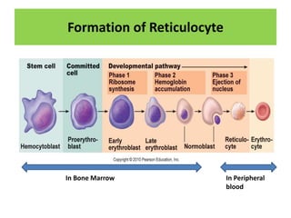
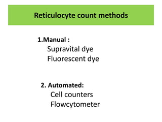
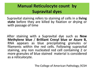
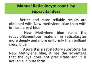
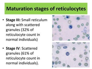
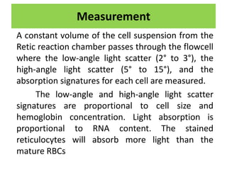
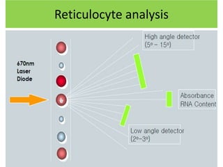
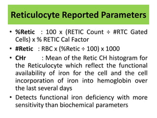
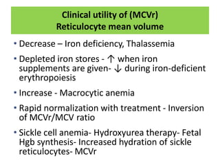
RETICULOCYTE
ASHA KUMARI
HAEMATOPATHOLOGY, TMH
What is Reticulocyte ?
Reticulocytes are young or juvenile ,
immature red cells released from
the bone marrow into the
bloodstream and that contain
remnants of ribonucleic acid (RNA),
protoporphyrin, ribosome's.
Formation of Reticulocyte
In Bone Marrow In Peripheral
blood
Why Reticulocyte count ?
• The number of Reticulocytes in the peripheral
blood is a fairly accurate reflection of
erythropoietic activity
• To diagnose anemia due to ineffective
erythropoiesis
• To assess response to specific therapy in iron
deficiency and megaloblastic anemia's
• Bone marrow recovery (after bone marrow
transplantation or intensive chemotherapy)
• To assess response to erythropoietin therapy
in anemia of chronic renal failure.
• Red cell production and regeneration
Reticulocyte count methods
1.Manual :
Supravital dye
Fluorescent dye
2. Automated:
Cell counters
Flowcytometer
Specimen collection and Storage
 K2EDTA is the anticoagulant of choice
 Venous or capillary blood
 At Room Temperature- within six hours after
blood collection.
 If sample analysis is delayed, the sample
should be refrigerated. Samples stored at 2 to
8 °C may be stable for up to 72 hours.
NCCLS guidelines H44-A2 Vol. 24 No. 8,2004
Manual Reticulocyte count by
Supravital dyes
Supravital staining refers to staining of cells in a living
state before they are killed by fixation or drying or
with passage of time
After staining with a Supravital dye such as New
Methylene blue / Brilliant Cresyl blue or Azure B,
RNA appears as blue precipitating granules or
filaments within the red cells. Following supravital
staining, any non nucleated red cell containing 2 or
more granules of blue stained material is considered
as a reticulocyte.
The College of American Pathology /ICSH
Manual Reticulocyte count by
Supravital dyes
Better and more reliable results are
obtained with New methylene blue than with
brilliant cresyl blue
New Methylene blue stains the
reticulofilamentous material in reticulocytes
more deeply and more uniformly than brilliant
cresyl blue
Azure B is a satisfactory substitute for
New Methylene blue, it has the advantage
that the dye does not precipitate and it is
available in pure form
PRINCIPLE
A equal volume of blood (collected in EDTA)
are incubated at 370C with Brilliant cresyl blue
solution which stains granules of RNA in red
cells. A thin smear is prepared on a glass slide
from the mixture and reticulocytes are
counted under the microscope. Number of
reticulocytes is expressed as a percentage of
red cells
Staining solution
1% Brilliant cresyl blue or 1% New methylene blue
• Dissolve 1gm of BCB /NMB in 100ml of iso-osmotic
phosphate buffer
• Iso osmotic phosphate buffer : pH 6.5
(A) NaH2PO4 2H2O (150mmol/L) 23.4gm/L
+
(B) Na2HPO4 (150mmol/L) 21.3gm/L
Mix 64 ml of A + 36 ml of B
• Check pH
• Filter with Whatmann’s No.1 filter
• Keep in dry bottle
• Stable for 4 – 6 weeks
Procedure
• Mix equal quantities of EDTA anticoagulated
blood and dye solution
• The exact volume of blood to be added to the
dye solution for optimal staining depends on
the RBC count.
• A larger proportion of anemic blood, and a
smaller proportion of polycythemic blood,
should be added than of normal blood
Procedure
 Incubate the mixture at 370C in water
bath for 15-20 mins
 Resuspend the red cells by gentle mixing
 Make films on glass slides in the usual way
 When dry, Examine the films under the
microscope using oil-immersion objective
 In a successful preparation, the
reticulofilamentous material stained deep
blue .
Reticulocyte
on Romanowsky stains:
Reticulocytes appear
as Polychromatophilic
(grey/blue) cells
Maturation stages of reticulocytes
• Stage I: Dense
cohesive reticulum in
non-nucleated red
cell (0.1% of
Reticulocyte count in
normal individuals)
• Stage II: Extensive
network of loose
reticulum
(0.7% of reticulocyte count
in normal individuals)
Heilmeyer and Westhaeuser, 1932
Maturation stages of reticulocytes
• Stage III: Small reticulum
along with scattered
granules (32% of
reticulocyte count in
normal individuals)
• Stage IV: Scattered
granules (61% of
reticulocyte count in
normal individuals).
Calculation
• There are two commonly used methods for
counting reticulocytes.
1. Standard counting procedure
2. Miller Reticle procedure
Standard counting procedure
 Dry smear is examined under oil immersion.
 Total of 1,000 RBCs are counted.
 Number of reticulocytes are recorded per
1,000 RBCs.
 Calculate the percentage of Reticulocyte and
absolute Reticulocyte count.
Miller Reticle procedure
Reticulocyte Count % =
Total reticulocytes counted in square A × 100
Total red cells counted in square B × 9
Miller ocular disk to facilitate
counting of reticulocytes.
The Miller ocular disk contains
two squares: A and B. Square
A is 9 times larger than square B.
Square B is used for counting
red cells, while reticulocytes are
counted in square A
• Reticulocyte count (%)= no. of Retic x100/1000 RBC
• Absolute Reticulocyte count (ARC) is the actual number
of reticulocytes in 1L of whole blood
Absolute Reticulocyte count = Retic % × RBC
• Reticulocyte Production Index (RPI) = Corrected Retic count
(%) ÷ # Days (Maturation time)
Reticulocyte Index(corrected
Reticulocyte count)
• This is a corrected reticulocyte count used in
anemias. In anemias, a falsely elevated reticulocyte
count can occur. A decrease in RBCs automatically
increases the reticulocyte count even if
erythropoiesis has not taken place because the
count represents a percentage in comparison to
RBCs. It is possible the appropriate increase of
erythropoiesis has not taken place in response to
the anaemia. This value, therefore, corrects for this
issue.
corrected reticulocyte count > 2% indicates reticulocyte release
appropriate for the degree of anemia.
If < 2%, reticulocyte release is inappropriate
Difference between Reticulocytes and other
red cell inclusions
Heinz bodies: These are precipitated globulin
Chains attached to the red cell membrane. They
stain light blue and are seen in glucose-6-phosphate
dehydrogenase deficiency following exposure to
oxidant stress.
Pappenheimer bodies:
These are iron-containing
granules which appear as
one or more small dots
in red cells.
Hb H inclusions: These are round,
golf ball like inclusion bodies seen in
α-thalassemias. Stain greenish blue
These can be easily differentiated
from reticulofilamentous material
• Howell-Jolly bodies: These are
nuclear remnants in
red cells seen in certain anemias
and following
splenectomy. As supravital dyes
stain both DNA
and RNA, these structures are also
stained.
• Granules of DYE: superimposed
on Red cells, if stain is not
filtered before use.
REFERENCE RANGE
Reticulocyte percentage:
Adults and Children : 0.5-2.5%
At birth or cord blood : 2 - 5 %
Dacie and Lewis Practical Hematology Twelfth edition 2017
Causes of increased reticulocyte
• High altitudes
• Hemolytic anemias
• Blood loss
• Following specific therapy of nutritional
anemia
(like iron in iron deficiency anemia, folate in
Folate deficiency anemia, Vit B12 in B12
deficiency anemia)
• Hemoglobinopathies, e.g. sickle cell anemia.
Causes of decreased reticulocyte
• Aplastic anemia and pure red cell aplasia
• Bone marrow infiltration (leukemia, lymphoma,
myelofibrosis, metastatic malignancy)
• Renal disease
• Anemia of chronic disease
• Alcoholism
• Myxedema
• Ineffective erythropoiesis: Megaloblastic anemia,
sideroblastic anemia, thalassemia, myelodysplasia
• Following blood transfusion
Fluorescence method
Reticulocytes can be counted manually by
fluorescence microscopy on appropriately stained
films.
Add 1 volume of acridine orange solution (50
mg/100 ml of 9 g/L NaCl) to 1 volume of blood. Mix
gently for 2 min; make films on glass slides, dry
rapidly and examine with a fluorescence microscope.
RNA gives an orange–red fluorescence, whereas
nuclear material (deoxyribonucleic acid, DNA)
fluoresces yellow
Although the amount of fluorescence
is proportional to the amount of RNA
Automated methods (Cell counters)
SIEMENS ADVIA 2120i
The ADVIA auto RETIC reagent
contains a zwitter ionic detergent
(surfactant) that isovolumetrically
spheres the red cells. It also
contains a cationic dye, Oxazine
750, that stains cells according to
their RNA content.
Measurement
A constant volume of the cell suspension from the
Retic reaction chamber passes through the flowcell
where the low-angle light scatter (2° to 3°), the
high-angle light scatter (5° to 15°), and the
absorption signatures for each cell are measured.
The low-angle and high-angle light scatter
signatures are proportional to cell size and
hemoglobin concentration. Light absorption is
proportional to RNA content. The stained
reticulocytes will absorb more light than the
mature RBCs
Reticulocyte analysis
The RETIC Scatter cytogram is the graphical representation of the
absorption and light-scatter measurements:
the high-gain, absorption (cell maturation) is plotted along the x axis
and the high-angle, low-gain light scatter (cell size) is
plotted along the y axis.
1 RTC Platelet threshold
2 RTC Coincidence threshold
3 RTC threshold
4 Low/Medium RTC threshold
5 Medium/High RTC threshold
A Mature RBCs
B Low absorption Retics
C Medium absorption Retics
D High absorption Retics
E Platelets
F Coincidence events
Reticulocyte Reported Parameters
• % RETIC
• # RETIC
• CHr
• MCVr
• CHCMr
Reticulocyte Reported Parameters
• %Retic : 100 x (RETIC Count ÷ #RTC Gated
Cells) x % RETIC Cal Factor
• #Retic : RBC x (%Retic ÷ 100) x 1000
• CHr : Mean of the Retic CH histogram for
the Reticulocyte which reflect the functional
availability of iron for the cell and the cell
incorporation of iron into hemoglobin over
the last several days
• Detects functional iron deficiency with more
sensitivity than biochemical parameters
Immature Reticulocyte Fraction (IRF)
• IRF helpful in evaluating marrow
erythropoietic response to anemia, in patient
receiving human recombinant
erythropoietin(rHuEPO) or iron therapy
• IRF earliest marker of marrow response it
increases before retic count ,hemoglobin and
hematocrit, Rbc count
• After marrow transplantation an increased IRF
has been observed as one of the first sign of
cell recovery
Clinical utility of (MCVr)
Reticulocyte mean volume
• Decrease – Iron deficiency, Thalassemia
• Depleted iron stores - ↑ when iron
supplements are given- ↓ during iron-deficient
erythropoiesis
• Increase - Macrocytic anemia
• Rapid normalization with treatment - Inversion
of MCVr/MCV ratio
• Sickle cell anemia- Hydroxyurea therapy- Fetal
Hgb synthesis- Increased hydration of sickle
reticulocytes- MCVr
BECKMEN COULTER DxH 800
• Reticulocyte analysis uses new methylene
blue stain to identify reticulated red cells by
precipitating the residual RNA.
• A portion of the blood sample is diluted and
treated with a hypo-osmotic ghosting solution
to clear the red cells of hemoglobin while
preserving the stained RNA contained within
reticulocytes.
Reticulocyte immaturity is related to cell volume
and light scatter. Since more immature
reticulocytes are larger, contain more RNA and
cause increased light scatter, the cell volume and
light scatter will increase with immaturity of the
cell
 Volume (V) is
plotted on the y-
axis,
 Linear light scatter
(llsn) is plotted on
the x-axis.
Measurement
The Dataplot shows mature red cells and
Reticulocytes
1.RBC
2.RETIC
3.OTHER
4.WBC
REFERENCE
• Dacie and Lewis Practical Hematology Twelfth
edition 2017
• S B McKenzie and J L Williams Clinical
Laboratory Hematology 3rd edition
• Siemens ADVIA 2120I Operator’s manual
• Beckman coulter DxH800 Operator’s manual
TANK YOU

reticulocyte.pptx

  • 1.
  • 2.
    What is Reticulocyte? Reticulocytes are young or juvenile , immature red cells released from the bone marrow into the bloodstream and that contain remnants of ribonucleic acid (RNA), protoporphyrin, ribosome's.
  • 3.
    Formation of Reticulocyte InBone Marrow In Peripheral blood
  • 4.
    Why Reticulocyte count? • The number of Reticulocytes in the peripheral blood is a fairly accurate reflection of erythropoietic activity • To diagnose anemia due to ineffective erythropoiesis • To assess response to specific therapy in iron deficiency and megaloblastic anemia's • Bone marrow recovery (after bone marrow transplantation or intensive chemotherapy) • To assess response to erythropoietin therapy in anemia of chronic renal failure. • Red cell production and regeneration
  • 5.
    Reticulocyte count methods 1.Manual: Supravital dye Fluorescent dye 2. Automated: Cell counters Flowcytometer
  • 6.
    Specimen collection andStorage  K2EDTA is the anticoagulant of choice  Venous or capillary blood  At Room Temperature- within six hours after blood collection.  If sample analysis is delayed, the sample should be refrigerated. Samples stored at 2 to 8 °C may be stable for up to 72 hours. NCCLS guidelines H44-A2 Vol. 24 No. 8,2004
  • 7.
    Manual Reticulocyte countby Supravital dyes Supravital staining refers to staining of cells in a living state before they are killed by fixation or drying or with passage of time After staining with a Supravital dye such as New Methylene blue / Brilliant Cresyl blue or Azure B, RNA appears as blue precipitating granules or filaments within the red cells. Following supravital staining, any non nucleated red cell containing 2 or more granules of blue stained material is considered as a reticulocyte. The College of American Pathology /ICSH
  • 8.
    Manual Reticulocyte countby Supravital dyes Better and more reliable results are obtained with New methylene blue than with brilliant cresyl blue New Methylene blue stains the reticulofilamentous material in reticulocytes more deeply and more uniformly than brilliant cresyl blue Azure B is a satisfactory substitute for New Methylene blue, it has the advantage that the dye does not precipitate and it is available in pure form
  • 9.
    PRINCIPLE A equal volumeof blood (collected in EDTA) are incubated at 370C with Brilliant cresyl blue solution which stains granules of RNA in red cells. A thin smear is prepared on a glass slide from the mixture and reticulocytes are counted under the microscope. Number of reticulocytes is expressed as a percentage of red cells
  • 10.
    Staining solution 1% Brilliantcresyl blue or 1% New methylene blue • Dissolve 1gm of BCB /NMB in 100ml of iso-osmotic phosphate buffer • Iso osmotic phosphate buffer : pH 6.5 (A) NaH2PO4 2H2O (150mmol/L) 23.4gm/L + (B) Na2HPO4 (150mmol/L) 21.3gm/L Mix 64 ml of A + 36 ml of B • Check pH • Filter with Whatmann’s No.1 filter • Keep in dry bottle • Stable for 4 – 6 weeks
  • 11.
    Procedure • Mix equalquantities of EDTA anticoagulated blood and dye solution • The exact volume of blood to be added to the dye solution for optimal staining depends on the RBC count. • A larger proportion of anemic blood, and a smaller proportion of polycythemic blood, should be added than of normal blood
  • 12.
    Procedure  Incubate themixture at 370C in water bath for 15-20 mins  Resuspend the red cells by gentle mixing  Make films on glass slides in the usual way  When dry, Examine the films under the microscope using oil-immersion objective  In a successful preparation, the reticulofilamentous material stained deep blue .
  • 13.
  • 14.
    on Romanowsky stains: Reticulocytesappear as Polychromatophilic (grey/blue) cells
  • 15.
    Maturation stages ofreticulocytes • Stage I: Dense cohesive reticulum in non-nucleated red cell (0.1% of Reticulocyte count in normal individuals) • Stage II: Extensive network of loose reticulum (0.7% of reticulocyte count in normal individuals) Heilmeyer and Westhaeuser, 1932
  • 16.
    Maturation stages ofreticulocytes • Stage III: Small reticulum along with scattered granules (32% of reticulocyte count in normal individuals) • Stage IV: Scattered granules (61% of reticulocyte count in normal individuals).
  • 17.
    Calculation • There aretwo commonly used methods for counting reticulocytes. 1. Standard counting procedure 2. Miller Reticle procedure Standard counting procedure  Dry smear is examined under oil immersion.  Total of 1,000 RBCs are counted.  Number of reticulocytes are recorded per 1,000 RBCs.  Calculate the percentage of Reticulocyte and absolute Reticulocyte count.
  • 18.
    Miller Reticle procedure ReticulocyteCount % = Total reticulocytes counted in square A × 100 Total red cells counted in square B × 9 Miller ocular disk to facilitate counting of reticulocytes. The Miller ocular disk contains two squares: A and B. Square A is 9 times larger than square B. Square B is used for counting red cells, while reticulocytes are counted in square A
  • 19.
    • Reticulocyte count(%)= no. of Retic x100/1000 RBC • Absolute Reticulocyte count (ARC) is the actual number of reticulocytes in 1L of whole blood Absolute Reticulocyte count = Retic % × RBC • Reticulocyte Production Index (RPI) = Corrected Retic count (%) ÷ # Days (Maturation time)
  • 20.
    Reticulocyte Index(corrected Reticulocyte count) •This is a corrected reticulocyte count used in anemias. In anemias, a falsely elevated reticulocyte count can occur. A decrease in RBCs automatically increases the reticulocyte count even if erythropoiesis has not taken place because the count represents a percentage in comparison to RBCs. It is possible the appropriate increase of erythropoiesis has not taken place in response to the anaemia. This value, therefore, corrects for this issue. corrected reticulocyte count > 2% indicates reticulocyte release appropriate for the degree of anemia. If < 2%, reticulocyte release is inappropriate
  • 21.
    Difference between Reticulocytesand other red cell inclusions Heinz bodies: These are precipitated globulin Chains attached to the red cell membrane. They stain light blue and are seen in glucose-6-phosphate dehydrogenase deficiency following exposure to oxidant stress.
  • 22.
    Pappenheimer bodies: These areiron-containing granules which appear as one or more small dots in red cells. Hb H inclusions: These are round, golf ball like inclusion bodies seen in α-thalassemias. Stain greenish blue These can be easily differentiated from reticulofilamentous material
  • 23.
    • Howell-Jolly bodies:These are nuclear remnants in red cells seen in certain anemias and following splenectomy. As supravital dyes stain both DNA and RNA, these structures are also stained. • Granules of DYE: superimposed on Red cells, if stain is not filtered before use.
  • 24.
    REFERENCE RANGE Reticulocyte percentage: Adultsand Children : 0.5-2.5% At birth or cord blood : 2 - 5 % Dacie and Lewis Practical Hematology Twelfth edition 2017
  • 25.
    Causes of increasedreticulocyte • High altitudes • Hemolytic anemias • Blood loss • Following specific therapy of nutritional anemia (like iron in iron deficiency anemia, folate in Folate deficiency anemia, Vit B12 in B12 deficiency anemia) • Hemoglobinopathies, e.g. sickle cell anemia.
  • 26.
    Causes of decreasedreticulocyte • Aplastic anemia and pure red cell aplasia • Bone marrow infiltration (leukemia, lymphoma, myelofibrosis, metastatic malignancy) • Renal disease • Anemia of chronic disease • Alcoholism • Myxedema • Ineffective erythropoiesis: Megaloblastic anemia, sideroblastic anemia, thalassemia, myelodysplasia • Following blood transfusion
  • 27.
    Fluorescence method Reticulocytes canbe counted manually by fluorescence microscopy on appropriately stained films. Add 1 volume of acridine orange solution (50 mg/100 ml of 9 g/L NaCl) to 1 volume of blood. Mix gently for 2 min; make films on glass slides, dry rapidly and examine with a fluorescence microscope. RNA gives an orange–red fluorescence, whereas nuclear material (deoxyribonucleic acid, DNA) fluoresces yellow Although the amount of fluorescence is proportional to the amount of RNA
  • 28.
    Automated methods (Cellcounters) SIEMENS ADVIA 2120i The ADVIA auto RETIC reagent contains a zwitter ionic detergent (surfactant) that isovolumetrically spheres the red cells. It also contains a cationic dye, Oxazine 750, that stains cells according to their RNA content.
  • 29.
    Measurement A constant volumeof the cell suspension from the Retic reaction chamber passes through the flowcell where the low-angle light scatter (2° to 3°), the high-angle light scatter (5° to 15°), and the absorption signatures for each cell are measured. The low-angle and high-angle light scatter signatures are proportional to cell size and hemoglobin concentration. Light absorption is proportional to RNA content. The stained reticulocytes will absorb more light than the mature RBCs
  • 30.
  • 31.
    The RETIC Scattercytogram is the graphical representation of the absorption and light-scatter measurements: the high-gain, absorption (cell maturation) is plotted along the x axis and the high-angle, low-gain light scatter (cell size) is plotted along the y axis. 1 RTC Platelet threshold 2 RTC Coincidence threshold 3 RTC threshold 4 Low/Medium RTC threshold 5 Medium/High RTC threshold A Mature RBCs B Low absorption Retics C Medium absorption Retics D High absorption Retics E Platelets F Coincidence events
  • 32.
    Reticulocyte Reported Parameters •% RETIC • # RETIC • CHr • MCVr • CHCMr
  • 33.
    Reticulocyte Reported Parameters •%Retic : 100 x (RETIC Count ÷ #RTC Gated Cells) x % RETIC Cal Factor • #Retic : RBC x (%Retic ÷ 100) x 1000 • CHr : Mean of the Retic CH histogram for the Reticulocyte which reflect the functional availability of iron for the cell and the cell incorporation of iron into hemoglobin over the last several days • Detects functional iron deficiency with more sensitivity than biochemical parameters
  • 34.
    Immature Reticulocyte Fraction(IRF) • IRF helpful in evaluating marrow erythropoietic response to anemia, in patient receiving human recombinant erythropoietin(rHuEPO) or iron therapy • IRF earliest marker of marrow response it increases before retic count ,hemoglobin and hematocrit, Rbc count • After marrow transplantation an increased IRF has been observed as one of the first sign of cell recovery
  • 35.
    Clinical utility of(MCVr) Reticulocyte mean volume • Decrease – Iron deficiency, Thalassemia • Depleted iron stores - ↑ when iron supplements are given- ↓ during iron-deficient erythropoiesis • Increase - Macrocytic anemia • Rapid normalization with treatment - Inversion of MCVr/MCV ratio • Sickle cell anemia- Hydroxyurea therapy- Fetal Hgb synthesis- Increased hydration of sickle reticulocytes- MCVr
  • 36.
    BECKMEN COULTER DxH800 • Reticulocyte analysis uses new methylene blue stain to identify reticulated red cells by precipitating the residual RNA. • A portion of the blood sample is diluted and treated with a hypo-osmotic ghosting solution to clear the red cells of hemoglobin while preserving the stained RNA contained within reticulocytes.
  • 37.
    Reticulocyte immaturity isrelated to cell volume and light scatter. Since more immature reticulocytes are larger, contain more RNA and cause increased light scatter, the cell volume and light scatter will increase with immaturity of the cell  Volume (V) is plotted on the y- axis,  Linear light scatter (llsn) is plotted on the x-axis. Measurement
  • 38.
    The Dataplot showsmature red cells and Reticulocytes 1.RBC 2.RETIC 3.OTHER 4.WBC
  • 39.
    REFERENCE • Dacie andLewis Practical Hematology Twelfth edition 2017 • S B McKenzie and J L Williams Clinical Laboratory Hematology 3rd edition • Siemens ADVIA 2120I Operator’s manual • Beckman coulter DxH800 Operator’s manual
  • 40.