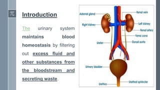
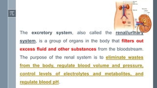
Excretory System
Ms. Devangi Sharma
Question quiz…
› Name the parts of excretory system
› Kidney weight is …………….. gm
› Length and width of kidney are ……….. & …………….
› Shape of kidney is …………
› Left superior associated organ is …………
› Right inferior associated organ is ……………
› Kidneys are located at posterior/anterior abdominal wall
› What includes the gross structure of kidney?
› Smallest functional unit of kidney is ………………..
› Difference between afferent and efferent arteriole
› Bowman’s capsule
Learning Objectives
1. Label structures of the urinary system
2. Characterize the roles of each of the parts of the urinary system
3. Illustrate the macroscopic and microscopic structures of the kidney
4. Describe the composition of urine
5. Trace the flow of blood through the kidney
6. Outline how blood is filtered in the kidney nephron
7. List some of the solutes filtered, secreted, and reabsorbed in different parts of the
nephron
8. Describe the role of a portal system in the kidney
9. Describe the regulation of major ions by the kidney
Introduction
The urinary system
maintains blood
homeostasis by filtering
out excess fluid and
other substances from
the bloodstream and
secreting waste.
The excretory system, also called the renal/urinary
system, is a group of organs in the body that filters out
excess fluid and other substances from the bloodstream.
The purpose of the renal system is to eliminate wastes
from the body, regulate blood volume and pressure,
control levels of electrolytes and metabolites, and
regulate blood pH.
Components of Urinary System
The renal system organs include:
1. The Pair of Kidney
2. The Pair of Ureters
3. The Urinary Bladder
4. The Urethra
Metabolic wastes and excess ions are filtered out of the
blood, along with water, and leave the body in the form of
urine.
The kidney
› Bean shaped
› 150gm weight
› 11cm long, 6cm wide and 3 cm thick
› Located on the posterior abdominal wall, on each side of vertebral column,
behind peritoneum and below diaphragm
› Right kidney is slightly lower than the left due to space occupied by the liver
› Extend from 12th thoracic vertebra to 3rd lumbar vertebra
› Protected by the ribcage
› Embeded in two layers of fat which give a cushion
Associated organs
POSITION RIGHT KIDNEY LEFT KIDNEY
Superiorly R. Adrenal Gland L. Adrenal Gland
Anteriorly R. Lobe of Liver,
duodenum and
hepatic flexure of the
colon
Spleen, stomach,
pancreas, jejunum
and splenic flexure of
the colon
Posteriorly Diaphragm and
posterior abdominal
wall muscles
Diaphragm and
posterior abdominal
wall muscles
Location and Relations of
the Kidney - 3D Anatomy
Tutorial.mp4
Anatomical structures of kidney
• Gross structure
• Microscopic structure
Gross structure of kidney
› The L.S of kidney is used to describe its gross
structure
› Kidney is surrounded by an outer fibrous capsule
› There are three major regions of the kidney:
1. Renal cortex
2. Renal medulla
3. Renal pelvis
› Urine formed is passes through the renal papilla at
the apex of the pyramid into a minor calyx →major
calyx → renal pelvis
RENAL CORTEX RENAL MEDULLA RENAL PELVIS
Reddish brown tissue It
is a space between the
medulla and the outer
capsule.
Contains the some parts
of nephron.
Innermost layer made
up of parenchymal cells
Contains pale conical
striations (renal
pyramids-containing a
dense network of
nephrons)
Funnel shaped
structure that connects
the kidney with the
circulatory and nervous
systems
Contains hilum
(concave part of the
bean-shape where blood
vessels and nerves enter
and exit the kidney)
Gross structure of kidney
Microscopic structure of kidney
› The renal structures that conduct the essential work of the kidney cannot
be seen by the naked eye.
› Only a light or electron microscope can reveal these structures.
› Thus, these structure contribute to form a microscopic structure in
kidney
› The nephron is the smallest functional unit of the kidney, also called
the microscopic structure of kidney
› Each kidney contains about 1-2 million nephrons and a much smaller
number of collecting ducts.
Collecting ducts → renal pyramids → minor calyx → major calyx → renal pelvis → ureters → urethra
The Nephron
› A tubule which is closed at one end
and open at the other connecting
to the collecting ducts
› The blind end of nephron forms a
cup-shaped structure called
glomerulus capsule/bowman’s
capsule.
› The capsule contains a coiled tuft
of tiny arterial capillaries (large
number) called glomerulus.
Continuing from the glomerular capsule the remaining
tubule/nephron is 3cm long and is divided into three parts:
1. The Proximal Convoluted Tubule (PCT)
2. The Medullary Loop (Loop Of Henle)
3. The Distal Convoluted Tubule (DCT), leading
to a collecting duct
The collecting ducts then unite to form a larger collecting
duct and empty up into the minor calyx
Bowman’s capsule:
› The Bowman’s capsule (also called the glomerular capsule) surrounds the
glomerulus.
› It is composed of visceral (simple squamous epithelial cells; inner) and parietal
(simple squamous epithelial cells; outer) layers.
› The visceral layer lies just beneath the thickened glomerular basement
membrane and only allows fluid and small molecules like glucose and ions
like sodium to pass through into the nephron.
› Red blood cells and large proteins, such as serum albumins, cannot pass
through the glomerulus under normal circumstances.
› In some injuries they may be able to pass through and can cause blood and
protein content to enter the urine, which is a sign of problems in the kidney.
Proximal Convoluted Tubule
› The proximal tubule is the first site of water reabsorption into the
bloodstream, and the site where the majority of water and salt
reabsorption takes place.
› Water reabsorption in the proximal convoluted tubule occurs due to both
passive diffusion across the membrane, and active transport from
Na+/K+/ATPase pumps that actively transports sodium across the
membrane.
› Approximately 2/3rds of water in the nephron and 100% of the glucose
in the nephron are reabsorbed in the proximal convoluted tubule.
The Loop of Henle
› The loop of Henle is a U-shaped tube that consists of a descending
limb and ascending limb.
› It transfers fluid from the proximal to the distal tubule.
› The descending(thin) limb is highly permeable to water but
completely impermeable to ions, causing a large amount of water to
be reabsorbed.
› In contrast, the ascending(thick) limb of Henle’s loop is impermeable
to water but highly permeable to ions, which causes a large drop in
the osmolarity of fluid passing through the loop.
Distal Convoluted Tubule
› The distal convoluted tubule and collecting duct is the final site of reabsorption in
the nephron.
› Anti-diuretic hormone (secreted from the pituitary gland as a part of
homeostasis) will act on the distal convoluted tubule to increase the
permeability of the tubule to water to increase water reabsorption.
› This example results in increased blood volume and increased blood pressure.
Many other hormones will induce other important changes in the distal convoluted
tubule that fulfill the other homeostatic functions of the kidney.
› The collecting duct is similar in function to the distal convoluted tubule and
generally responds the same way to the same hormone stimuli.
Blood supply to kidney
› The renal veins drain the kidney and the renal arteries supply blood to the kidney.
› Renal blood supply starts with the branching of the aorta into the renal arteries (which
are each named based on the region of the kidney they pass through) and ends with
the exiting of the renal veins to join the inferior vena cava.
Renal Artery
› The renal arteries split into several segmental arteries upon entering the kidneys, which
then split into several arterioles.
› These afferent arterioles branch into the glomerular capillaries, which facilitate fluid
transfer to the nephrons inside the Bowman’s capsule, while efferent arterioles take
blood away from the glomerulus, and into the interlobular capillaries, which provide
tissue oxygenation to the parenchyma of the kidney.
Renal Veins
› The renal veins are the veins that
drain the kidneys and connect them to
the inferior vena cava.
› The renal vein drains blood from
venules that arise from the
interlobular capillaries inside the
parenchyma of the kidney.
.
› Blood enters the kidney by renal artery, at the hilum it divides
into smaller arteries and arterioles.
› In the cortex, afferent arteriole enters each glomerular
capsule and gets divided into cluster of tiny capillaries forming
glomerulus.
› The blood vessel leaving out the glomerulus is called the
efferent arteriole.
› The afferent arteriole has a larger diameter than that of
efferent arteriole, which helps to increase pressure inside
the glomerulus and promotes filtration across the
glomerular capillary walls.
ENTRY OF RENAL BLOOD
VESSELS
AFFERENT AND EFFERENT
ARTERY
› The efferent arteriole divides into a second peritubular
capillary network which wraps around the remaining
tubule, allowing exchange between the fluid in the tubule
and the blood stream, which helps in maintaining the local
supply of oxygen and nutrients and removes the waste
products.
Peritubular
Capillary
› Venous blood drained from this
capillary bed eventually leaves
the kidney in the renal vein,
which further gets empty into
the inferior vena cava.
› Blood supply of Kidney ( Renal
artery ) - Gross anatomy
animated Usmle Lecture.mp4
Nerve supply to kidney
› The renal plexus are the source of nervous tissue innervation within the kidney,
which surround and primarily alter the size of the arterioles within the renal cortex.
› Input from the sympathetic nervous system triggers vasoconstriction of the
arterioles in the kidney, thereby reducing renal blood flow into the glomerulus.
› The kidney also receives input from the parasympathetic nervous system, by way
of the renal branches of the vagus nerve (cranial nerve X), which causes
vasodilation and increased blood flow of the afferent arterioles.
› Due to this mechanism, sympathetic nervous stimulation will decrease urine
production, while parasympathetic nervous stimulation will increase urine
production.
Kidney Functions in human body - video for
kids.mp4
Physiology of kidney
› Urine is a waste byproduct formed from excess water and metabolic waste
molecules during the process of renal system filtration.
› The primary function of the renal system is to regulate blood volume and
plasma osmolarity, and waste removal via urine
› Kidneys form urine, by removing the nitrogenous waste from the blood and thus
purify the blood.
› Formation of urine occurs in three steps:
1. Filtration (glomerulus)
2. Selective reabsorption
3. Secretion
PCT & Medullary loop
› Filtration involves the transfer of soluble components, such as
water and waste, from the blood into the glomerulus.
› Reabsorption involves the absorption of molecules, ions, and
water that are necessary for the body to maintain homeostasis
from the glomerular filtrate back into the blood.
› Secretion involves the transfer of hydrogen ions, creatinine,
drugs, and urea from the blood into the collecting duct, and is
primarily made of water.
Blood and glucose are not normally found in urine
Filtration
Blood enters the afferent arteriole and flows
into the glomerulus where filterable blood
components, such as water and nitrogenous
waste, will move towards the inside of the
glomerulus through the semipermeable walls
of the glomerulus and glomerular capsule
and non filterable components, such as
blood cells, plasma and serum albumins, will
exit via the efferent arteriole.
These filterable components accumulate in the
glomerulus to form the glomerular filtrate.
› Filtration takes place due to the pressure difference between
the blood pressure in the glomerulus and the pressure of
filtrate in the glomerular capsule.
› Because of the afferent and efferent arteriole diameter
difference the capillary hydrostatic pressure(CHP) i.e around
7.3kPa (55mmHg) pressure gets builded in the glomerulus
during filtration.
› The CHP is then opposed by the osmotic pressure of the
blood (provided mainly by plasma proteins ) , about 4kPa
(30mmHg) and by filtrate hydrostatic pressure of about 2kPa
(15mmHg) in the glomerular capsule.
› The net filtration pressure is:
7.3-(4+2) = 1.3kPa
OR
55-(30+15) = 10mmHg
› The volume of filtrate formed by both the kidneys in each
minute is called as glomerulus filtration rate (GFR).
› GFR in adults is about 125ml/min (180litres filtrate is
produced per day)
› Normally, about 20% of the total blood pumped by the
heart each minute will enter the kidneys to undergo
filtration; this is called the filtration fraction.
› The remaining 80% of the blood flows through the rest of
the body to facilitate tissue perfusion and gas exchange.
Selective reabsorption
› Reabsorption from the filtrate back into the blood takes place in the
PCT , as its wall are lined by the microvilli to increase the surface area
for the absorption.
› Substances such as water, electrolytes and glucose are absorbed in
PCT.
› Reabsorption is a passive process but some substance like glucose
are actively transported.
› Active transport takes place at carrier sites in the epithelial membrane,
using chemical energy (ATP) to transport substance against
concentration gradient.
› Ions e.g. sodium and chloride are absorbed both
actively and passively depending upon the site of the
nephron.
› Components of filtrate e.g. glucose and amino acids are
not found in urine as it is completely reabsorbed.
› Nitrogenous waste products e.g. urea, uric acid and
creatinine are reabsorbed in very limited
concentration and thus excreted through the urine.
› After selective reabsorption, only 60-70% of filtrate reaches
the medullary loop.
› Water , Sodium and chloride is reabsorbed in the loop.
› Thus, remaining 15-20% of original filtrate reach the DCT
which is in a diluted form.
Site of
reabsorption of
fluid and
electrolytes
› The maximum reabsorption capacity of kidneys’ is called
as renal threshold.
e.g. normal blood glucose level is 3.5-8mmol/L and if it rises
above 9mmol/L glucose molecules are found in urine as,
maximum glucose is reabsorbed and the carrier transport
sites are overloaded.
Hormones influencing selective reabsorption are:
1. Parathyroid hormone
2. Anti-diuretic hormone (ADH)
3. Aldosterone
4. Atrial natriuretic peptide, ANP
Secretion/ Tubular Secretion
› Tubular secretion occurs from the epithelial cells that line
the renal tubules and collecting ducts.
› It is the tubular secretion of H+ and NH4
+ from the blood into
the tubular fluid that helps to keep blood pH at its normal
level.
› Renal secretion is different from reabsorption as it deals with
filtering and cleaning substances from the blood, rather
than retaining them.
› Hydrogen, creatinine, and drugs are removed from the
blood and into the collecting duct through the
peritubular capillary network.
› Substances not required and foreign materials e.g. drugs
which is not completely filtered out of the blood as it
remains in the glomerulus .
› Such substances are cleared by secretion from the
peritubular capillaries into the filtrate within the
convoluted tubules.
› The tubular secretion of H+ and NH4+ from the blood into
the tubular fluid is involved in blood pH regulation.
› The movement of these ions also helps to conserve
sodium bicarbonate (NaHCO3).
› The typical pH of urine is about 6.0.
Urine formation and nephron filtration.mp4
Urine and its composition
› Urine is a sterile waste product composed of water soluble nitrogen
products.
› It is a liquid excrement consisting of water, salts, and urea, which is
made in the kidneys then released out through the urethra.
› Healthy adult passes almost 1000-1500 ml urine per day
› Urinalysis is the process of analyzing (physical characteristics) and
detecting chemicals excreted in urine.
› Physical characteristics of urine include its color, smell, pH, density
and turbidity.
PHYSICAL CHARACTERISTICS OF URINE
Characteristic Normal values
Color Pale yellow to deep amber (clear)
Odor Odorless
Volume 750–2000 mL/24 hour
pH 4.5–8.0 (appx. 6)
Specific gravity 1.003–1.032 (1020 to 1030)
Osmolarity 40–1350 mOsmol/kg
Urobilinogen 0.2–1.0 mg/100 mL
White blood cells 0–2 HPF (per high-power field of microscope)
Protein None or trace
Bilirubin <0.3 mg/100 mL
Ketones None
Nitrites None
Blood None
Glucose No
COMPOSITION
OF
URINE
CONSTITUENT PERCENTANGE
Water 96%
Urea 2%
Uric Acid
2%
Creatinine
Amonia
Sodium
Potassium
Chlorides
2%
Phosphates
Sulphates
Oxalates
Conclusion
1) Human urinary system.
2) Kidney.
3) Renal pelvis.
4) Ureter.
5) Urinary bladder.
6) Urethra (left side with frontal
section).
7) Adrenal gland vessels.
8) Renal artery and vein.
9) Inferior vena cava.
10) Abdominal aorta.
11) Common iliac artery and vein
with transparency.
12) Liver.
13) Large intestine.
14) Pelvis.
Dimensions of the kidney
Characteristic Normal values
Shape
Weight
Length
Width
Location
Protection
Position Anterior /posterior
Associated organs
POSITION RIGHT KIDNEY LEFT KIDNEY
Superiorly
Anteriorly
Posteriorly
PHYSICAL CHARACTERISTICS
OF URINE
Characteristic Normal values
Color
Odor
Volume
pH
Specific gravity
Bilirubin
Blood Present/absent
Glucose Present/absent
Fluid & Electrolyte Balance
› Water from the body is excreted as a main constituent of urine,
in expired air, faeces and through skin as sweat.
› Amount lost in expired air and faeces is almost constant
› Sweat production depends upon the environmental and body
temperatures
› The balance between fluid intake and output is controlled by the
kidneys
› The minimum urine output per day is about 500mL
› Fluid can leave the body in three ways:
1. Urination
2. Feces
3. Perspiration (sweating)
› The majority of fluid output occurs from urination, at
approximately 1500 ml/day in a normal adult at resting state.
› Some fluid is lost through perspiration and as water vapor in
expired air is in very minor amount
› The body’s homeostatic control mechanisms maintain a constant internal
environment to ensure that a balance between fluid gain and fluid loss is
maintained.
› Urinary volume is controlled by ADH released in the blood by posterior pituitary
gland and aldosterone hormone created by the renin–angiotensin system.
› If the body is becoming fluid deficient, there will be an increase in the
secretion of these hormones that causes water to be retained by the kidneys
through increased tubular reabsorption and urine output to be reduced.
› Conversely, if fluid levels are excessive, the secretion of these hormones is
suppressed and results in less retention of fluid by the kidneys and a
subsequent increase in the volume of urine produced, due to reduced fluid
retention.
› The change in the osmotic pressure(OP)
of blood is detected by the sensory nerve
cells and thus the osmo-receptors from
the hypothalamus stimulate the posterior
pituitary to release the ADH
› When OP is raised, blood is more
concentrated→ADH secretion is
increased → water reabsorption by DCT
and collecting tubule is increased ;
reduction the blood OP and ADH output.
› This negative feedback mechanism
maintains the blood OP(sodium &
potassium concentration) within its
normal limit.
Blood Osmotic Pressure
Increased
Stimulate the posterior
pituitary gland
Increased reabsorption of
water by the kidney
Blood osmotic pressure
lowered
Osmo-receptors in
hypothalamus
Increases the ADH
secretion
Regulation of body temperature
Electrolyte Balance
› Concentration of electrolytes from the body fluid changes
due to:
1. Body water content
2. Electrolyte levels
› Mechanisms such as sodium-potassium balance and
calcium balance maintain the fluid electrolyte
concentration in the body
Sodium & Potassium Balance
› Na+ is a common cation in ECF; K + is a common anion in ICF.
› Sodium is a common food additive which becomes excess in body
than the body’s need
› It is excreted through urine and sweat.
› Amount of sodium excreted by sweating is insignificant unless
sweating is excessive (occur during fever, physical exercise or high
environmental temperature)
› Normally sodium-potassium concentration in its
physiological limits is maintained by a mechanism called renin-
angiotensin-aldosterone system.
› Sodium & potassium occur in high concentration in digestive
juices:
• Sodium- Gastric Juice
• Potassium- Pancreatic And Intestinal Juice
› These ions are normally reabsorbed by the colon
› But during acute and prolonged diarrhea they are secreted in
large quantity causing electrolyte imbalance
Renin-angiotensin-aldosterone system
› The system can be activated when there is a loss of blood
volume or a drop in blood pressure (such as
in hemorrhage or dehydration).
› Na+ is a normal constituent of urine and its excretion is regulated by
the aldosterone hormone secreted by adrenal cortex.
› Cells in the afferent artery (juxtaglomerular cells) release a
enzyme ,renin in response to the sympathetic stimulation i.e. low
blood volume/ low blood pressure.
Low Blood Volume, Low BP & Low Blood Sodium Level
ANGIOTENSIN IRENIN ACE
Stimulate
Adrenal Gland
Angiotensin
Converting Enzyme
(lungs. PCT & Tissues)
RENIN & ↑ K +
Juxtaglomerular
Cells
ALDOSTERONE
REDUCES
RENIN
SECRETION
Negative Feedback
Mechanism
ANGIOTENSIN
(peptide hormone,
vasoconstriction and
produced in liver)
ANGIOTENSIN II
(vasoconstrictor
,increases thirst &
increases blood
pressure)
REGULATE
THE
SODIUM
LEVEL
WATER IS
REABSORBED
WITH SODIUM
IN DCT
INCREASE
BLOOD
VOLUME
pH Balance in kidney
› To maintain the blood pH (acid-base balance) the proximal
convoluted tubule secrete hydrogen ions into the filtrate
and they combine with the buffers (chemical which neutralize
the pH fluctuations)
• Bicarbonate combines with hydrogen ion to form Carbonic
Acid
• Ammonia combines with hydrogen ion to form Ammonium
Ions
• Hydrogen phosphate combine with hydrogen ion to
Dihydrogen Phosphate
› Hydrogen ions are excreted in urine as ammonium salts
and hydrogen phosphate
› If blood pH falls hydrogen ion excretion increases and
bicarbonate is conserved.
› If blood pH rises hydrogen ion is conserved and
bicarbonate is excreted.
The Ureter
› The ureters are two tubes that drain urine from each of the
kidneys into the bladder.
› The ureters are tubes made of smooth muscle fibers that
propel urine from the kidneys to the urinary bladder.
› In the adult, the ureters are usually 25–30 cm (10–12 in)
long and 3–4 mm in diameter.
› The ureter is one of the essential organs of urinary tract that
controls urine transport.
› Ureter passes downwards through the abdominal
cavity, behind the peritoneum, in front of the psoas
muscle into the pelvic cavity and obliquely through the
posterior wall of bladder.
› This arrangement of the ureter, as urine accumulates and
the pressure in the bladder rises, the ureters are
compressed and the openings into the bladder are
occluded prevents the reflux/backflow of urine from the
bladder to the ureter.
› Walls of the ureters consist of three layers:
1. Outer Covering –Fibrous Tissue
2. Middle Layer – Muscular Layer
3. Inner layer – mucosal layer
› Muscles in the walls of the ureters send the urine in small
spurts (sudden burst) into the bladder, in a process called
peristalsis.
› After the urine enters the bladder from the ureters, small folds
in the bladder mucosa act like valves to prevent the backward
flow of the urine; these are called the ureteral valves.
› Ureters act as urine transport organ in the renal system
which transports urine from kidneys to the urinary bladder.
› Urine is passed through the ureters by peristaltic
movement with the help of the smooth muscle (intrinsic
property for peristalsis) layer in the ureters.
› Peristaltic waves occur several times per minute,
increasing in frequency with the volume of urine produced
and send to the bladder in little spurts.
The Urinary Bladder
› Urinary bladder is a reservoir for urine.
› It is a hollow, muscular, and distendible or elastic organ.
› Located on the pelvic floor/cavity.
› Size and position vary depending upon the volume of urine it
contains; when distended it rises into the abdominal cavity.
› The bladder plays a minor role in temperature regulation.
› The bladder is made of transitional epithelium and has a trigone.
› A normal bladder empties completely upon a complete discharge,
otherwise it is a sign that its elasticity is compromised.
› When the bladder becomes half full, stretch receptors send nerve
impulses to the spinal cord, which then sends a reflex nerve impulse
back to the sphincter (muscular valve) at the neck of the bladder,
causing it to relax and allow the flow of urine into the urethra.
› The internal urethral sphincter is involuntary and the external
sphincter is voluntary.
› The bladder has both mechanical and sensory innervation through
the sympathetic and parasympathetic nervous systems.
Organs Associated With Male & Female Urethra
POSITION FEMALE MALE
Anterior Pubis Symphysis Pubis Symphysis
Posteriorly Uterus & Upper Part of Vagina Rectum & Seminal Vesicles
Superiorly Small Intestine Small Intestine
Inferiorly Urethra & the Muscles forming the
Pelvic floor
Urethra & Prostate Gland
Structure of Bladder
› Roughly inverted pear shaped,
turns to balloon shaped as it fills
up with urine
› Posterior surface of bladder is
its base
› Bladder opens into urethra and
this point is its neck
› Bladder wall is composed of 3
tissue layers:
1. Outer loose connective tissue layer, containing blood and
lymphatic vessels, nerves.
2. Middle smooth muscle fibres and elastic tissue layer
arranged loosely in three subsequent layers. This is called a
destrusor muscle and when it contracts it empties up the
bladder.
3. Inner mucosal layer composed of transitional epithelium
layer which helps in bladder distension when it gets filled up
with urine.
› Capacity of the bladder is upto
600mL.
› Bladder has 3 orifices in its wall
forming a triangle known as
trigone.
› Upper two orifice are at the posterior
region and are the opening of the two
ureters and lower one is the opening
towards the urethra.
› Internal urethral sphincter controls
the urine outflow from bladder
The Urethra
› Urine exits from the body through the urethra.
› Urethra is a muscular tube extending from the neck of the bladder to the
exterior, at the external urethral orifice which removes urine from the body.
› The external urethral sphincter is a striated muscle that allows voluntary
control over urination by controlling the flow of urine from the bladder into the
urethra.
› Voiding is regulated by an involuntary autonomic nervous system-
controlled internal urinary sphincter, consisting of smooth muscle and
voluntary skeletal muscle that forms the external urinary sphincter below it.
› Urethra is longer in the male than in the female.
The Female Urethra
› The urethra is shorter; 4cm long and 6mm in diameter
› Emerges from above the vaginal opening ;composed primarily of non-keratinized
stratified squamous epithelium that becomes transitional near the bladder.
› The urethra consists of three layers of tissues:
1. The muscular layer is a continuation of the smooth muscle of the
bladder.
2. The erectile urethral tissue is specialized tissue that may become
engorged with blood during sexual arousal.
3. The mucuous urethral tissue are mucous membranes that line the
interior of the urethra.
› Urethra runs downwards and forward behind the symphysis
pubis and opens at the external urethral orifice
› The urethral sphincter separates the bladder from the urethra.
› Somatic (conscious) innervation of the external urethral
sphincter is supplied by the pudendal nerve, which allows
the sphincter to open and close.
› As the urethra is shorter in females relative to males, they are
more vulnerable to bacterial urinary tract infections.
The Male Urethra
› The male urethra passes through the prostate gland
immediately inferior to the bladder before passing below the
pubic symphysis.
› It is around 20 cm long
› Mucous glands are found along much of the length of the
urethra and protect the urethra from extremes of urine pH.
› The mucus neutralizes the usually acidic environment and
lubricates the urethra, decreasing the resistance to ejaculation.
It is divided into four regions:
1. The Preprostatic Urethra (short and incorporated into the bladder
wall)
2. The Prostatic Urethra (passes through the prostate gland, during
sexual intercourse, it receives sperm via the ejaculatory ducts and
secretions from the seminal vesicles)
3. The Membranous Urethra (passes through the deep muscles of
the perineum, where it is invested by the overlying urethral
sphincters)
4. The Spongy Or Penile Urethra (exits at the tip i.e. external urethral
orifice, of the penis after passing through the corpus spongiosum )
Micturition
› Micturition, also known as urination, is the ejection of urine
from the urinary bladder through the urethra to the outside
of the body.
› In healthy humans the process of urination is under
voluntary control.
› In infants, elderly individuals, and those with neurological
injury, urination may occur as an involuntary reflex.
Micturition consists of two phases:
1. The storage phase: A relaxed bladder in which urine slowly fills the bladder.
2. The voiding phase: A contracted bladder that forces the external sphincter open and
discharges urine through the urethra.
› The muscles controlling micturition are controlled by the autonomic and somatic nervous
systems, which open the two sphincters during the voiding phase of micturition.
› During the storage phase the internal urethral sphincter is tense and the detrusor muscle
is relaxed by sympathetic stimulation.
› During the voiding phase of micturition, parasympathetic stimulation causes the
internal urethral sphincter to relax.
› The external urethral sphincter (sphincter urethrae) is under somatic control and is
consciously relaxed (and thus opened) during micturition.
The Skin
› Skin is the largest organ in the body which has a surface
area of about 1.5-2 m2 in adults
› There are two main layers: Epidermis & Dermis
› Color of skin is due to the presence of melanin pigment
› Accessory structures such as: glands, hair and nails are
associated with skin
Functions of skin
› Protection
› Regulation of the body temperature
› Formation of vitamin D
› Cutaneous sensation
› Absorption
› Excretion
› Wound healing
Excretion
› Skin has sweat glands that secrete a fluid waste called perspiration.
› Perspiration, or sweat, is a fluid consisting primarily of water, as well
as various dissolved solid wastes, that is excreted by the sweat
glands.
› The sweat glands remove a bit of excess water and salts, and also
serve the function of cooling the body during thermoregulation.
› Excess sweating may lead to low blood sodium levels
Effects of Ageing on the Urinary System
› There is a decrease in the number of nephrons with age, glomerular
filtration rate falls down and the renal tubules functioning efficiency also
decreases
› These changes make older people to more sensitive to fluid balance and
problems associated with fluid overload or dehydration
› Elimination of drugs also becomes less efficient which may accumulate in
kidney developing toxicity
› Ability of detrusor muscle to contract also decreases which leads to renal
incontinency
› Nocturia also increases
Implications In Nursing
› We need to understand how the body works
› Nurse's primary roles of health promoter and detective.
› In multiple assessments
› Give a plethora of medications, and if we didn't understand normal physiology and
the effects of those medications, we could cause irreversible harm.
› We need to understand normal physiology, so that we can identify when things are
wrong. While we may not be diagnosing a patient, bur frequent assessments
have become the physician's first inkling of something worsening.
› Unless we understand the human body, we cannot know how to
improve its condition. .
› Even with an understanding of anatomy, physiology,
pathophysiology, pharmacology, chemistry, and mathematics, there
are still mysteries throughout a patient's stay.
› That knowledge base enables us to educate our patients, advocate
for their health, and ensure the care we provide is the most
appropriate.
Abnormal conditions
› Glomerulonephritis (GN)
› Nephrotic Syndrome
› Acute Pyelonephritis
› Renal Failure
› Diabetic Nephropathy
› Renal Calculi
› Kidney & Bladder Tumors
› Urinary Tract Infections
› Congenital Renal Abnormalities
ANATOMY PHYSIOLOGY
FLUID &
ELECTROLYTE
BALANCE
IMPLICATIONS
IN NURSING
SKIN
EXCRETION
RENAL
DISORDERS
EXCRETORY
SYSTEM
URINE
FORMATION
KIDNEY
URETER
URINARY
BLADDER
URETHRA
THANK YOU

Excretory system

  • 1.
  • 2.
    Question quiz… › Namethe parts of excretory system › Kidney weight is …………….. gm › Length and width of kidney are ……….. & ……………. › Shape of kidney is ………… › Left superior associated organ is ………… › Right inferior associated organ is …………… › Kidneys are located at posterior/anterior abdominal wall › What includes the gross structure of kidney? › Smallest functional unit of kidney is ……………….. › Difference between afferent and efferent arteriole › Bowman’s capsule
  • 3.
    Learning Objectives 1. Labelstructures of the urinary system 2. Characterize the roles of each of the parts of the urinary system 3. Illustrate the macroscopic and microscopic structures of the kidney 4. Describe the composition of urine 5. Trace the flow of blood through the kidney 6. Outline how blood is filtered in the kidney nephron 7. List some of the solutes filtered, secreted, and reabsorbed in different parts of the nephron 8. Describe the role of a portal system in the kidney 9. Describe the regulation of major ions by the kidney
  • 4.
    Introduction The urinary system maintainsblood homeostasis by filtering out excess fluid and other substances from the bloodstream and secreting waste.
  • 5.
    The excretory system,also called the renal/urinary system, is a group of organs in the body that filters out excess fluid and other substances from the bloodstream. The purpose of the renal system is to eliminate wastes from the body, regulate blood volume and pressure, control levels of electrolytes and metabolites, and regulate blood pH.
  • 6.
    Components of UrinarySystem The renal system organs include: 1. The Pair of Kidney 2. The Pair of Ureters 3. The Urinary Bladder 4. The Urethra Metabolic wastes and excess ions are filtered out of the blood, along with water, and leave the body in the form of urine.
  • 7.
    The kidney › Beanshaped › 150gm weight › 11cm long, 6cm wide and 3 cm thick › Located on the posterior abdominal wall, on each side of vertebral column, behind peritoneum and below diaphragm › Right kidney is slightly lower than the left due to space occupied by the liver › Extend from 12th thoracic vertebra to 3rd lumbar vertebra › Protected by the ribcage › Embeded in two layers of fat which give a cushion
  • 8.
    Associated organs POSITION RIGHTKIDNEY LEFT KIDNEY Superiorly R. Adrenal Gland L. Adrenal Gland Anteriorly R. Lobe of Liver, duodenum and hepatic flexure of the colon Spleen, stomach, pancreas, jejunum and splenic flexure of the colon Posteriorly Diaphragm and posterior abdominal wall muscles Diaphragm and posterior abdominal wall muscles Location and Relations of the Kidney - 3D Anatomy Tutorial.mp4
  • 9.
    Anatomical structures ofkidney • Gross structure • Microscopic structure
  • 10.
    Gross structure ofkidney › The L.S of kidney is used to describe its gross structure › Kidney is surrounded by an outer fibrous capsule › There are three major regions of the kidney: 1. Renal cortex 2. Renal medulla 3. Renal pelvis › Urine formed is passes through the renal papilla at the apex of the pyramid into a minor calyx →major calyx → renal pelvis
  • 11.
    RENAL CORTEX RENALMEDULLA RENAL PELVIS Reddish brown tissue It is a space between the medulla and the outer capsule. Contains the some parts of nephron. Innermost layer made up of parenchymal cells Contains pale conical striations (renal pyramids-containing a dense network of nephrons) Funnel shaped structure that connects the kidney with the circulatory and nervous systems Contains hilum (concave part of the bean-shape where blood vessels and nerves enter and exit the kidney)
  • 12.
  • 13.
    Microscopic structure ofkidney › The renal structures that conduct the essential work of the kidney cannot be seen by the naked eye. › Only a light or electron microscope can reveal these structures. › Thus, these structure contribute to form a microscopic structure in kidney › The nephron is the smallest functional unit of the kidney, also called the microscopic structure of kidney › Each kidney contains about 1-2 million nephrons and a much smaller number of collecting ducts. Collecting ducts → renal pyramids → minor calyx → major calyx → renal pelvis → ureters → urethra
  • 14.
    The Nephron › Atubule which is closed at one end and open at the other connecting to the collecting ducts › The blind end of nephron forms a cup-shaped structure called glomerulus capsule/bowman’s capsule. › The capsule contains a coiled tuft of tiny arterial capillaries (large number) called glomerulus.
  • 15.
    Continuing from theglomerular capsule the remaining tubule/nephron is 3cm long and is divided into three parts: 1. The Proximal Convoluted Tubule (PCT) 2. The Medullary Loop (Loop Of Henle) 3. The Distal Convoluted Tubule (DCT), leading to a collecting duct The collecting ducts then unite to form a larger collecting duct and empty up into the minor calyx
  • 16.
    Bowman’s capsule: › TheBowman’s capsule (also called the glomerular capsule) surrounds the glomerulus. › It is composed of visceral (simple squamous epithelial cells; inner) and parietal (simple squamous epithelial cells; outer) layers. › The visceral layer lies just beneath the thickened glomerular basement membrane and only allows fluid and small molecules like glucose and ions like sodium to pass through into the nephron. › Red blood cells and large proteins, such as serum albumins, cannot pass through the glomerulus under normal circumstances. › In some injuries they may be able to pass through and can cause blood and protein content to enter the urine, which is a sign of problems in the kidney.
  • 17.
    Proximal Convoluted Tubule ›The proximal tubule is the first site of water reabsorption into the bloodstream, and the site where the majority of water and salt reabsorption takes place. › Water reabsorption in the proximal convoluted tubule occurs due to both passive diffusion across the membrane, and active transport from Na+/K+/ATPase pumps that actively transports sodium across the membrane. › Approximately 2/3rds of water in the nephron and 100% of the glucose in the nephron are reabsorbed in the proximal convoluted tubule.
  • 18.
    The Loop ofHenle › The loop of Henle is a U-shaped tube that consists of a descending limb and ascending limb. › It transfers fluid from the proximal to the distal tubule. › The descending(thin) limb is highly permeable to water but completely impermeable to ions, causing a large amount of water to be reabsorbed. › In contrast, the ascending(thick) limb of Henle’s loop is impermeable to water but highly permeable to ions, which causes a large drop in the osmolarity of fluid passing through the loop.
  • 19.
    Distal Convoluted Tubule ›The distal convoluted tubule and collecting duct is the final site of reabsorption in the nephron. › Anti-diuretic hormone (secreted from the pituitary gland as a part of homeostasis) will act on the distal convoluted tubule to increase the permeability of the tubule to water to increase water reabsorption. › This example results in increased blood volume and increased blood pressure. Many other hormones will induce other important changes in the distal convoluted tubule that fulfill the other homeostatic functions of the kidney. › The collecting duct is similar in function to the distal convoluted tubule and generally responds the same way to the same hormone stimuli.
  • 20.
    Blood supply tokidney › The renal veins drain the kidney and the renal arteries supply blood to the kidney. › Renal blood supply starts with the branching of the aorta into the renal arteries (which are each named based on the region of the kidney they pass through) and ends with the exiting of the renal veins to join the inferior vena cava. Renal Artery › The renal arteries split into several segmental arteries upon entering the kidneys, which then split into several arterioles. › These afferent arterioles branch into the glomerular capillaries, which facilitate fluid transfer to the nephrons inside the Bowman’s capsule, while efferent arterioles take blood away from the glomerulus, and into the interlobular capillaries, which provide tissue oxygenation to the parenchyma of the kidney.
  • 21.
    Renal Veins › Therenal veins are the veins that drain the kidneys and connect them to the inferior vena cava. › The renal vein drains blood from venules that arise from the interlobular capillaries inside the parenchyma of the kidney. .
  • 22.
    › Blood entersthe kidney by renal artery, at the hilum it divides into smaller arteries and arterioles. › In the cortex, afferent arteriole enters each glomerular capsule and gets divided into cluster of tiny capillaries forming glomerulus. › The blood vessel leaving out the glomerulus is called the efferent arteriole. › The afferent arteriole has a larger diameter than that of efferent arteriole, which helps to increase pressure inside the glomerulus and promotes filtration across the glomerular capillary walls.
  • 23.
    ENTRY OF RENALBLOOD VESSELS AFFERENT AND EFFERENT ARTERY
  • 24.
    › The efferentarteriole divides into a second peritubular capillary network which wraps around the remaining tubule, allowing exchange between the fluid in the tubule and the blood stream, which helps in maintaining the local supply of oxygen and nutrients and removes the waste products. Peritubular Capillary
  • 25.
    › Venous blooddrained from this capillary bed eventually leaves the kidney in the renal vein, which further gets empty into the inferior vena cava. › Blood supply of Kidney ( Renal artery ) - Gross anatomy animated Usmle Lecture.mp4
  • 26.
    Nerve supply tokidney › The renal plexus are the source of nervous tissue innervation within the kidney, which surround and primarily alter the size of the arterioles within the renal cortex. › Input from the sympathetic nervous system triggers vasoconstriction of the arterioles in the kidney, thereby reducing renal blood flow into the glomerulus. › The kidney also receives input from the parasympathetic nervous system, by way of the renal branches of the vagus nerve (cranial nerve X), which causes vasodilation and increased blood flow of the afferent arterioles. › Due to this mechanism, sympathetic nervous stimulation will decrease urine production, while parasympathetic nervous stimulation will increase urine production.
  • 27.
    Kidney Functions inhuman body - video for kids.mp4
  • 28.
    Physiology of kidney ›Urine is a waste byproduct formed from excess water and metabolic waste molecules during the process of renal system filtration. › The primary function of the renal system is to regulate blood volume and plasma osmolarity, and waste removal via urine › Kidneys form urine, by removing the nitrogenous waste from the blood and thus purify the blood. › Formation of urine occurs in three steps: 1. Filtration (glomerulus) 2. Selective reabsorption 3. Secretion PCT & Medullary loop
  • 29.
    › Filtration involvesthe transfer of soluble components, such as water and waste, from the blood into the glomerulus. › Reabsorption involves the absorption of molecules, ions, and water that are necessary for the body to maintain homeostasis from the glomerular filtrate back into the blood. › Secretion involves the transfer of hydrogen ions, creatinine, drugs, and urea from the blood into the collecting duct, and is primarily made of water. Blood and glucose are not normally found in urine
  • 30.
    Filtration Blood enters theafferent arteriole and flows into the glomerulus where filterable blood components, such as water and nitrogenous waste, will move towards the inside of the glomerulus through the semipermeable walls of the glomerulus and glomerular capsule and non filterable components, such as blood cells, plasma and serum albumins, will exit via the efferent arteriole. These filterable components accumulate in the glomerulus to form the glomerular filtrate.
  • 31.
    › Filtration takesplace due to the pressure difference between the blood pressure in the glomerulus and the pressure of filtrate in the glomerular capsule. › Because of the afferent and efferent arteriole diameter difference the capillary hydrostatic pressure(CHP) i.e around 7.3kPa (55mmHg) pressure gets builded in the glomerulus during filtration. › The CHP is then opposed by the osmotic pressure of the blood (provided mainly by plasma proteins ) , about 4kPa (30mmHg) and by filtrate hydrostatic pressure of about 2kPa (15mmHg) in the glomerular capsule.
  • 32.
    › The netfiltration pressure is: 7.3-(4+2) = 1.3kPa OR 55-(30+15) = 10mmHg › The volume of filtrate formed by both the kidneys in each minute is called as glomerulus filtration rate (GFR). › GFR in adults is about 125ml/min (180litres filtrate is produced per day)
  • 33.
    › Normally, about20% of the total blood pumped by the heart each minute will enter the kidneys to undergo filtration; this is called the filtration fraction. › The remaining 80% of the blood flows through the rest of the body to facilitate tissue perfusion and gas exchange.
  • 34.
    Selective reabsorption › Reabsorptionfrom the filtrate back into the blood takes place in the PCT , as its wall are lined by the microvilli to increase the surface area for the absorption. › Substances such as water, electrolytes and glucose are absorbed in PCT. › Reabsorption is a passive process but some substance like glucose are actively transported. › Active transport takes place at carrier sites in the epithelial membrane, using chemical energy (ATP) to transport substance against concentration gradient.
  • 35.
    › Ions e.g.sodium and chloride are absorbed both actively and passively depending upon the site of the nephron. › Components of filtrate e.g. glucose and amino acids are not found in urine as it is completely reabsorbed. › Nitrogenous waste products e.g. urea, uric acid and creatinine are reabsorbed in very limited concentration and thus excreted through the urine.
  • 36.
    › After selectivereabsorption, only 60-70% of filtrate reaches the medullary loop. › Water , Sodium and chloride is reabsorbed in the loop. › Thus, remaining 15-20% of original filtrate reach the DCT which is in a diluted form.
  • 37.
  • 38.
    › The maximumreabsorption capacity of kidneys’ is called as renal threshold. e.g. normal blood glucose level is 3.5-8mmol/L and if it rises above 9mmol/L glucose molecules are found in urine as, maximum glucose is reabsorbed and the carrier transport sites are overloaded.
  • 39.
    Hormones influencing selectivereabsorption are: 1. Parathyroid hormone 2. Anti-diuretic hormone (ADH) 3. Aldosterone 4. Atrial natriuretic peptide, ANP
  • 40.
    Secretion/ Tubular Secretion ›Tubular secretion occurs from the epithelial cells that line the renal tubules and collecting ducts. › It is the tubular secretion of H+ and NH4 + from the blood into the tubular fluid that helps to keep blood pH at its normal level. › Renal secretion is different from reabsorption as it deals with filtering and cleaning substances from the blood, rather than retaining them.
  • 41.
    › Hydrogen, creatinine,and drugs are removed from the blood and into the collecting duct through the peritubular capillary network. › Substances not required and foreign materials e.g. drugs which is not completely filtered out of the blood as it remains in the glomerulus .
  • 42.
    › Such substancesare cleared by secretion from the peritubular capillaries into the filtrate within the convoluted tubules. › The tubular secretion of H+ and NH4+ from the blood into the tubular fluid is involved in blood pH regulation. › The movement of these ions also helps to conserve sodium bicarbonate (NaHCO3). › The typical pH of urine is about 6.0. Urine formation and nephron filtration.mp4
  • 43.
    Urine and itscomposition › Urine is a sterile waste product composed of water soluble nitrogen products. › It is a liquid excrement consisting of water, salts, and urea, which is made in the kidneys then released out through the urethra. › Healthy adult passes almost 1000-1500 ml urine per day › Urinalysis is the process of analyzing (physical characteristics) and detecting chemicals excreted in urine. › Physical characteristics of urine include its color, smell, pH, density and turbidity.
  • 44.
    PHYSICAL CHARACTERISTICS OFURINE Characteristic Normal values Color Pale yellow to deep amber (clear) Odor Odorless Volume 750–2000 mL/24 hour pH 4.5–8.0 (appx. 6) Specific gravity 1.003–1.032 (1020 to 1030) Osmolarity 40–1350 mOsmol/kg Urobilinogen 0.2–1.0 mg/100 mL White blood cells 0–2 HPF (per high-power field of microscope) Protein None or trace Bilirubin <0.3 mg/100 mL Ketones None Nitrites None Blood None Glucose No
  • 45.
    COMPOSITION OF URINE CONSTITUENT PERCENTANGE Water 96% Urea2% Uric Acid 2% Creatinine Amonia Sodium Potassium Chlorides 2% Phosphates Sulphates Oxalates
  • 46.
  • 47.
    1) Human urinarysystem. 2) Kidney. 3) Renal pelvis. 4) Ureter. 5) Urinary bladder. 6) Urethra (left side with frontal section). 7) Adrenal gland vessels. 8) Renal artery and vein. 9) Inferior vena cava. 10) Abdominal aorta. 11) Common iliac artery and vein with transparency. 12) Liver. 13) Large intestine. 14) Pelvis.
  • 48.
    Dimensions of thekidney Characteristic Normal values Shape Weight Length Width Location Protection Position Anterior /posterior
  • 51.
    Associated organs POSITION RIGHTKIDNEY LEFT KIDNEY Superiorly Anteriorly Posteriorly
  • 52.
    PHYSICAL CHARACTERISTICS OF URINE CharacteristicNormal values Color Odor Volume pH Specific gravity Bilirubin Blood Present/absent Glucose Present/absent
  • 53.
    Fluid & ElectrolyteBalance › Water from the body is excreted as a main constituent of urine, in expired air, faeces and through skin as sweat. › Amount lost in expired air and faeces is almost constant › Sweat production depends upon the environmental and body temperatures › The balance between fluid intake and output is controlled by the kidneys › The minimum urine output per day is about 500mL
  • 54.
    › Fluid canleave the body in three ways: 1. Urination 2. Feces 3. Perspiration (sweating) › The majority of fluid output occurs from urination, at approximately 1500 ml/day in a normal adult at resting state. › Some fluid is lost through perspiration and as water vapor in expired air is in very minor amount
  • 55.
    › The body’shomeostatic control mechanisms maintain a constant internal environment to ensure that a balance between fluid gain and fluid loss is maintained. › Urinary volume is controlled by ADH released in the blood by posterior pituitary gland and aldosterone hormone created by the renin–angiotensin system. › If the body is becoming fluid deficient, there will be an increase in the secretion of these hormones that causes water to be retained by the kidneys through increased tubular reabsorption and urine output to be reduced. › Conversely, if fluid levels are excessive, the secretion of these hormones is suppressed and results in less retention of fluid by the kidneys and a subsequent increase in the volume of urine produced, due to reduced fluid retention.
  • 56.
    › The changein the osmotic pressure(OP) of blood is detected by the sensory nerve cells and thus the osmo-receptors from the hypothalamus stimulate the posterior pituitary to release the ADH › When OP is raised, blood is more concentrated→ADH secretion is increased → water reabsorption by DCT and collecting tubule is increased ; reduction the blood OP and ADH output. › This negative feedback mechanism maintains the blood OP(sodium & potassium concentration) within its normal limit. Blood Osmotic Pressure Increased Stimulate the posterior pituitary gland Increased reabsorption of water by the kidney Blood osmotic pressure lowered Osmo-receptors in hypothalamus Increases the ADH secretion
  • 57.
    Regulation of bodytemperature
  • 58.
    Electrolyte Balance › Concentrationof electrolytes from the body fluid changes due to: 1. Body water content 2. Electrolyte levels › Mechanisms such as sodium-potassium balance and calcium balance maintain the fluid electrolyte concentration in the body
  • 59.
    Sodium & PotassiumBalance › Na+ is a common cation in ECF; K + is a common anion in ICF. › Sodium is a common food additive which becomes excess in body than the body’s need › It is excreted through urine and sweat. › Amount of sodium excreted by sweating is insignificant unless sweating is excessive (occur during fever, physical exercise or high environmental temperature)
  • 60.
    › Normally sodium-potassiumconcentration in its physiological limits is maintained by a mechanism called renin- angiotensin-aldosterone system. › Sodium & potassium occur in high concentration in digestive juices: • Sodium- Gastric Juice • Potassium- Pancreatic And Intestinal Juice › These ions are normally reabsorbed by the colon › But during acute and prolonged diarrhea they are secreted in large quantity causing electrolyte imbalance
  • 61.
    Renin-angiotensin-aldosterone system › Thesystem can be activated when there is a loss of blood volume or a drop in blood pressure (such as in hemorrhage or dehydration). › Na+ is a normal constituent of urine and its excretion is regulated by the aldosterone hormone secreted by adrenal cortex. › Cells in the afferent artery (juxtaglomerular cells) release a enzyme ,renin in response to the sympathetic stimulation i.e. low blood volume/ low blood pressure.
  • 62.
    Low Blood Volume,Low BP & Low Blood Sodium Level ANGIOTENSIN IRENIN ACE Stimulate Adrenal Gland Angiotensin Converting Enzyme (lungs. PCT & Tissues) RENIN & ↑ K + Juxtaglomerular Cells ALDOSTERONE REDUCES RENIN SECRETION Negative Feedback Mechanism ANGIOTENSIN (peptide hormone, vasoconstriction and produced in liver) ANGIOTENSIN II (vasoconstrictor ,increases thirst & increases blood pressure) REGULATE THE SODIUM LEVEL WATER IS REABSORBED WITH SODIUM IN DCT INCREASE BLOOD VOLUME
  • 63.
    pH Balance inkidney › To maintain the blood pH (acid-base balance) the proximal convoluted tubule secrete hydrogen ions into the filtrate and they combine with the buffers (chemical which neutralize the pH fluctuations) • Bicarbonate combines with hydrogen ion to form Carbonic Acid • Ammonia combines with hydrogen ion to form Ammonium Ions • Hydrogen phosphate combine with hydrogen ion to Dihydrogen Phosphate
  • 64.
    › Hydrogen ionsare excreted in urine as ammonium salts and hydrogen phosphate › If blood pH falls hydrogen ion excretion increases and bicarbonate is conserved. › If blood pH rises hydrogen ion is conserved and bicarbonate is excreted.
  • 65.
    The Ureter › Theureters are two tubes that drain urine from each of the kidneys into the bladder. › The ureters are tubes made of smooth muscle fibers that propel urine from the kidneys to the urinary bladder. › In the adult, the ureters are usually 25–30 cm (10–12 in) long and 3–4 mm in diameter. › The ureter is one of the essential organs of urinary tract that controls urine transport.
  • 66.
    › Ureter passesdownwards through the abdominal cavity, behind the peritoneum, in front of the psoas muscle into the pelvic cavity and obliquely through the posterior wall of bladder. › This arrangement of the ureter, as urine accumulates and the pressure in the bladder rises, the ureters are compressed and the openings into the bladder are occluded prevents the reflux/backflow of urine from the bladder to the ureter.
  • 67.
    › Walls ofthe ureters consist of three layers: 1. Outer Covering –Fibrous Tissue 2. Middle Layer – Muscular Layer 3. Inner layer – mucosal layer › Muscles in the walls of the ureters send the urine in small spurts (sudden burst) into the bladder, in a process called peristalsis. › After the urine enters the bladder from the ureters, small folds in the bladder mucosa act like valves to prevent the backward flow of the urine; these are called the ureteral valves.
  • 68.
    › Ureters actas urine transport organ in the renal system which transports urine from kidneys to the urinary bladder. › Urine is passed through the ureters by peristaltic movement with the help of the smooth muscle (intrinsic property for peristalsis) layer in the ureters. › Peristaltic waves occur several times per minute, increasing in frequency with the volume of urine produced and send to the bladder in little spurts.
  • 69.
    The Urinary Bladder ›Urinary bladder is a reservoir for urine. › It is a hollow, muscular, and distendible or elastic organ. › Located on the pelvic floor/cavity. › Size and position vary depending upon the volume of urine it contains; when distended it rises into the abdominal cavity. › The bladder plays a minor role in temperature regulation.
  • 70.
    › The bladderis made of transitional epithelium and has a trigone. › A normal bladder empties completely upon a complete discharge, otherwise it is a sign that its elasticity is compromised. › When the bladder becomes half full, stretch receptors send nerve impulses to the spinal cord, which then sends a reflex nerve impulse back to the sphincter (muscular valve) at the neck of the bladder, causing it to relax and allow the flow of urine into the urethra. › The internal urethral sphincter is involuntary and the external sphincter is voluntary. › The bladder has both mechanical and sensory innervation through the sympathetic and parasympathetic nervous systems.
  • 71.
    Organs Associated WithMale & Female Urethra POSITION FEMALE MALE Anterior Pubis Symphysis Pubis Symphysis Posteriorly Uterus & Upper Part of Vagina Rectum & Seminal Vesicles Superiorly Small Intestine Small Intestine Inferiorly Urethra & the Muscles forming the Pelvic floor Urethra & Prostate Gland
  • 72.
    Structure of Bladder ›Roughly inverted pear shaped, turns to balloon shaped as it fills up with urine › Posterior surface of bladder is its base › Bladder opens into urethra and this point is its neck › Bladder wall is composed of 3 tissue layers:
  • 73.
    1. Outer looseconnective tissue layer, containing blood and lymphatic vessels, nerves. 2. Middle smooth muscle fibres and elastic tissue layer arranged loosely in three subsequent layers. This is called a destrusor muscle and when it contracts it empties up the bladder. 3. Inner mucosal layer composed of transitional epithelium layer which helps in bladder distension when it gets filled up with urine.
  • 74.
    › Capacity ofthe bladder is upto 600mL. › Bladder has 3 orifices in its wall forming a triangle known as trigone. › Upper two orifice are at the posterior region and are the opening of the two ureters and lower one is the opening towards the urethra. › Internal urethral sphincter controls the urine outflow from bladder
  • 76.
    The Urethra › Urineexits from the body through the urethra. › Urethra is a muscular tube extending from the neck of the bladder to the exterior, at the external urethral orifice which removes urine from the body. › The external urethral sphincter is a striated muscle that allows voluntary control over urination by controlling the flow of urine from the bladder into the urethra. › Voiding is regulated by an involuntary autonomic nervous system- controlled internal urinary sphincter, consisting of smooth muscle and voluntary skeletal muscle that forms the external urinary sphincter below it. › Urethra is longer in the male than in the female.
  • 77.
    The Female Urethra ›The urethra is shorter; 4cm long and 6mm in diameter › Emerges from above the vaginal opening ;composed primarily of non-keratinized stratified squamous epithelium that becomes transitional near the bladder. › The urethra consists of three layers of tissues: 1. The muscular layer is a continuation of the smooth muscle of the bladder. 2. The erectile urethral tissue is specialized tissue that may become engorged with blood during sexual arousal. 3. The mucuous urethral tissue are mucous membranes that line the interior of the urethra.
  • 78.
    › Urethra runsdownwards and forward behind the symphysis pubis and opens at the external urethral orifice › The urethral sphincter separates the bladder from the urethra. › Somatic (conscious) innervation of the external urethral sphincter is supplied by the pudendal nerve, which allows the sphincter to open and close. › As the urethra is shorter in females relative to males, they are more vulnerable to bacterial urinary tract infections.
  • 79.
    The Male Urethra ›The male urethra passes through the prostate gland immediately inferior to the bladder before passing below the pubic symphysis. › It is around 20 cm long › Mucous glands are found along much of the length of the urethra and protect the urethra from extremes of urine pH. › The mucus neutralizes the usually acidic environment and lubricates the urethra, decreasing the resistance to ejaculation.
  • 80.
    It is dividedinto four regions: 1. The Preprostatic Urethra (short and incorporated into the bladder wall) 2. The Prostatic Urethra (passes through the prostate gland, during sexual intercourse, it receives sperm via the ejaculatory ducts and secretions from the seminal vesicles) 3. The Membranous Urethra (passes through the deep muscles of the perineum, where it is invested by the overlying urethral sphincters) 4. The Spongy Or Penile Urethra (exits at the tip i.e. external urethral orifice, of the penis after passing through the corpus spongiosum )
  • 82.
    Micturition › Micturition, alsoknown as urination, is the ejection of urine from the urinary bladder through the urethra to the outside of the body. › In healthy humans the process of urination is under voluntary control. › In infants, elderly individuals, and those with neurological injury, urination may occur as an involuntary reflex.
  • 83.
    Micturition consists oftwo phases: 1. The storage phase: A relaxed bladder in which urine slowly fills the bladder. 2. The voiding phase: A contracted bladder that forces the external sphincter open and discharges urine through the urethra. › The muscles controlling micturition are controlled by the autonomic and somatic nervous systems, which open the two sphincters during the voiding phase of micturition. › During the storage phase the internal urethral sphincter is tense and the detrusor muscle is relaxed by sympathetic stimulation. › During the voiding phase of micturition, parasympathetic stimulation causes the internal urethral sphincter to relax. › The external urethral sphincter (sphincter urethrae) is under somatic control and is consciously relaxed (and thus opened) during micturition.
  • 84.
    The Skin › Skinis the largest organ in the body which has a surface area of about 1.5-2 m2 in adults › There are two main layers: Epidermis & Dermis › Color of skin is due to the presence of melanin pigment › Accessory structures such as: glands, hair and nails are associated with skin
  • 85.
    Functions of skin ›Protection › Regulation of the body temperature › Formation of vitamin D › Cutaneous sensation › Absorption › Excretion › Wound healing
  • 86.
    Excretion › Skin hassweat glands that secrete a fluid waste called perspiration. › Perspiration, or sweat, is a fluid consisting primarily of water, as well as various dissolved solid wastes, that is excreted by the sweat glands. › The sweat glands remove a bit of excess water and salts, and also serve the function of cooling the body during thermoregulation. › Excess sweating may lead to low blood sodium levels
  • 87.
    Effects of Ageingon the Urinary System › There is a decrease in the number of nephrons with age, glomerular filtration rate falls down and the renal tubules functioning efficiency also decreases › These changes make older people to more sensitive to fluid balance and problems associated with fluid overload or dehydration › Elimination of drugs also becomes less efficient which may accumulate in kidney developing toxicity › Ability of detrusor muscle to contract also decreases which leads to renal incontinency › Nocturia also increases
  • 88.
    Implications In Nursing ›We need to understand how the body works › Nurse's primary roles of health promoter and detective. › In multiple assessments › Give a plethora of medications, and if we didn't understand normal physiology and the effects of those medications, we could cause irreversible harm. › We need to understand normal physiology, so that we can identify when things are wrong. While we may not be diagnosing a patient, bur frequent assessments have become the physician's first inkling of something worsening.
  • 89.
    › Unless weunderstand the human body, we cannot know how to improve its condition. . › Even with an understanding of anatomy, physiology, pathophysiology, pharmacology, chemistry, and mathematics, there are still mysteries throughout a patient's stay. › That knowledge base enables us to educate our patients, advocate for their health, and ensure the care we provide is the most appropriate.
  • 90.
    Abnormal conditions › Glomerulonephritis(GN) › Nephrotic Syndrome › Acute Pyelonephritis › Renal Failure › Diabetic Nephropathy › Renal Calculi › Kidney & Bladder Tumors › Urinary Tract Infections › Congenital Renal Abnormalities
  • 91.
    ANATOMY PHYSIOLOGY FLUID & ELECTROLYTE BALANCE IMPLICATIONS INNURSING SKIN EXCRETION RENAL DISORDERS EXCRETORY SYSTEM URINE FORMATION KIDNEY URETER URINARY BLADDER URETHRA
  • 92.