Lymphatic System
By
Yogeshwary M. Bhongade
Assistant Professor
Kamla Neharu College of Pharmacy, Butibori, Nagpur
Lymphatic System
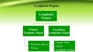
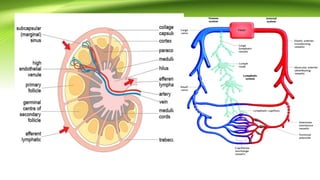
• The lymphatic system is a network of tissues and organs that help rid the
body of toxins, waste and other unwanted materials.
• The primary function of the lymphatic system is to transport lymph, a
fluid containing infection-fighting white blood cells, throughout the
body.
• The lymphatic system is part of the immune system. It also maintains
fluid balance and plays a role in absorbing fats and fat-soluble nutrients.
Lymphatic Tissues and Organs
• Lymphatic cells are organized into tissues and organs based on how tightly
the lymphatic cells are arranged and whether the tissue is encapsulated by a
layer of connective tissue.
• Three general categories exist:
1. Diffuse, unencapsulated bundles of lymphatic cells. This kind of
lymphatic tissue consists of lymphocytes and macrophages associated
with a reticular fiber network. It occurs in the lamina propria (middle
layer) of the mucus membranes (mucosae) that line the respiratory and
gastrointestinal tracts.
2. Discrete, unencapsulated bundles of lymphatic cells, called lymphatic
nodules (follicles). These bundles have clear boundaries that separate
them from neighboring cells. Nodules occur within the lamina propria
of the mucus membranes that line the gastrointestinal, respiratory,
reproductive, and urinary tracts. They are referred to as
mucosa‐associated lymphoid tissue (MALT). The nodules contain
lymphocytes and macrophages that protect against bacteria and other
pathogens that may enter these passages with food, air, or urine.
Nodules occur as solitary nodules, or they cluster as patches or
aggregates. Here are the major clusters of nodules:
a. Peyer's patches are clusters of lymphatic nodules that occur in the mucosa
that lines the ileum of the small intestine.
b. The tonsils are aggregates of lymphatic nodules that occur in the mucosa
that lines the pharynx (throat). Each of the seven tonsils that form a ring
around the pharynx are named for their specific region: a single
pharyngeal tonsil ( adenoid) in the rear wall of the nasopharynx, two
palatine tonsils on each side wall of the oral cavity at its entrance in the
throat, two lingual tonsils at the base of the tongue, and two small tubal
tonsils in the pharynx at the entrance to the auditory tubes.
c. The appendix, a small fingerlike attachment to the beginning of the large
intestine, is lined with aggregates of lymph nodules.
3. Encapsulated organs contain lymphatic nodules and diffuse
lymphatic cells surrounded by a capsule of dense connective tissue.
The three lymphatic organs are discussed in the following sections.
Lymphatic Organs
Lymphatic
Organs
Primary
Lympatic Organ
1. Red Bone Marrow
2. Thymus
Secondary
Lymphatic Organ
1. Lymph Node
2. Spleen
3. Lymphatic Nodule
Lymph nodes
• Lymph nodes are small, oval, or bean‐shaped bodies that occur along
lymphatic vessels.
• They are abundant where lymphatic vessels merge to form trunks,
especially in the inguinal (groin), axillary (armpit), and mammary gland
areas.
• Lymph flows into a node through afferent lymphatic vessels that enter
the convex side of a node.
• It exits the node at the hilus, the indented region on the opposite,
concave side of the node, through efferent lymphatic vessels.
• Efferent vessels contain valves that restrict lymph to movement in one
direction out of the lymph node.
• The number of efferent vessels leaving the lymph node is fewer than the
number of afferent vessels entering, slowing the flow of lymph through
the node.
Structure of a lymph node
• Lymph nodes are encapsulated organs that are strategically placed along the
lymphatic network. Here they can trap foreign material (antigens), which are
presented to the lymphocytes by antigen-presenting cells, to initiate an
immune response.
• The lymphocytes are densely packed in the lymph node, and the tissue is
organised both to facilitate the interactions needed to generate an immune
response against the antigen and to promote rapid division of the responding
lymphocytes.
• Lymph nodes vary in size (from a few millimetres to 1–2 cm), and are
distributed in different areas of the body. They are linked in chains by
lymphatic ducts, so that fluid flowing out of one lymph node via the efferent
lymphatic vessel becomes the inflow to the next in line, via the afferent
lymphatics.
• The fluid in question is called lymph, and is derived from the tissue that
carries cells and foreign material to the lymph nodes. Eventually, lymph
returns to the bloodstream via one of the body’s two major lymphatic
ducts.
• Secondary lymphoid organs can be thought of as guard posts that are
strategically placed to intercept any infectious agent that enters an area of
the body. So, for example, the lymph nodes in the axilla of the arm (the
armpit) will intercept infections which enter that part of the body.
• Lymphocytes located in the local lymph nodes are responsible for the
initial recognition of the infection and the development of the immune
response.
• Once the immune response has developed, the cells will migrate out from
the lymph node to the blood and, ultimately, cells will move to the site of
infection to combat the pathogen there.
• Lymph nodes have a well defined structure with different sub-regions. Different
types of leukocyte are localised within the regions so that they can interact with
each other appropriately to initiate and develop the immune response. Antigens
and cells enter the node through afferent lymphatics, and cells and fluid leave
through the efferent lymphatic.
• Cells (lymphocytes) can also enter the node from the blood by migrating
across the specialised high endothelial venules. Within the node, cells
distribute themselves to distinct zones. B cells proliferate and develop
within the follicles of the cortex, while T cells are primarily located in the
paracortex.
• The capsule, medullary cords and hilus are fixed structural elements of the
tissue.
• There is a capsule of dense connective tissue that surrounds the lymph
node.
• Trabeculae are projections of the capsule that extend into the node,
forming compartments. The trabeculae support reticular fibers that
form a network that supports lymphocytes.
• The cortex is the dense, outer region of the node. It contains lymphatic
nodules where B cells and macrophages proliferate.
• The medulla is the center of the node. Less dense than the surrounding
cortex, the medulla primarily contains T cells.
• Medullary cords are strands of reticular fibers with lymphocytes and
macrophages that extend from the cortex toward the hilus.
• Sinuses are passageways through the cortex and medulla through which
lymph moves toward the hilus.
Lymph nodes perform three functions:
• hey filter the lymph, preventing the spread of microorganisms and toxins
that enter interstitial fluids.
• They destroy bacteria, toxins, and particulate matter through the
phagocytic action of macrophages.
• They produce antibodies through the activity of B cells.
Function of Lymph Node
Lymph Circulation
• Lymph enters the convex side of lymph node through the number of
afferent lymphatic vessels.
• Then it moves threough the large bag like sinus, the subcapsular synus
into a number of smaller sinuses that cut through the cortex (Cortical
Sinuses) and enters into the midulla ( Medulary Sinuses).
• The lymph meander through these sinuses and finally exist the node at
its hylum.
• Efferent vessels draining the node the allow lymphocytes and
macrophages to carry protective function.
Lymphoid Organ
Thymus
• The thymus is a bilobed organ located in the upper chest region between the
lungs, posterior to the sternum. It grows during childhood and reaches its
maximum size of 40 g at puberty. It then slowly decreases in size as it is replaced
by adipose and areolar connective tissue. By age 65, it weighs about 6 g.
• Each lobe of the thymus is surrounded by a capsule of connective tissue. Lobules
produced by trabeculae (inward extensions of the capsule) are characterized by
an outer cortex and inner medulla. The following cells are present:
• Lymphocytes consist almost entirely of T cells.
• Epithelial‐reticular cells resemble reticular cells, but do not form reticular fibers.
Instead, these star‐shaped cells form a reticular network by interlocking their
slender cellular processes (extensions).
• These processes are held together by desmosomes, cell junctions
formed by protein fibers.
• Epithelial‐reticular cells produce thymosin and other hormones
believed to promote the maturation of T cells.
Function of Thymus
• The function of the thymus is to promote the maturation of T lymphocytes.
Immature T cells migrate through the blood from the red bone marrow to the
thymus. Within the thymus, the immature T cells concentrate in the cortex,
where they continue their development. Mature T cells leave the thymus by
way of blood vessels or efferent lymphatic vessels, migrating to other
lymphatic tissues and organs where they become active (immunocompetent) in
immune responses. The thymus does not provide a filtering function similar to
lymph nodes (there are no afferent lymphatic vessels leading into the thymus),
and unlike all other centers of lymphatic tissues, the thymus does not play a
direct role in immune responses.
• Blood vessels that permeate the thymus are surrounded by epithelial‐reticular
cells. These cells establish a protective blood‐thymus barrier that prevents the
entrance of antigens from the blood and into the thymus where T cells are
maturing. Thus, an antigen‐free environment is maintained for the development
of T cells.
Spleen
• Measuring about 12 cm (5 inches) in length, the spleen is the largest
lymphatic organ. It is located on the left side of the body, inferior to the
diaphragm and at the left edge of the stomach. Like other lymphatic organs,
the spleen is surrounded by a capsule whose extensions into the spleen
form trabeculae. The splenic artery, splenic vein, nerves, and efferent
lymphatic vessels pass through the hilus of the spleen located on its slightly
concave, upper surface. There are two distinct areas within the spleen:
• White pulp consists of reticular fibers and lymphocytes in nodules that
resemble the nodules of lymph nodes.
• Red pulp consists of venous sinuses filled with blood. Splenic cords
consisting of reticular connective tissue, macrophages, and lymphocytes
form a mesh between the venous sinuses and act as a filter as blood passes
between arterial vessels and the sinuses.
Functon of Slpeen
• The functions of the spleen include the following:
• The spleen filters the blood. Macrophages in the spleen remove bacteria and other
pathogens, cellular debris, and aged blood cells. There are no afferent lymphatic vessels,
and unlike lymph nodes, the spleen does not filter lymph.
• The spleen destroys old red blood cells and recycles their parts. It removes the iron from
heme groups and binds the iron to the storage protein.
• The spleen provides a reservoir of blood. The diffuse nature of the red pulp retains large
quantities of blood, which can be directed to the circulation when necessary. One third of
the blood platelets are stored in the spleen.
• The spleen is active in immune responses. T cells proliferate in the white pulp before
returning to the blood to attack nonself cells when necessary. B cells proliferate in the
white pulp, producing plasma cells and antibodies that return to the blood to inactivate
antigens.
• The spleen produces blood cells. Red and white blood cells are produced in the spleen
during fetal development.
MCQ’s
1. Which of the following is not directly associated with the lymphatic pathway?
a. Lymphatic trunk
b. Collectingduct
c. Subclavian vein
d. Carotid arteries
2. The thymus is responsiblefor secreting _____ from epithelialcells.
a. Thymosin
b. Growth hormone
c. Macrophages
d. Plasma cells
3. Which of the following types of cytokines is responsiblefor the growth and maturation of B cells?
a. Interleukin-1
b. Interleukin-2
c. Interleukin-4
d. Interleukin-7
Spleen
4. Which of the following types of immunoglobulinsis the most responsiblefor promoting allergic
reactions?
a. IgA
b. IgM
c. IgD
d. IgE
5. Which of the following types of immunoglobulinsis located on the surface of most B-lymphocytes?
a. IgA
b. IgM
c. IgD
d. IgE
6. Which of the following statements regarding the lymphatic system is FALSE?
a. Lymph originates from excess cellularfluid
b. Lymph nodes trap bacteria
c. Swelling of the lymph nodes indicates dysfunction of the lymphatic system
d. Swelling of the lymph nodes indicates properfunctioningof the lymphatic system
7. Which of the following is not an autoimmune disease?
a. Graves disease
b. Myasthenia gravis
c. Insulin-dependentdiabetes mellitus
d. Alzheimer’s disease
8. T-cell activation requires a/an _______cell.
a. Activation
b. Accessory
c. Plasma
d. Helper
9. The thymus is located with the _______.
a. Mediastinum
b. Peristinum
c. Epistinum
d. Endostinum
10. Which of the following statements is false regarding the spleen?
a. Divided up into lobules
b. Similar to a large lymph node
c. Containsmacrophages
d. Limited bloodwithin the lobules
11. Which of the following is not considered a central location of lymph nodes?
a. Cervical
b. Axillary
c. Inguinal
d. Tibial
12. Lymphocytes that reach the thymus become _____.
a. T-cells
b. B-cells
c. Plasma cells
d. Beta cells
13. Lymphocytes that do not reach the thymus become _____.
a. T-cells
b. B-cells
c. Plasma cells
d. Beta cells
14. Which of the following is associated with a B cell deficiency?
a. Job’s syndrome
b. Chronicgranulomatousdisease
c. Bruton’sagammaglobulinemia
d. Wiskott-Aldrich syndrome
15. Which of the following is the autoantibodyfor systemic lupus?
a. Anti-microsomal
b. Antinuclearantibodies
c. Anti-gliadin
d. Anti-histone
16. The TB skin test is an example of ______.
a. Delayed hypersensitivity
b. Serum sickness
c. Cytotoxic reaction
d. Arthus reaction
17. Which of the following types of cytokines is secreted by macrophages?
a. IL-1
b. IL-2
c. IL-3
d. IL-4
18. Which of the following types of immunoglobulins binds complement?
a. IgA
b. IgD
c. IgE
d. IgG
19. Which of the following is a key component of cytotoxicT cells?
a. CD2
b. CD4
c. CD8
d. CD10
20. Which of the following is not a primary target group of T cells?
a. Viruses
b. Toxins
c. Fungi
d. TB
Ans. Key
1. D
2. A
3. C
4. D
5. C
6. C
7. D
8. B
9. A
10. D
11. D
12. A
13. B
14. C
15. B
16. A
17. A
18. D
19. C
20. B

5. lymphatic system

  • 1.
    Lymphatic System By Yogeshwary M.Bhongade Assistant Professor Kamla Neharu College of Pharmacy, Butibori, Nagpur
  • 2.
    Lymphatic System • Thelymphatic system is a network of tissues and organs that help rid the body of toxins, waste and other unwanted materials. • The primary function of the lymphatic system is to transport lymph, a fluid containing infection-fighting white blood cells, throughout the body. • The lymphatic system is part of the immune system. It also maintains fluid balance and plays a role in absorbing fats and fat-soluble nutrients.
  • 3.
    Lymphatic Tissues andOrgans • Lymphatic cells are organized into tissues and organs based on how tightly the lymphatic cells are arranged and whether the tissue is encapsulated by a layer of connective tissue. • Three general categories exist: 1. Diffuse, unencapsulated bundles of lymphatic cells. This kind of lymphatic tissue consists of lymphocytes and macrophages associated with a reticular fiber network. It occurs in the lamina propria (middle layer) of the mucus membranes (mucosae) that line the respiratory and gastrointestinal tracts.
  • 4.
    2. Discrete, unencapsulatedbundles of lymphatic cells, called lymphatic nodules (follicles). These bundles have clear boundaries that separate them from neighboring cells. Nodules occur within the lamina propria of the mucus membranes that line the gastrointestinal, respiratory, reproductive, and urinary tracts. They are referred to as mucosa‐associated lymphoid tissue (MALT). The nodules contain lymphocytes and macrophages that protect against bacteria and other pathogens that may enter these passages with food, air, or urine. Nodules occur as solitary nodules, or they cluster as patches or aggregates. Here are the major clusters of nodules:
  • 5.
    a. Peyer's patchesare clusters of lymphatic nodules that occur in the mucosa that lines the ileum of the small intestine. b. The tonsils are aggregates of lymphatic nodules that occur in the mucosa that lines the pharynx (throat). Each of the seven tonsils that form a ring around the pharynx are named for their specific region: a single pharyngeal tonsil ( adenoid) in the rear wall of the nasopharynx, two palatine tonsils on each side wall of the oral cavity at its entrance in the throat, two lingual tonsils at the base of the tongue, and two small tubal tonsils in the pharynx at the entrance to the auditory tubes. c. The appendix, a small fingerlike attachment to the beginning of the large intestine, is lined with aggregates of lymph nodules.
  • 6.
    3. Encapsulated organscontain lymphatic nodules and diffuse lymphatic cells surrounded by a capsule of dense connective tissue. The three lymphatic organs are discussed in the following sections.
  • 7.
    Lymphatic Organs Lymphatic Organs Primary Lympatic Organ 1.Red Bone Marrow 2. Thymus Secondary Lymphatic Organ 1. Lymph Node 2. Spleen 3. Lymphatic Nodule
  • 8.
    Lymph nodes • Lymphnodes are small, oval, or bean‐shaped bodies that occur along lymphatic vessels. • They are abundant where lymphatic vessels merge to form trunks, especially in the inguinal (groin), axillary (armpit), and mammary gland areas. • Lymph flows into a node through afferent lymphatic vessels that enter the convex side of a node. • It exits the node at the hilus, the indented region on the opposite, concave side of the node, through efferent lymphatic vessels. • Efferent vessels contain valves that restrict lymph to movement in one direction out of the lymph node. • The number of efferent vessels leaving the lymph node is fewer than the number of afferent vessels entering, slowing the flow of lymph through the node.
  • 9.
    Structure of alymph node • Lymph nodes are encapsulated organs that are strategically placed along the lymphatic network. Here they can trap foreign material (antigens), which are presented to the lymphocytes by antigen-presenting cells, to initiate an immune response. • The lymphocytes are densely packed in the lymph node, and the tissue is organised both to facilitate the interactions needed to generate an immune response against the antigen and to promote rapid division of the responding lymphocytes. • Lymph nodes vary in size (from a few millimetres to 1–2 cm), and are distributed in different areas of the body. They are linked in chains by lymphatic ducts, so that fluid flowing out of one lymph node via the efferent lymphatic vessel becomes the inflow to the next in line, via the afferent lymphatics.
  • 10.
    • The fluidin question is called lymph, and is derived from the tissue that carries cells and foreign material to the lymph nodes. Eventually, lymph returns to the bloodstream via one of the body’s two major lymphatic ducts. • Secondary lymphoid organs can be thought of as guard posts that are strategically placed to intercept any infectious agent that enters an area of the body. So, for example, the lymph nodes in the axilla of the arm (the armpit) will intercept infections which enter that part of the body. • Lymphocytes located in the local lymph nodes are responsible for the initial recognition of the infection and the development of the immune response. • Once the immune response has developed, the cells will migrate out from the lymph node to the blood and, ultimately, cells will move to the site of infection to combat the pathogen there.
  • 11.
    • Lymph nodeshave a well defined structure with different sub-regions. Different types of leukocyte are localised within the regions so that they can interact with each other appropriately to initiate and develop the immune response. Antigens and cells enter the node through afferent lymphatics, and cells and fluid leave through the efferent lymphatic.
  • 12.
    • Cells (lymphocytes)can also enter the node from the blood by migrating across the specialised high endothelial venules. Within the node, cells distribute themselves to distinct zones. B cells proliferate and develop within the follicles of the cortex, while T cells are primarily located in the paracortex. • The capsule, medullary cords and hilus are fixed structural elements of the tissue.
  • 13.
    • There isa capsule of dense connective tissue that surrounds the lymph node. • Trabeculae are projections of the capsule that extend into the node, forming compartments. The trabeculae support reticular fibers that form a network that supports lymphocytes. • The cortex is the dense, outer region of the node. It contains lymphatic nodules where B cells and macrophages proliferate. • The medulla is the center of the node. Less dense than the surrounding cortex, the medulla primarily contains T cells. • Medullary cords are strands of reticular fibers with lymphocytes and macrophages that extend from the cortex toward the hilus. • Sinuses are passageways through the cortex and medulla through which lymph moves toward the hilus.
  • 14.
    Lymph nodes performthree functions: • hey filter the lymph, preventing the spread of microorganisms and toxins that enter interstitial fluids. • They destroy bacteria, toxins, and particulate matter through the phagocytic action of macrophages. • They produce antibodies through the activity of B cells. Function of Lymph Node
  • 15.
    Lymph Circulation • Lymphenters the convex side of lymph node through the number of afferent lymphatic vessels. • Then it moves threough the large bag like sinus, the subcapsular synus into a number of smaller sinuses that cut through the cortex (Cortical Sinuses) and enters into the midulla ( Medulary Sinuses). • The lymph meander through these sinuses and finally exist the node at its hylum. • Efferent vessels draining the node the allow lymphocytes and macrophages to carry protective function.
  • 17.
    Lymphoid Organ Thymus • Thethymus is a bilobed organ located in the upper chest region between the lungs, posterior to the sternum. It grows during childhood and reaches its maximum size of 40 g at puberty. It then slowly decreases in size as it is replaced by adipose and areolar connective tissue. By age 65, it weighs about 6 g. • Each lobe of the thymus is surrounded by a capsule of connective tissue. Lobules produced by trabeculae (inward extensions of the capsule) are characterized by an outer cortex and inner medulla. The following cells are present: • Lymphocytes consist almost entirely of T cells. • Epithelial‐reticular cells resemble reticular cells, but do not form reticular fibers. Instead, these star‐shaped cells form a reticular network by interlocking their slender cellular processes (extensions).
  • 18.
    • These processesare held together by desmosomes, cell junctions formed by protein fibers. • Epithelial‐reticular cells produce thymosin and other hormones believed to promote the maturation of T cells.
  • 19.
    Function of Thymus •The function of the thymus is to promote the maturation of T lymphocytes. Immature T cells migrate through the blood from the red bone marrow to the thymus. Within the thymus, the immature T cells concentrate in the cortex, where they continue their development. Mature T cells leave the thymus by way of blood vessels or efferent lymphatic vessels, migrating to other lymphatic tissues and organs where they become active (immunocompetent) in immune responses. The thymus does not provide a filtering function similar to lymph nodes (there are no afferent lymphatic vessels leading into the thymus), and unlike all other centers of lymphatic tissues, the thymus does not play a direct role in immune responses. • Blood vessels that permeate the thymus are surrounded by epithelial‐reticular cells. These cells establish a protective blood‐thymus barrier that prevents the entrance of antigens from the blood and into the thymus where T cells are maturing. Thus, an antigen‐free environment is maintained for the development of T cells.
  • 20.
    Spleen • Measuring about12 cm (5 inches) in length, the spleen is the largest lymphatic organ. It is located on the left side of the body, inferior to the diaphragm and at the left edge of the stomach. Like other lymphatic organs, the spleen is surrounded by a capsule whose extensions into the spleen form trabeculae. The splenic artery, splenic vein, nerves, and efferent lymphatic vessels pass through the hilus of the spleen located on its slightly concave, upper surface. There are two distinct areas within the spleen: • White pulp consists of reticular fibers and lymphocytes in nodules that resemble the nodules of lymph nodes. • Red pulp consists of venous sinuses filled with blood. Splenic cords consisting of reticular connective tissue, macrophages, and lymphocytes form a mesh between the venous sinuses and act as a filter as blood passes between arterial vessels and the sinuses.
  • 21.
    Functon of Slpeen •The functions of the spleen include the following: • The spleen filters the blood. Macrophages in the spleen remove bacteria and other pathogens, cellular debris, and aged blood cells. There are no afferent lymphatic vessels, and unlike lymph nodes, the spleen does not filter lymph. • The spleen destroys old red blood cells and recycles their parts. It removes the iron from heme groups and binds the iron to the storage protein. • The spleen provides a reservoir of blood. The diffuse nature of the red pulp retains large quantities of blood, which can be directed to the circulation when necessary. One third of the blood platelets are stored in the spleen. • The spleen is active in immune responses. T cells proliferate in the white pulp before returning to the blood to attack nonself cells when necessary. B cells proliferate in the white pulp, producing plasma cells and antibodies that return to the blood to inactivate antigens. • The spleen produces blood cells. Red and white blood cells are produced in the spleen during fetal development.
  • 22.
    MCQ’s 1. Which ofthe following is not directly associated with the lymphatic pathway? a. Lymphatic trunk b. Collectingduct c. Subclavian vein d. Carotid arteries 2. The thymus is responsiblefor secreting _____ from epithelialcells. a. Thymosin b. Growth hormone c. Macrophages d. Plasma cells 3. Which of the following types of cytokines is responsiblefor the growth and maturation of B cells? a. Interleukin-1 b. Interleukin-2 c. Interleukin-4 d. Interleukin-7 Spleen
  • 23.
    4. Which ofthe following types of immunoglobulinsis the most responsiblefor promoting allergic reactions? a. IgA b. IgM c. IgD d. IgE 5. Which of the following types of immunoglobulinsis located on the surface of most B-lymphocytes? a. IgA b. IgM c. IgD d. IgE 6. Which of the following statements regarding the lymphatic system is FALSE? a. Lymph originates from excess cellularfluid b. Lymph nodes trap bacteria c. Swelling of the lymph nodes indicates dysfunction of the lymphatic system d. Swelling of the lymph nodes indicates properfunctioningof the lymphatic system
  • 24.
    7. Which ofthe following is not an autoimmune disease? a. Graves disease b. Myasthenia gravis c. Insulin-dependentdiabetes mellitus d. Alzheimer’s disease 8. T-cell activation requires a/an _______cell. a. Activation b. Accessory c. Plasma d. Helper 9. The thymus is located with the _______. a. Mediastinum b. Peristinum c. Epistinum d. Endostinum
  • 25.
    10. Which ofthe following statements is false regarding the spleen? a. Divided up into lobules b. Similar to a large lymph node c. Containsmacrophages d. Limited bloodwithin the lobules 11. Which of the following is not considered a central location of lymph nodes? a. Cervical b. Axillary c. Inguinal d. Tibial 12. Lymphocytes that reach the thymus become _____. a. T-cells b. B-cells c. Plasma cells d. Beta cells
  • 26.
    13. Lymphocytes thatdo not reach the thymus become _____. a. T-cells b. B-cells c. Plasma cells d. Beta cells 14. Which of the following is associated with a B cell deficiency? a. Job’s syndrome b. Chronicgranulomatousdisease c. Bruton’sagammaglobulinemia d. Wiskott-Aldrich syndrome 15. Which of the following is the autoantibodyfor systemic lupus? a. Anti-microsomal b. Antinuclearantibodies c. Anti-gliadin d. Anti-histone
  • 27.
    16. The TBskin test is an example of ______. a. Delayed hypersensitivity b. Serum sickness c. Cytotoxic reaction d. Arthus reaction 17. Which of the following types of cytokines is secreted by macrophages? a. IL-1 b. IL-2 c. IL-3 d. IL-4 18. Which of the following types of immunoglobulins binds complement? a. IgA b. IgD c. IgE d. IgG
  • 28.
    19. Which ofthe following is a key component of cytotoxicT cells? a. CD2 b. CD4 c. CD8 d. CD10 20. Which of the following is not a primary target group of T cells? a. Viruses b. Toxins c. Fungi d. TB
  • 29.
    Ans. Key 1. D 2.A 3. C 4. D 5. C 6. C 7. D 8. B 9. A 10. D 11. D 12. A 13. B 14. C 15. B 16. A 17. A 18. D 19. C 20. B