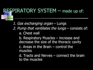
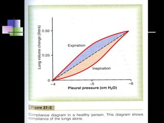
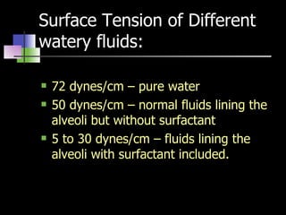
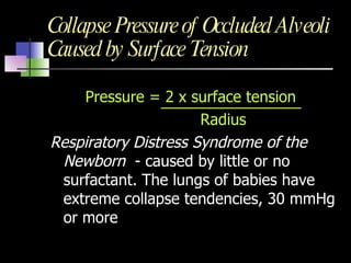
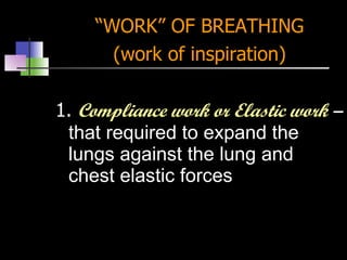
RESPIRATION Dr. Victoria G. Giango Chair, Dept. of Physiology
GOALS OF RESPIRATION 1. To provide oxygen to the tissues 2. To remove carbon dioxide MAJOR FUNCTIONAL EVENTS OF RESPIRATION 1.  Pulmonary Ventilation –  inflow and outflow of air between the atmosphere and lung alveoli
2.  Diffusion  of oxygen and carbon dioxide between the alveoli and the blood 3.  Transport  of oxygen and carbon dioxide in the blood and body fluids to and from the cells. 4.  Regulation of Ventilation
 
 
RESPIRATORY SYSTEM –  made up of: 1. Gas exchanging organ  – Lungs 2. Pump that ventilates the lungs  – consists of: a. Chest wall b. Respiratory Muscles – increase and  decrease the size of the thoracic cavity c. Areas in the Brain – control the  muscles d. Tracts and Nerves – connect the brain  to the muscles
MUSCLES OF INSPIRATION Diaphragm – 75% External intercostals Sternocleidomastoid Serratus (anterior) Scaleni MUSCLES OF EXPIRATION 1. Internal intercostals 2. Abdominal recti
COMPLIANCE The extent to which the lungs expand for each unit increase in transpulmonary pressure Both lungs  – 200 ml of air per cm of water transpulmonary pressure Thorax and lungs together  – 110 ml per cm of water transpulmonary pressure
FACTORS THAT DETERMINE COMPLIANCE: 1. Elastic forces of the lung tissue itself  (elastin and collagen fibers interwoven among the lung parenchyma 2.  Elastic forces caused by surface tension of the fluid that lines the inside walls of the alveoli  and other lung spaces Surface Tension accounts for 2/3  of total elastic forces in normal lung
 
 
SURFACTANT - Surface active agent, when it spreads over the surface of a fluid, it  reduces the surface tension -  Secreted by  type II alveolar epithelial cells - Complex mixture of phospholipids, proteins and ions - dipalmitoylphosphatidylcholine - surfactant apoproteins - calcium ions
SURFACTANT Lowers surface tension Stabilizes the size of the alveoli Prevents the accumulation of fluid
Surface Tension of Different watery fluids: 72 dynes/cm – pure water 50 dynes/cm – normal fluids lining the alveoli but without surfactant 5 to 30 dynes/cm – fluids lining the alveoli with surfactant included.
Stabilize the sizes of the alveoli - inversely affected by radius of the alveolus - begin to be secreted between the 6 th  and 7 th  month of gestation
Collapse Pressure of Occluded Alveoli Caused by Surface Tension Pressure = 2 x surface tension   Radius Respiratory Distress Syndrome of the Newborn  - caused by little or no surfactant. The lungs of babies have extreme collapse tendencies, 30 mmHg or more
“ WORK” OF BREATHING (work of inspiration) 1.  Compliance work or Elastic work  –  that required to expand the lungs against the lung and chest elastic forces
2.  Tissue resistance work  – required to overcome the viscosity of the lung and chest wall structures 3.  Airway resistance work  –  required to overcome airway resistance to movement of air into the lungs
Compliance and Tissue resistance work  – increased by diseases that cause  fibrosis of the lungs  as in  tuberculosis Airway resistance work  – increased by diseases  that obstruct the airways as in asthma 3 to 5%  of the total energy expended by the body is required to energize the pulmonary ventilatory process
LUNG VOLUMES Tidal Volume  ( Vt) – is the volume of air inspired or expired with each normal breath - 500 ml Inspiratory Reserve Volume  (IRV) – the extra volume of air that can be inspired over and above the normal tidal volume when the person inspires with full force – 3000ml
3. Expiratory Reserve Volume (ERV) –  is the maximum extra volume of air that can be expired by forceful expiration after the end of the normal tidal expiration - 1100 ml 4. Residual Volume (RV) –  the volume of air remaining in the lungs after the most forceful expiration – 1200ml
 
 
CAPACITIES Inspiratory Capacity (IC)  I C = Vt + IRV = 3, 500 ml 2. Functional Residual Capacity (FRC) = 2,300 ml FRC = ERV + RV 3. Vital Capacity (VC)  VC = IRV + Vt + ERV   = IC + ERV 4. Total Lung Capacity (TLC) =  5, 800ml TLC = Vt + IRV + ERV + RV = IC + FRC  = VC + RV
HELIUM DILUTION METHOD  – determination Functional Residual capacity, Residual volume, Total Lung Capacity FRC =  Initial conc. Of Helium in spirometer Final conc. Of Helium FRC =  Ci He __ - 1  Vi Spir Cf He RV = FRC – REV TLC = FRC + IC
FACTORS AFFECTING LUNG VOLUMES AND VITAL CAPACITY Body build or physique Position of the body Strength of respiratory muscles Pulmonary compliance
Minute Respiratory Volume is the total amount of new moved into the respiratory passages each minute  = Tidal volume x Respiratory rate/minute = 500 x 12 Alveolar Ventilation Total volume of new air entering the the alveoli and adjacent gas exchange areas each minute V  A = Freq  . (V T  – V D  )
Respiratory Dead Space – space in the conducting zone of the airways occupied by gas that does not exchange with the blood in the pulmonary vessels Vital Capacity  – the largest volume of air that can be expired after a maximal inspiratory effort. It is  frequently measured clinically as an index of pulmonary function . It gives useful information about the  strength of the respiratory muscles and other pulmonary functions. Maximal Voluntary Ventilation – (MVV) or Maximal breathing Capacity – largest volume of gas that can be moved into and out of the lungs in 1 minute by voluntary effort – 125 – 170 L/min.
ANATOMIC DEAD SPACE  – space of the respiratory system besides the alveoli and other gas exchange areas Dead space air = 15 0 m PHYSIOLOGIC DEAD SPACE – alveolar dead space and anatomic dead space In normal person the Anatomic and Physiologic dead spaces are nearly equal In person with partially functional or nonfunctional alveoli the Physiologic dead space is much as 10 x the volume of Anatomic dead space
 
FUNCTIONS OF THE RESPIRATORY PASSAGEWAYS ANATOMY: Trachea, Bronchi, Bronchioles between trachea and alveolar sacs airways divide 23 times – first 16 generations made  up of bronchi, bronchioles, and terminal bronchioles. Last 7 generations are made up of  respiratory bronchioles, alveolar ducts, and alveoli
Muscular Wall of the Bronchi and Bronchioles and its Control Resistance to Airflow in the Bronchial Tree Nervous and Local Control of the Bronchiolar Musculature –  “Sympathetic”  Dilatation of the Bronchioles Parasympathetic constriction of the Bronchioles Local Secretory Factors Often Cause Bronchiolar Constriction - Histamine - Slow Reacting Substance of Anaphylaxis
FUNCTIONS: Mucous Lining of the Respiratory Passageways and Action of Cilia to Clear the Passageways Cough Reflex – Vagus Nerve a. Irritation b. Inspiration  – 2.5 liters of air are rapidly inspired. Epiglottis closes and the vocal cords shut tightly to entrap the air within the lungs. c. Compression  – abdominal muscles contract forcefully. Pushing against the diaphragm. Pressure rises to 100 mmHg or more. d. Expulsion  – air under high pressure in the lungs explude outward
 
3. Sneeze Reflex – Trigeminal Nerve Uvula  is depressed so large amounts of  air pass directly through the nose helping  to clear the nasal passages of foreign  matter, 4. Normal Respiratory Functions of the Nose – Air Conditioning Function of the Upper Respiratory Passages a. Air is warmed b. Air is Humidified c. Air is Filtered - turbulent precipitation - gravitational precipitation
5. Vocalization Phonation  – larynx is adapted to act as vibrator (vibrating element is the vocal cord) Vocalization and resonance  – organs of articulation are the  lips, tongue, and soft palate

Respiration Ventilation

  • 1.
    RESPIRATION Dr. VictoriaG. Giango Chair, Dept. of Physiology
  • 2.
    GOALS OF RESPIRATION1. To provide oxygen to the tissues 2. To remove carbon dioxide MAJOR FUNCTIONAL EVENTS OF RESPIRATION 1. Pulmonary Ventilation – inflow and outflow of air between the atmosphere and lung alveoli
  • 3.
    2. Diffusion of oxygen and carbon dioxide between the alveoli and the blood 3. Transport of oxygen and carbon dioxide in the blood and body fluids to and from the cells. 4. Regulation of Ventilation
  • 4.
  • 5.
  • 6.
    RESPIRATORY SYSTEM – made up of: 1. Gas exchanging organ – Lungs 2. Pump that ventilates the lungs – consists of: a. Chest wall b. Respiratory Muscles – increase and decrease the size of the thoracic cavity c. Areas in the Brain – control the muscles d. Tracts and Nerves – connect the brain to the muscles
  • 7.
    MUSCLES OF INSPIRATIONDiaphragm – 75% External intercostals Sternocleidomastoid Serratus (anterior) Scaleni MUSCLES OF EXPIRATION 1. Internal intercostals 2. Abdominal recti
  • 8.
    COMPLIANCE The extentto which the lungs expand for each unit increase in transpulmonary pressure Both lungs – 200 ml of air per cm of water transpulmonary pressure Thorax and lungs together – 110 ml per cm of water transpulmonary pressure
  • 9.
    FACTORS THAT DETERMINECOMPLIANCE: 1. Elastic forces of the lung tissue itself (elastin and collagen fibers interwoven among the lung parenchyma 2. Elastic forces caused by surface tension of the fluid that lines the inside walls of the alveoli and other lung spaces Surface Tension accounts for 2/3 of total elastic forces in normal lung
  • 10.
  • 11.
  • 12.
    SURFACTANT - Surfaceactive agent, when it spreads over the surface of a fluid, it reduces the surface tension - Secreted by type II alveolar epithelial cells - Complex mixture of phospholipids, proteins and ions - dipalmitoylphosphatidylcholine - surfactant apoproteins - calcium ions
  • 13.
    SURFACTANT Lowers surfacetension Stabilizes the size of the alveoli Prevents the accumulation of fluid
  • 14.
    Surface Tension ofDifferent watery fluids: 72 dynes/cm – pure water 50 dynes/cm – normal fluids lining the alveoli but without surfactant 5 to 30 dynes/cm – fluids lining the alveoli with surfactant included.
  • 15.
    Stabilize the sizesof the alveoli - inversely affected by radius of the alveolus - begin to be secreted between the 6 th and 7 th month of gestation
  • 16.
    Collapse Pressure ofOccluded Alveoli Caused by Surface Tension Pressure = 2 x surface tension Radius Respiratory Distress Syndrome of the Newborn - caused by little or no surfactant. The lungs of babies have extreme collapse tendencies, 30 mmHg or more
  • 17.
    “ WORK” OFBREATHING (work of inspiration) 1. Compliance work or Elastic work – that required to expand the lungs against the lung and chest elastic forces
  • 18.
    2. Tissueresistance work – required to overcome the viscosity of the lung and chest wall structures 3. Airway resistance work – required to overcome airway resistance to movement of air into the lungs
  • 19.
    Compliance and Tissueresistance work – increased by diseases that cause fibrosis of the lungs as in tuberculosis Airway resistance work – increased by diseases that obstruct the airways as in asthma 3 to 5% of the total energy expended by the body is required to energize the pulmonary ventilatory process
  • 20.
    LUNG VOLUMES TidalVolume ( Vt) – is the volume of air inspired or expired with each normal breath - 500 ml Inspiratory Reserve Volume (IRV) – the extra volume of air that can be inspired over and above the normal tidal volume when the person inspires with full force – 3000ml
  • 21.
    3. Expiratory ReserveVolume (ERV) – is the maximum extra volume of air that can be expired by forceful expiration after the end of the normal tidal expiration - 1100 ml 4. Residual Volume (RV) – the volume of air remaining in the lungs after the most forceful expiration – 1200ml
  • 22.
  • 23.
  • 24.
    CAPACITIES Inspiratory Capacity(IC) I C = Vt + IRV = 3, 500 ml 2. Functional Residual Capacity (FRC) = 2,300 ml FRC = ERV + RV 3. Vital Capacity (VC) VC = IRV + Vt + ERV = IC + ERV 4. Total Lung Capacity (TLC) = 5, 800ml TLC = Vt + IRV + ERV + RV = IC + FRC = VC + RV
  • 25.
    HELIUM DILUTION METHOD – determination Functional Residual capacity, Residual volume, Total Lung Capacity FRC = Initial conc. Of Helium in spirometer Final conc. Of Helium FRC = Ci He __ - 1 Vi Spir Cf He RV = FRC – REV TLC = FRC + IC
  • 26.
    FACTORS AFFECTING LUNGVOLUMES AND VITAL CAPACITY Body build or physique Position of the body Strength of respiratory muscles Pulmonary compliance
  • 27.
    Minute Respiratory Volumeis the total amount of new moved into the respiratory passages each minute = Tidal volume x Respiratory rate/minute = 500 x 12 Alveolar Ventilation Total volume of new air entering the the alveoli and adjacent gas exchange areas each minute V A = Freq . (V T – V D )
  • 28.
    Respiratory Dead Space– space in the conducting zone of the airways occupied by gas that does not exchange with the blood in the pulmonary vessels Vital Capacity – the largest volume of air that can be expired after a maximal inspiratory effort. It is frequently measured clinically as an index of pulmonary function . It gives useful information about the strength of the respiratory muscles and other pulmonary functions. Maximal Voluntary Ventilation – (MVV) or Maximal breathing Capacity – largest volume of gas that can be moved into and out of the lungs in 1 minute by voluntary effort – 125 – 170 L/min.
  • 29.
    ANATOMIC DEAD SPACE – space of the respiratory system besides the alveoli and other gas exchange areas Dead space air = 15 0 m PHYSIOLOGIC DEAD SPACE – alveolar dead space and anatomic dead space In normal person the Anatomic and Physiologic dead spaces are nearly equal In person with partially functional or nonfunctional alveoli the Physiologic dead space is much as 10 x the volume of Anatomic dead space
  • 30.
  • 31.
    FUNCTIONS OF THERESPIRATORY PASSAGEWAYS ANATOMY: Trachea, Bronchi, Bronchioles between trachea and alveolar sacs airways divide 23 times – first 16 generations made up of bronchi, bronchioles, and terminal bronchioles. Last 7 generations are made up of respiratory bronchioles, alveolar ducts, and alveoli
  • 32.
    Muscular Wall ofthe Bronchi and Bronchioles and its Control Resistance to Airflow in the Bronchial Tree Nervous and Local Control of the Bronchiolar Musculature – “Sympathetic” Dilatation of the Bronchioles Parasympathetic constriction of the Bronchioles Local Secretory Factors Often Cause Bronchiolar Constriction - Histamine - Slow Reacting Substance of Anaphylaxis
  • 33.
    FUNCTIONS: Mucous Liningof the Respiratory Passageways and Action of Cilia to Clear the Passageways Cough Reflex – Vagus Nerve a. Irritation b. Inspiration – 2.5 liters of air are rapidly inspired. Epiglottis closes and the vocal cords shut tightly to entrap the air within the lungs. c. Compression – abdominal muscles contract forcefully. Pushing against the diaphragm. Pressure rises to 100 mmHg or more. d. Expulsion – air under high pressure in the lungs explude outward
  • 34.
  • 35.
    3. Sneeze Reflex– Trigeminal Nerve Uvula is depressed so large amounts of air pass directly through the nose helping to clear the nasal passages of foreign matter, 4. Normal Respiratory Functions of the Nose – Air Conditioning Function of the Upper Respiratory Passages a. Air is warmed b. Air is Humidified c. Air is Filtered - turbulent precipitation - gravitational precipitation
  • 36.
    5. Vocalization Phonation – larynx is adapted to act as vibrator (vibrating element is the vocal cord) Vocalization and resonance – organs of articulation are the lips, tongue, and soft palate