SlideShare now has a player specifically designed for infographics. Upload your infographics now and see them take off! Need advice on creating infographics? This presentation includes tips for producing stand-out infographics. Read more about the new SlideShare infographics player here: http://wp.me/p24NNG-2ay
This infographic was designed by Column Five: http://columnfivemedia.com/
No need to wonder how the best on SlideShare do it. The Masters of SlideShare provides storytelling, design, customization and promotion tips from 13 experts of the form. Learn what it takes to master this type of content marketing yourself.
10 Ways to Win at SlideShare SEO & Presentation OptimizationOneupweb
Thank you, SlideShare, for teaching us that PowerPoint presentations don't have to be a total bore. But in order to tap SlideShare's 60 million global users, you must optimize. Here are 10 quick tips to make your next presentation highly engaging, shareable and well worth the effort.
For more content marketing tips: http://www.oneupweb.com/blog/
Are you new to SlideShare? Are you looking to fine tune your channel plan? Are you using SlideShare but are looking for ways to enhance what you're doing? How can you use SlideShare for content marketing tactics such as lead generation, calls-to-action to other pieces of your content, or thought leadership? Read more from the CMI team in their latest SlideShare presentation on SlideShare.
Each month, join us as we highlight and discuss hot topics ranging from the future of higher education to wearable technology, best productivity hacks and secrets to hiring top talent. Upload your SlideShares, and share your expertise with the world!
Not sure what to share on SlideShare?
SlideShares that inform, inspire and educate attract the most views. Beyond that, ideas for what you can upload are limitless. We’ve selected a few popular examples to get your creative juices flowing.
How to Make Awesome SlideShares: Tips & TricksSlideShare
Turbocharge your online presence with SlideShare. We provide the best tips and tricks for succeeding on SlideShare. Get ideas for what to upload, tips for designing your deck and more.
Acute respiratory distress syndrome (ARDS) occurs when fluid builds up in the tiny, elastic air sacs (alveoli) in your lungs. The fluid keeps your lungs from filling with enough air, which means less oxygen reaches your bloodstream. This deprives your organs of the oxygen they need to function.
Updates on Acute respiratory distress syndromeHamdi Turkey
These lecture notes were made by Dr. Hamdi Turkey (Pulmonologist at Taiz university)
** Contents:
- Historical view on ARDS
- New definition of ARDS
- Precipitating risk factors
- Pathophysiology of ARDS
- Clinical picture, Diagnosis, Management and Prognosis
Do Not Forget To Visit Our Pages On Facebook on the following Links:
https://www.facebook.com/groups/569435236444761/
AND
https://www.facebook.com/groups/690331650977113/
Chronic obstructive pulmonary disease..It is one of the most affecting lung disease.. In detailed explanation of disease is there and including its ayurvedic aspect of management is also there...
#Ayurveda#Emphysema#Chronic brochitis
Ozempic: Preoperative Management of Patients on GLP-1 Receptor Agonists Saeid Safari
Preoperative Management of Patients on GLP-1 Receptor Agonists like Ozempic and Semiglutide
ASA GUIDELINE
NYSORA Guideline
2 Case Reports of Gastric Ultrasound
Prix Galien International 2024 Forum ProgramLevi Shapiro
June 20, 2024, Prix Galien International and Jerusalem Ethics Forum in ROME. Detailed agenda including panels:
- ADVANCES IN CARDIOLOGY: A NEW PARADIGM IS COMING
- WOMEN’S HEALTH: FERTILITY PRESERVATION
- WHAT’S NEW IN THE TREATMENT OF INFECTIOUS,
ONCOLOGICAL AND INFLAMMATORY SKIN DISEASES?
- ARTIFICIAL INTELLIGENCE AND ETHICS
- GENE THERAPY
- BEYOND BORDERS: GLOBAL INITIATIVES FOR DEMOCRATIZING LIFE SCIENCE TECHNOLOGIES AND PROMOTING ACCESS TO HEALTHCARE
- ETHICAL CHALLENGES IN LIFE SCIENCES
- Prix Galien International Awards Ceremony
Tom Selleck Health: A Comprehensive Look at the Iconic Actor’s Wellness Journeygreendigital
Tom Selleck, an enduring figure in Hollywood. has captivated audiences for decades with his rugged charm, iconic moustache. and memorable roles in television and film. From his breakout role as Thomas Magnum in Magnum P.I. to his current portrayal of Frank Reagan in Blue Bloods. Selleck's career has spanned over 50 years. But beyond his professional achievements. fans have often been curious about Tom Selleck Health. especially as he has aged in the public eye.
Follow us on: Pinterest
Introduction
Many have been interested in Tom Selleck health. not only because of his enduring presence on screen but also because of the challenges. and lifestyle choices he has faced and made over the years. This article delves into the various aspects of Tom Selleck health. exploring his fitness regimen, diet, mental health. and the challenges he has encountered as he ages. We'll look at how he maintains his well-being. the health issues he has faced, and his approach to ageing .
Early Life and Career
Childhood and Athletic Beginnings
Tom Selleck was born on January 29, 1945, in Detroit, Michigan, and grew up in Sherman Oaks, California. From an early age, he was involved in sports, particularly basketball. which played a significant role in his physical development. His athletic pursuits continued into college. where he attended the University of Southern California (USC) on a basketball scholarship. This early involvement in sports laid a strong foundation for his physical health and disciplined lifestyle.
Transition to Acting
Selleck's transition from an athlete to an actor came with its physical demands. His first significant role in "Magnum P.I." required him to perform various stunts and maintain a fit appearance. This role, which he played from 1980 to 1988. necessitated a rigorous fitness routine to meet the show's demands. setting the stage for his long-term commitment to health and wellness.
Fitness Regimen
Workout Routine
Tom Selleck health and fitness regimen has evolved. adapting to his changing roles and age. During his "Magnum, P.I." days. Selleck's workouts were intense and focused on building and maintaining muscle mass. His routine included weightlifting, cardiovascular exercises. and specific training for the stunts he performed on the show.
Selleck adjusted his fitness routine as he aged to suit his body's needs. Today, his workouts focus on maintaining flexibility, strength, and cardiovascular health. He incorporates low-impact exercises such as swimming, walking, and light weightlifting. This balanced approach helps him stay fit without putting undue strain on his joints and muscles.
Importance of Flexibility and Mobility
In recent years, Selleck has emphasized the importance of flexibility and mobility in his fitness regimen. Understanding the natural decline in muscle mass and joint flexibility with age. he includes stretching and yoga in his routine. These practices help prevent injuries, improve posture, and maintain mobilit
HOT NEW PRODUCT! BIG SALES FAST SHIPPING NOW FROM CHINA!! EU KU DB BK substit...GL Anaacs
Contact us if you are interested:
Email / Skype : kefaya1771@gmail.com
Threema: PXHY5PDH
New BATCH Ku !!! MUCH IN DEMAND FAST SALE EVERY BATCH HAPPY GOOD EFFECT BIG BATCH !
Contact me on Threema or skype to start big business!!
Hot-sale products:
NEW HOT EUTYLONE WHITE CRYSTAL!!
5cl-adba precursor (semi finished )
5cl-adba raw materials
ADBB precursor (semi finished )
ADBB raw materials
APVP powder
5fadb/4f-adb
Jwh018 / Jwh210
Eutylone crystal
Protonitazene (hydrochloride) CAS: 119276-01-6
Flubrotizolam CAS: 57801-95-3
Metonitazene CAS: 14680-51-4
Payment terms: Western Union,MoneyGram,Bitcoin or USDT.
Deliver Time: Usually 7-15days
Shipping method: FedEx, TNT, DHL,UPS etc.Our deliveries are 100% safe, fast, reliable and discreet.
Samples will be sent for your evaluation!If you are interested in, please contact me, let's talk details.
We specializes in exporting high quality Research chemical, medical intermediate, Pharmaceutical chemicals and so on. Products are exported to USA, Canada, France, Korea, Japan,Russia, Southeast Asia and other countries.
These simplified slides by Dr. Sidra Arshad present an overview of the non-respiratory functions of the respiratory tract.
Learning objectives:
1. Enlist the non-respiratory functions of the respiratory tract
2. Briefly explain how these functions are carried out
3. Discuss the significance of dead space
4. Differentiate between minute ventilation and alveolar ventilation
5. Describe the cough and sneeze reflexes
Study Resources:
1. Chapter 39, Guyton and Hall Textbook of Medical Physiology, 14th edition
2. Chapter 34, Ganong’s Review of Medical Physiology, 26th edition
3. Chapter 17, Human Physiology by Lauralee Sherwood, 9th edition
4. Non-respiratory functions of the lungs https://academic.oup.com/bjaed/article/13/3/98/278874
TEST BANK for Operations Management, 14th Edition by William J. Stevenson, Ve...kevinkariuki227
TEST BANK for Operations Management, 14th Edition by William J. Stevenson, Verified Chapters 1 - 19, Complete Newest Version.pdf
TEST BANK for Operations Management, 14th Edition by William J. Stevenson, Verified Chapters 1 - 19, Complete Newest Version.pdf
Couples presenting to the infertility clinic- Do they really have infertility...Sujoy Dasgupta
Dr Sujoy Dasgupta presented the study on "Couples presenting to the infertility clinic- Do they really have infertility? – The unexplored stories of non-consummation" in the 13th Congress of the Asia Pacific Initiative on Reproduction (ASPIRE 2024) at Manila on 24 May, 2024.
Title: Sense of Smell
Presenter: Dr. Faiza, Assistant Professor of Physiology
Qualifications:
MBBS (Best Graduate, AIMC Lahore)
FCPS Physiology
ICMT, CHPE, DHPE (STMU)
MPH (GC University, Faisalabad)
MBA (Virtual University of Pakistan)
Learning Objectives:
Describe the primary categories of smells and the concept of odor blindness.
Explain the structure and location of the olfactory membrane and mucosa, including the types and roles of cells involved in olfaction.
Describe the pathway and mechanisms of olfactory signal transmission from the olfactory receptors to the brain.
Illustrate the biochemical cascade triggered by odorant binding to olfactory receptors, including the role of G-proteins and second messengers in generating an action potential.
Identify different types of olfactory disorders such as anosmia, hyposmia, hyperosmia, and dysosmia, including their potential causes.
Key Topics:
Olfactory Genes:
3% of the human genome accounts for olfactory genes.
400 genes for odorant receptors.
Olfactory Membrane:
Located in the superior part of the nasal cavity.
Medially: Folds downward along the superior septum.
Laterally: Folds over the superior turbinate and upper surface of the middle turbinate.
Total surface area: 5-10 square centimeters.
Olfactory Mucosa:
Olfactory Cells: Bipolar nerve cells derived from the CNS (100 million), with 4-25 olfactory cilia per cell.
Sustentacular Cells: Produce mucus and maintain ionic and molecular environment.
Basal Cells: Replace worn-out olfactory cells with an average lifespan of 1-2 months.
Bowman’s Gland: Secretes mucus.
Stimulation of Olfactory Cells:
Odorant dissolves in mucus and attaches to receptors on olfactory cilia.
Involves a cascade effect through G-proteins and second messengers, leading to depolarization and action potential generation in the olfactory nerve.
Quality of a Good Odorant:
Small (3-20 Carbon atoms), volatile, water-soluble, and lipid-soluble.
Facilitated by odorant-binding proteins in mucus.
Membrane Potential and Action Potential:
Resting membrane potential: -55mV.
Action potential frequency in the olfactory nerve increases with odorant strength.
Adaptation Towards the Sense of Smell:
Rapid adaptation within the first second, with further slow adaptation.
Psychological adaptation greater than receptor adaptation, involving feedback inhibition from the central nervous system.
Primary Sensations of Smell:
Camphoraceous, Musky, Floral, Pepperminty, Ethereal, Pungent, Putrid.
Odor Detection Threshold:
Examples: Hydrogen sulfide (0.0005 ppm), Methyl-mercaptan (0.002 ppm).
Some toxic substances are odorless at lethal concentrations.
Characteristics of Smell:
Odor blindness for single substances due to lack of appropriate receptor protein.
Behavioral and emotional influences of smell.
Transmission of Olfactory Signals:
From olfactory cells to glomeruli in the olfactory bulb, involving lateral inhibition.
Primitive, less old, and new olfactory systems with different path
- Video recording of this lecture in English language: https://youtu.be/lK81BzxMqdo
- Video recording of this lecture in Arabic language: https://youtu.be/Ve4P0COk9OI
- Link to download the book free: https://nephrotube.blogspot.com/p/nephrotube-nephrology-books.html
- Link to NephroTube website: www.NephroTube.com
- Link to NephroTube social media accounts: https://nephrotube.blogspot.com/p/join-nephrotube-on-social-media.html
Pulmonary Thromboembolism - etilogy, types, medical- Surgical and nursing man...VarunMahajani
Disruption of blood supply to lung alveoli due to blockage of one or more pulmonary blood vessels is called as Pulmonary thromboembolism. In this presentation we will discuss its causes, types and its management in depth.
17. Capnography in Bronchospastic Conditions Classification of Asthma Source: Edmond S. D. 1998. 1997 National Asthma Education and Prevention Program Guidelines: A Practical Summary for Emergency Physicians. Annals of Emergency Medicine 31: 5: 579-594 Adopted from the NIH Guidelines for the Diagnosis and Management of Asthma <91% 91-95% >95% SaCO 2 Sitting upright Prefers sitting Can lie down Position > 42mmHg <42mmHg <42mmHg PaCO 2 Brady >120 100-120 <100 Pulse Absent Loud: I/E Loud; Exp Mod; EE Wheeze Paradox Usually Commonly No Accessory >30/min Increased Increased Resp Rate Drowsy Agitated Agitated Agitated? Alertness Words Phrases Sentences Talks in Resting Talking Walking Breathless Arrest Imminent Severe Moderate Mild Symptoms
18.
19.
20.
21. Capnography in Bronchospastic Conditions Capnogram of Asthma Source: Krauss B., et al . 2003. FEV1 in Restrictive Lung Disease Does Not Predict the Shape of the Capnogram. Oral presentation. Annual Meeting, American Thoracic Society, May, Seattle, WA Changes in dCO 2 /dt seen with increasing bronchospasm Bronchospasm Normal
45. Capnography in Low Perfusion Case Scenario Low EtCO 2 seen in low cardiac output Ventilation controlled
46.
47.
48. Capnography in Pulmonary Embolus Case Scenario Strong radial pulse Low EtCO 2 seen in decreased alveolar perfusion
49.
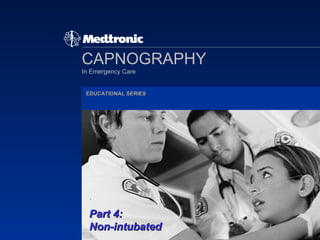
50. Part 4: The Non-intubated Patient Ready to take capnography for a run?
Editor's Notes
Part 4: Capnography on the non-intubated patient builds on the information presented in parts one, two and three. It may be presented as part of the series or in as a separate independent class. This PowerPoint program is designed for initial training on capnography in EMS. It is strictly an introduction and all information be adapted to your local protocols. The program is not product specific and qualifies for continuing education credits through individual CE providers and/or the Center for Healthcare Education. Information on the Center for Healthcare Education and the CE process is contained on this CD. You may also contact the Center at 1-800-888-8700 or their website http://www.healthcareeducation.org All contents are under the copyright of Medtronic Physio-Control Corp.
Capnography has been available and used for the intubated patient for several years. Now, new technology with nasal/oral cannulas provide easy ventilation monitoring of your non-intubated patients.
The learning objectives for part four are for you to be able to: List three non-intubated applications Identify four characteristic patterns seen in: - Bronchospasm such as Asthma COPD - Hypoventilation states - Hyperventilation - Low-perfusion states
Neighbors called 911 when they found an elderly woman sitting in her front yard dressed in pajamas. They reported her to be “a bit confused” and complaining of “some chest or breathing problems”.
Familiar? What comes to mind?
Capnography can be useful in this fairly typical EMS situation in: - Identifying the problem and underlying pathology - Assessing the patient’s status and - Helping you to anticipate sudden changes
Applications for capnography in your non-intubated patients include: Identification and monitoring of bronchospasm as in: - Asthma - COPD Assessing and monitoring: - Hypoventilation states - Hyperventilation - Low-perfusion states
Capnography reflects changes in: Ventilation - the movement of gases in and out of the lungs Diffusion - the exchange of gases between the air-filled alveoli and the pulmonary circulation Perfusion - circulation of blood through the arterial and venous systems
Patients can have several alterations in their ventilatory status. One that is of frequent concern in EMS is: Airway obstruction such as in: Smooth muscle contraction Bronchospasm Airway narrowing Uneven emptying of the alveoli Mucous plugs Capnography is useful in monitoring your patient’s airway.
Since capnography monitors the amount of carbon dioxide in the airway it is affected by changes in the diffusion of gases across into the alveoli. Decreased diffusion occurs with pulmonary changes found in bronchospastic diseases that produce: Airway inflammation Retained secretions Fibrosis Decreased compliance of alveoli walls Chronic airway modeling which is seen in COPD Reversible airway disease that occurs with asthma
Bronchospasm interferes with the normally smooth flow of air as the degree and timing of spasm varies throughout the pulmonary tree. The alveoli are unevenly ventilated on inspiration and empty asynchronously during expiration. This uneven emptying dilutes the carbon dioxide which results in a slower rise in CO 2 concentration during exhalation.
These spasmodic alterations in air flow affect phase II (ascending phase) and phase III (the plateau) of the capnography waveform and produce a characteristic pattern often referred to as the “shark fin”.
Asthma is a bronchospastic disease often seen in EMS. Asthma is increasing in the US. • 20.3 million citizens report having the disease and • Prevalence increased 75% from 1980-1994 • There are over two million ED visits each year for asthma • It is the most common chronic health problem in children Unfortunately, there is an increase in deaths due to asthma. • From 1987 to 1995, the death rate doubled to 5600 per year Sources: Delbridge T., et al . 2003 Prehospital Asthma Management. Prehospital Emergency Care 7 (1) 42-47 Asthmatic Statistics. American Academy of Allergies, Asthma and Immunology. http.//www.aaaai.org
An asthma attack may occur suddenly such as in reaction to an allergen or it may be progressive over days to weeks which may occur with an infection. The underlying pathology is in the airway with: • Increased responsiveness (or hyper-reactivity) of the airway • Bronchospasm resulting in reversible obstruction • and an inflammation process
The asthmatic patient’s response to either intrinsic or extrinsic triggers is: The release of inflammatory mediators - histamine, bradykinin and prostaglandins produces reactions in the bronchial wall that include: • Spasm of bronchial smooth muscle • Vasodilatation with swelling of bronchial mucous membranes and • Increased mucous production
In addition to the bronchi, this chemical release triggers responses in other systems. Symptoms produced by these reactions include: • Tachycardia • Tachypnea • Wheezing • Cough • Chest tightness • Use of accessory muscles (retractions) • Anxiety • Diaphoresis
The National Institute of Health has issued “Guidelines for the Diagnosis and Management of Asthma”. This slide is a summary of the classification of asthma. As shown here, if your initial exam finds your asthmatic patient to be not agitated, not wheezing and not bradycardic, do not be lulled into thinking that everything is going okay. Respiratory arrest may be imminent. Source: Edmond S. D. 1998. 1997 National Asthma Education and Prevention Program Guidelines: A Practical Summary for Emergency Physicians. Annals of Emergency Medicine 31: 5: 579-594
As we can see from the previous slide, your patient’s symptoms and your observations are primarily subjective. The severity of symptoms and your patient’s perception may not accurately reflect severity of condition. More objective data is needed to assess the situation. Source: Teeter J.G., et al. 1998. “Relationship Between Airway Obstruction and Respiratory Symptoms in Adult Asthmatics. CHEST .113:5:272-277
Capnography may be able to provide that objective data. This study by Yaron compared the capnograms of 28 normal volunteers and those of 20 asthma patients. The study objectives were to determine whether the slope of the expiratory capnogram (dCO 2 /dt) can detect bronchospasm in adult asthma patients in the ED and to assess the correlation between the plateau and the peak expiratory flow rate (PEFR). Their conclusion: • The slope value correlated with PEFR • The “dCo 2 /dt is an effort independent, rapid noninvasive measure that indicates significant bronchospasm” Source: Yaron M. 1996. Utility of the Expiratory Capnogram in the Assessment of Bronchospasm. Annals of Emergency Medicine 28: 4
The change in phase two is thought to be due to the uneven empting of the alveolar gas. Bronchospasm of varying degrees traps the carbon dioxide in alveoli throughout the lungs. This results in an uneven flow of the gas. Waveform examples show increasing change in the plateau with increasing obstruction. Source: Hall J.B., Acute Asthma, Assessment and Management, McGraw-Hill, New York.
These capnograms show the changes in the dCO 2 /dt seen with increasing bronchospasm. Source: Krauss B., et al . 2003. FEV1 in Restrictive Lung Disease Does Not Predict the Shape of the Capnogram. Oral presentation. Annual Meeting, American Thoracic Society, May, Seattle, WA
More research is needed on assessing and treating nonintubated patients. Research is now underway on the correlation of capnographic changes to patient’s respiratory status. Future studies will also look at the impact this technology will have patient care, outcomes and healthcare costs.
Here’s another case scenario using the findings in the some of the research: 16 year old female - a cheerleader who has become dyspneic while cheering in the year’s first football game. C/O “having difficulty breathing” • Visible distress • History of asthma, physical exertion, and “a cold this week” • Patient has used her “puffer” 8 times over the last two hours • Pulse 136, BP 148/80, RR 34 • Wheezing noted on expiration
Her initial capnogram is shown here with the “shark fin” on phase two. Note the change in the slope following therapy. Training Note: Ask about local protocol and what actions may be taken if the waveform changes as shown and what actions may be considered if the waveform does not change. Discuss what changes in patient symptoms and physical signs would be expected to be seen with these changes .
Another respiratory condition that you may frequently encounter is chronic obstructive lung disease. COPD is increasing in the United States. In fact it is now the: • Fourth leading cause of death in adults • In 1996, there were 16 million cases of COPD in this country. We are seeing an increase in deaths due to COPD • There were an estimated 110,000 deaths in 1999 and the • number of deaths has doubled in the past 25 years Source: Boyle, A.H. 2000. Recommendations of the National Lung Health Education Program, Heart & Lung 29: 6: 446-449
Although COPD is actually a group of diseases, the pathology includes several commonalities. The term COPD is used to describe a chronic, progressive disease process that usually has a history of one or more: • Major risk factors: smoking, exposure to dusts and fumes, history of frequent respiratory infections This spectrum of diseases includes: • Chronic bronchitis • Emphysema • Bronchiectisis
The COPD disease process is progressive and may be partially reversible. Airways are obstructed by: • Hyperplasia of mucous glands and smooth muscle • Excess mucous production, especially during an exacerbation • There is often some hyper-responsiveness here
The small airways are the: • Main sites of airway obstruction in COPD • There is usually chronic inflammation with • Fibrosis and narrowing and • Damage to alveoli • There is hyper-expansion of the alveoli due to air trapping which causes • Impaired gas exchange
Patients with COPD live with daily respiratory limitations and symptoms and call EMS when there is an increase in their chronic symptoms which include: • Increased SOB - often they wait until this is severe • Frequent cough • More wheezing - which may be audible without your stethoscope • Greater use of accessory muscles • Sputum of increased volume, tenacity and purulence • Higher anxiety and restlessness - they may be sleep deprived at this point • Diaphoresis • and chest tightness.
Patients with an acute escalation of their COPD may also have: • Fever from an underlying infection They frequently have other chronic conditions including: • Congestive heart failure • Acute coronary syndrome • Diabetes mellitus • Hypertension Many of these co-morbities have similar risk factors and presenting symptoms.
The symptoms and observations of COPD are primarily subjective. Your patient’s “baseline” may not be “normal”. Patients living with this chronic condition may have difficulty differentiating the specific change in their symptoms and the degree of change. Severity of symptoms and your patient’s perception may not accurately reflect severity of condition. Objective data is often needed in order to differentiate the most problematic diagnosis and assess each patient.
Correlating capnograms to patient status would provide objective data. Arterial CO 2 in COPD patients is known to: • Increase as the disease progresses • Blood gases require frequent arterial punctures for ABGs and the procedure is not available in EMS Correlating capnograph to the COPD patient status can be helpful. As with PaCO 2 : • Ascending phase and plateau is altered by uneven emptying of gases
Here’s a scenario of a dyspneic patient on capnography. • 72 year old male • C/O difficulty breathing • History of CAD, CHF, smoking and COPD • Productive cough, recent respiratory infection • Pulse 90, BP 158/82 RR 27
If his initial capnogram is similar to A. What would direction would your assessment take? If his initial capnogram is similar to B. How would your assessment change?
In a study presented at the 2003 International Conference of the American Thoracic Society, the difference in the capnogram of patients with obstructive diseases such as COPD was compared with those of patients with restrictive diseases such as CHF. They studied 207 patients in a pulmonary function lab. • 61 with obstructive disease (OD); 34 with restrictive disease (RD) Change in the exhalation plateau: • C/O severe difficulty breathing • 80% of OD had elevations >4°; 5% of RD had elevations >4° P<0.0001 Source: Krauss B., et al. 2003. FEV1in Restrictive Lung Disease Does Not Predict the Shape of the Capnogram. Oral presentation. Annual Meeting, American Thoracic Society, May, Seattle, WA. Teaching Note : Obstructive diseases are conditions that increase resistance to airflow (a) inside the airway (obstruction by a foreign body, excessive secretions), (b) by altering the airway wall (bronchitis, bronchospasm) or (c) through narrowing the airway (emphysema). Restrictive diseases impair lung expansion secondary to alterations in the lung, pleura, chest wall or neuromuscular impairment. RD conditions include pulmonary pneumothorax, pleural effusion, and pulmonary edema.
Here is a scenario based on a CHF report: • 78 year old male • C/O: Short of breath • H/O: COPD, MI X 2, on oxygen at 2 L/m • Pulse 66, BP 114/76/p, RR 36 labored and shallow, skin cool and diaphoretic, 2+ pedal edema • Initial SpO 2 69%; EtCO 2 17mmHG
In the initial assessment, the medics noted that his capnogram had a normal pattern and no evidence of acute bronchospasm. Based on this, they followed their medical protocol for CHF rather than COPD. He was placed on a non-rebreather mask with 100% oxygen at 15 L/m and given an IV diuretic and SL nitroglycerin. Ten minutes after treatment his SpO 2 went from 69% to 99%. The patient’s his EtCO 2 increased from 17mmHG to 35mmHG as his circulatory and respiratory status improved.
In hypoventilation, patients retain carbon dioxide with EtCO 2 >50mmHg. This can occur in an altered mental status and abnormal breathing such as in: • Sedation • Alcohol intoxication • Drug Ingestion • Postictal states • CNS infections • Head injury
Observer called 911. 76 year old male sleeping and unresponsive on sidewalk, “gash on his head”. Known history of hypertension, EtOH intoxication, Pulse 100, BP 188/82, RR 10, Pulse oximetry is 96% on room air.
This is his capnogram. Note the shape and height of the waveform.
Hypoventilation may also be exhibited differently if the patient’s breathing becomes more shallow. Shallow breathing often involves such low exhaled volumes that the gas deep inside the lung, “alveolar gas”, may not flow all the way to the mouth to be sampled. Instead, some of the gas in the trachea, called dead space gas, that does not contain CO2 may mix with the alveolar gas and dilute it. In this scenario, even though blood and alveolar CO2 are elevated, end tidal CO2 will appear to decrease. When the patient takes a deep breath, the carbon dioxide in the airway system is exhaled and the EtCO 2 level is elevated.
Capnography can also show alteration in perfusion both in the pulmonary tree and total body system. As we discussed in Part 3, it provides a noninvasive measure of cardiac output.
Let’s look at a case scenario of a patient with low perfusion. • 57 year old male • Auto crash with injury to chest • History of atrial fib, anticoagulant • Unresponsive • Pulse 100 irregular, BP 88/p • Intubated on scene
With his airway and ventilation controlled, capnography his perfusion status. The shape of the waveform is the normal rectangular box and the low EtCO 2 is an indication of his low cardiac output. Capnography provides another parameter to monitor the state of this critical patient.
New applications for capnography are being reported as research and the use of the technology expands. These new applications include: • Pulmonary emboli • CHF • DKA • Bioterrorism • Others?
Here is a scenario for a patient with a possible pulmonary embolus. • 72 year old female • CC: Sharp chest pain, short of breath • History: Legs swollen and pain in right calf following flight from Alaska • Pulse 98 and regular, RR 22, BP 158/88, SpO 2 95%
In this case, the patient has a strong pulse and adequate blood pressure. Capnography indicates a low perfusion state. If the systemic circulation is adequate, the more likely reason for this reading is the diminished pulmonary perfusion. As always, this is to be added to the other information on the patient.
In summary, capnography is now available in an easy-to-use cannula that provides for many applications in your non-intubated patients including: • Identification and monitoring bronchospasm as seen in asthma and COPD • Assessing and monitoring: - hypoventilation states - hyperventilation - low perfusion states - others that we have discussed.