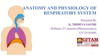
7. Functional anatomy of the respiratory system:
Conducting Zone
• Rigid conduits for air to reach site
of gas exchange
-nose
-nasal cavity
-pharynx
-larynx
-trachea
-bronchi
Respiratory Zone
• site of gas exchange
-respiratory bronchioles
-alveolar ducts
7
8. Conducting Zone:
• Nose
-airway
-moistens and warms air
-filters inspired air
-resonating chamber for speech
-olfaction
• Paranasal sinuses
-frontal, sphenoid, ethmoid and
maxillary bones
-warm and moisten air
8
9. • Pharynx
-connects the nasal cavity and mouth to the larynx and oesophagus
-common pathway for food and air (throat)
-nasopharynx
-oropharynx
-laryngopharynx
9
10. • Laryngopharynx – common passage way for food and air
• Larynx – voice box
10
12. Bronchi
• Bronchial tree
-left and right primary bronchi
-formed by divisions of the trachea
-secondary bronchi (lobar)
-inside the lungs
-3 on the right
-2 on the left
-tertiary bronchi (segmental)
-fourth-order
-fifth-order
-23 orders of branching air ways
-bronchioles (under 1 mm in
diameter
12
16. Respiratory membrane (air-blood barrier) or (Alveolar-capillary membrane) is
composed of:
-simple squamous epithelial cells (Type I cells)
-cobweb of pulmonary
capillaries
Primary function is gas exchange
-Type II cells (cuboidal) surfactant
-elastic fibers
-alveolar pores allow for pressure
equalization between alveoli
-alveolar macrophages (dust cells)
16
18. Secretion of Surfactant
by Alveoli
Pulmonary surfactant is a mixture of lipids and proteins which is
secreted by the epithelial type II cells into the alveolar space. Its
main function is to reduce the surface tension at the air/liquid
interface in the lung. This is achieved by forming a surface film that
consists of a monolayer which is highly enriched in
dipalmitoylphosphatidylcholine and bilayer lipid/protein structures
closely attached to it.
18
19. • Pleural Coverings:
-double layered serosa
-parietal pleura lines the thoracic
wall
-pulmonary or visceral pleura
which covers the lung surface
-pleural cavity is the space
between the two layers
-pleural fluid fills the cavity
19
20. • Blood supply:
-Pulmonary circulation
-Bronchial circulation
Pulmonary arteries from the right side of the heart supply blood to the lungs.
-pulmonary arteries branch profusely along with the bronchi
-pulmonary capillary networks surrounding alveoli
-pulmonary veins form post alveoli to carry oxygenated blood back to the heart
Bronchial arteries come from the aorta and enter the lung at the hilus
-the bronchial arteries run along the branching bronchi and supply lung tissue except the alveoli
-bronchial veins drain the bronchi but most moves into the pulmonary circulation
20
23. Pulmonary Vessels. The pulmonary artery extends only 5 centimeters
beyond the apex of the right ventricle and then divides into right and left main
branches that supply blood to the two respective lungs.
The pulmonary artery is thin, with a wall thickness one third that of the
aorta. The pulmonary arterial branches are very short, and all the pulmonary
arteries, even the smaller arteries and arterioles, have larger diameters than their
counterpart systemic arteries. This, combined with the fact that the vessels are thin
and distensible, gives the pulmonary arterial tree a large compliance, averaging
almost 7 ml/mm Hg, which is similar to that of the entire systemic arterial tree.
This large compliance allows the pulmonary arteries to accommodate the stroke
volume output of the right ventricle.
The pulmonary veins, like the pulmonary arteries, are also short. They
immediately empty their effluent blood into the left atrium, to be pumped by the
left heart through the systemic circulation.
23
24. Bronchial Vessels. Blood also flows to the lungs through small bronchial
arteries that originate from the systemic circulation, amounting to about 1 to 2
per cent of the total cardiac output. This bronchial arterial blood is oxygenated
blood, in contrast to the partially deoxygenated blood in the pulmonary arteries.
It supplies the supporting tissues of the lungs, including the connective tissue,
septa, and large and small bronchi. After this bronchial and arterial blood has
passed through the supporting tissues, it empties into the pulmonary veins and
enters the left atrium, rather than passing back to the right atrium. Therefore, the
flow into the left atrium and the left ventricular output are about 1 to 2 per cent
greater than the right ventricular output.
24
25. Lymphatics. Lymph vessels are present in all the supportive tissues of
the lung, beginning in the connective tissue spaces that surround the
terminal bronchioles, coursing to the hilum of the lung, and thence
mainly into the right thoracic lymph duct.
Particulate matter entering the alveoli is partly removed by way of
these channels, and plasma protein leaking from the lung capillaries is
also removed from the lung tissues, thereby helping to prevent
pulmonary edema.
25
26. • Innervation:
-parasympathetic motor fibers (some sympathetic fibers)
-visceral sensory fibers
• Enter the lung through the pulmonary plexus on the lung root
• parasympathetic fibers – constrict the air tubes
• sympathetic fibers – dilate air tubes
26
28. Respiratory Mechanisms
• The lungs can be expanded
and contracted in two ways:
• (1) By downward and
upward movement of the
diaphragm to lengthen or
shorten the chest cavity,
• (2) By elevation and
depression of the ribs to
increase and decrease the
anteroposterior diameter of
the chest cavity.
28
29. INSPIRATION: Inspiration is the active part of the breathing process, which is
initiated by the respiratory control center in medulla oblongata (Brain stem).
• Activation of medulla causes a contraction of the diaphragm and intercostal
muscles leading to an expansion of thoracic cavity and a decrease in the
pleural space pressure.
• When it contracts, it moves downward and because it is attached to the lower
ribs it also rotates the ribs toward the horizontal plane, and thereby further
expands the chest cavity.
29
30. • The external intercostal muscles connect adjacent ribs. When they
contract the ribs are pulled upward and forward causing further
increase in the volume of the thoracic cavity. As a result fresh air flows
along the branching airways into the alveoli until the alveolar pressure
equals to the pressure at the airway opening.
• EXPIRATION: Expiration is a passive event due to elastic recoil of
the lungs. However, when a great deal of air has to be removed
quickly, as in exercise, or when the airways narrow excessively during
expiration, as in asthma, the internal intercostal muscles and the
anterior abdominal muscles contract and accelerate expiration by
raising pleural pressure.
30
33. • Airway Resistance-
-friction or drag along the respiratory
passageway
-maximum resistance in medium size
bronchi then drops as cross sectional area
increases
-bronchiole smooth muscle very sensitive
to parasympathetic stimulation
33
34. Lung Volumes
• Tidal volume (TV): Volume of air inhaled or exhaled with each
breath during normal breathing (0.5 L).
• Inspiratory reserve volume (IRV): Maximal volume of air inhaled at
the end of a normal inspiration (3 L)
• Expiratory reserve volume (ERV): Maximal volume of air exhaled
at the end of a tidal volume (1.2 L).
• Inspiratory capacity (IC): Maximal volume of air inhaled after a
normal expiration (3.6 L) (TV+IRV)
• Functional Residual Capacity (FRC): The volume of gas that
remains in the lung at the end of a passive expiration. (2-2.5 L or 40 %
of the maximal lung volume) (ERV+RV).
• Residual Volume (RV): The volume of gas remains in the lung after
maximal expiration. (1-1.2 L) 34
35. • Total Lung Capacity (TLC): The maximal lung volume that can be
achieved voluntarily. (5-6 L) (IRV+ERV+TV+RV)
• Vital capacity (VC): The volume of air moved between TLC and RV.
(4-5 L) (IRV+ERV+TV).
• Multiplying the tidal volume at rest by the number of breaths per
minute gives the total minute volume (6 L/min). During exercise the
tidal volume and the number of breaths per minute increase to produce
a total minute volume as high as 100 to 200 L/min.
35
37. Regulation and Control
of Breathing
Basic elements of the respiratory
control system are
(1) Strategically placed sensors
(2) Central controller
(3) Respiratory muscles.
37
38. Sensors
1.Mechanoreceptors: These receptors are placed in the walls of
bronchi and bronchioles of the lung and the main function of these
receptors is to prevent the overinflation of the lungs. Inflation of the
lungs activates these receptors and activation of the stretch receptors in
turn inhibits the neurones in inspiratory centre via vagus nerve.
2.Chemoreceptors: The respiratory system maintains concentrations of
O2, CO2 and the pH of the body fluids within the normal range of
values. Any deviation from these values has a marked influence on the
respiration. Chemoreceptors are specialized neurons activated by
changes in O2 or CO2 levels in the blood and the brain tissue,
respectively.
38
40. Central Controller
Breathing is mainly controlled at the level of brainstem. The normal
automatic and periodic nature of breathing is triggered and controlled by
the respiratory centres located in the pons and medulla. These centers
are not located in a special nucleus or a group of nuclei but they are
rather poor defined collection of neurons.
40
41. 1. Medullary respiratory centre:
• -Dorsal medullary respiratory neurons are associated with
inspiration.
• -Ventral medullary respiratory neurons are associated with
expiration.
2.Apneustic Centre: It is located in the lower pons.
3.Pneumotaxic center: It is located in the upper pons. This center is a
group of neurons that have an inhibitory effect on the both inspiratory
and apneustic centers.
41
42. Gas Exchange
• After the alveoli are ventilated with
fresh air, the next step in the
respiratory process is diffusion of
oxygen from the alveoli into the
pulmonary blood and diffusion of
carbon dioxide in the opposite
direction, out of the blood.
42
44. Partial pressures (pp) of individual gases
The pressure of a gas acting on the surfaces of the respiratory
passages and alveoli is proportional to the summated force of impact
of all the molecules of that gas striking the surface at any given
instant.
This means that the pressure is directly proportional to the concentration
of the gas molecules.
44
45. Air is composed of
• 79% nitrogen
• 21% oxygen.
The total pressure at sea level averages 760 mm Hg
-79 % of the 760 mm Hg is caused by nitrogen PN2 (600 mm
Hg)
-21 % by oxygen ,PO2 (160 mm Hg).
45
46. Factors determine the partial pressure of a gas dissolved in a fluid
The partial pressure of a gas in a solution is
Determined by:
• concentration
• solubility coefficient of the gas.
• Henry's Law - solubility of a gas in a liquid depends on temperature,
the partial pressure of the gas over the liquid, the nature of the solvent
and the nature of the gas.
46
47. Diffusion of gases between the gas phase in the alveoli and
the dissolved phase in the pulmonary blood
The rate at which each gas diffused is directly proportional to their
partial pressure in the blood tends to force molecules of that gas into
solution in the blood of the alveolar capillaries.
47
48. Oxygen concentration and partial pressure in the alveoli
• Oxygen is continually being absorbed from the alveoli into the blood of the lungs,
and new oxygen is continually being breathed into the alveoli from the
atmosphere.
• oxygen concentration in the alveoli, and its p p , is controlled by
(1) the rate of absorption of oxygen into the blood and
(2) the rate of entry of new oxygen into the lungs by the ventilatory process.
• The more rapidly oxygen is absorbed, the lower its concentration in the alveoli
becomes conversely, the more rapidly new oxygen is breathed into the alveoli
from the atmosphere, the higher its concentration becomes.
48
49. Carbon dioxide concentration and partial pressure in the alveoli
Carbon dioxide is continually being formed in the body and then
carried in the blood to the alveoli; it is continually being removed
from the alveoli by ventilation.
normal rate of CO2 excretion of 200 ml/min.
At the normal rate of alveolar ventilation of 4.2 L/min alveolar PCO2
is, 40 mm Hg.
49
50. - The alveolar PCO2 increases directly in proportion to the
rate of carbon dioxide excretion,
- The alveolar PCO2 decreases in inverse proportion to
alveolar ventilation.
- Therefore, the concentrations and pp of both O2 and CO2
in the alveoli are determined by :
*The rates of absorption or excretion of the two gases.
*by the amount of alveolar ventilation.
50
51. Expired Air
Expired air is combination of dead
space & alveolar air
• Dead space air is first portion
which consists of humidified air
• Second portion is mixture of
both
• Alveolar air is expired at end of
exhalation
so it determined by
(1) the amount of the expired air
that is dead space air and
(2) the amount that is alveolar air.
51
52. Layers of the respiratory membrane:
1. A layer of fluid lining the alveolus
and containing surfactant
2. The alveolar epithelium composed of
thin epithelial cells
3. An epithelial basement membrane
4. A thin interstitial space between the
alveolar epithelium and the capillary
membrane
5. A capillary basement membrane that
in many
places fuses with the alveolar epithelial
basement membrane
6. The capillary endothelial membrane
52
53. Factors affect gas diffusion
The factors that determine gas diffusion through the
membrane are
(1) the thickness of the membrane,
(2) the surface area of the membrane,
(3) the diffusion coefficient of the gas in the substance
of the membrane, and
(4) the partial pressure difference of the gas between
the two sides of the membrane
53
54. 54
• Diffusion of Oxygen from
the Alveoli to the Pulmonary
Capillary Blood
56. 56
• Diffusion of Oxygen from the Peripheral Capillaries into the Tissue Fluid
57. 57
Diffusion of Carbon Dioxide from the Peripheral
Tissue Cells into the Capillaries and from the Pulmonary Capillaries
into the Alveoli
a. When oxygen is used by the cells, virtually all of it becomes carbon dioxide,
and thus increases the intracellular PCO2
b. Carbon dioxide diffuses about 20x as rapidly as oxygen
61. Factors That Shift the Oxygen-Hb Dissociation Curve
61
• pH; acidic it shifts to the right and if basic, it shifts to the left
• Increased carbon dioxide concentration
• Increased blood temperature
• Increased BPG (2,3 biphosphoglycerate), metabolic compound found in the
blood
63. Bohr Effect- a shift of the dissociation curve to the right due to
increased CO2 and H ions enhances the release of oxygen from the
blood into the tissues and enhances the oxygenation of the blood in the
lungs
As blood passes through the tissues, carbon dioxide diffuses from
the tissue cells into the blood. This increases PCO2 which in turn raises
the blood H2CO3(carbonic acid) and the hydrogen ion concentration.
This forces oxygen away from Hb and delivers increased amounts
to the tissues
• Exactly the opposite happens in the lungs
63
65. When Oxygen Binds with Hb, Carbon Dioxide
is Released to Increase CO2 (Haldane Effect)
Binding of oxygen with Hb tends to displace carbon dioxide from the blood
(more important than the Bohr Effect)
Oxygen plus Hb in the lungs causes Hb to become a stronger acid
65