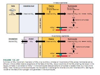
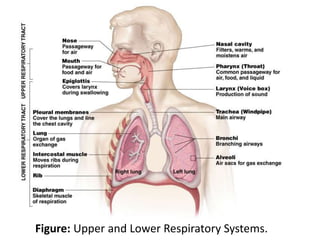
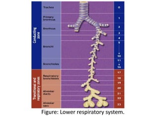
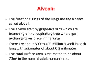
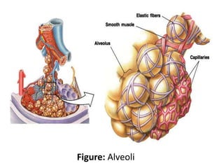
SlideShare a Scribd company logo
Respiratory System
Respiration:
Respiration: It refers to interchange of gases between the
atmosphere and the cells of the body.
– External respiration: Gas exchange (O2 & CO2) between the air
and blood.
– Internal respiration: Gas exchange (O2 & CO2) between the
blood and other tissues and O2 utilization by the tissues of the
body.
Pulmonary Ventilation (breathing): is transport of air to
and from the lungs. Pulmonary ventilation (breathing)
consists of two phases:
– Inspiration (Inhalation) – air in
– Expiration (Exhalation) – air out
Functions of the respiratory system:
1. Gas exchange: Uptake of O2 by cells, and the release of CO2 to the
lungs.
2. Acid base balance: CO2 +H2O←→ H2CO3 ←→ H+ + HCO3-
3. Pulmonary defense
4. Respiratory heat loss by evaporation of water: It removes heat and
water from body by warming and saturation the inhalated air with
water vapor.
5. Pulmonary metabolism: The lungs converts substances that pass
through the pulmonary blood vessels, for e.g. Angiotensin I →
angiotensin II.
6. It fascilitates venous return to the heart (Respiratory pump).
7. Production of sound (Vocalization): The larynx contains the paired
vocal folds (vocal cords).
8. Site for olfactory sensation (Smelling)
Physiological anatomy of the respiratory system:
The respiratory system is divided into:
1. Upper respiratory system:
- Nose
- Pharynx
2. Lower respiratory system:
- Larynx (Voice box)
- Trachea (Windpipe)
- Bronchi
- Bronchioles
- Alveoli
Figure: Upper and Lower Respiratory Systems.
Nose and Pharynx:
• Nose:
– External nose
– Nasal cavity
• Functions:
– Passageway for air
– Filters and cleans the air
– Humidifies, warms air
– Smell
– Along with paranasal
sinuses are resonating
chambers for speech
• Pharynx:
– Common opening for
digestive and respiratory
systems.
– Three regions:
• Nasopharynx
• Oropharynx
• Laryngopharynx
Figure: Nasal Cavity and Pharynx.
Larynx:
• Functions:
– The air passageway between pharynx and trachea.
– The epiglottis prevents food and liquid from entering the larynx
and directed through the pharynx into the esophagus.
– Vocal folds (vocal cords) are primary source of sound production.
Figure: Vocal folds (Vocal cord).
Trachea:
- It is a flexible tube, extends from the larynx to the
primary bronchi. It transports air to and from the
lungs.
- The wall of the trachea contains 16-20 C-shaped
rings of hyaline cartilage, which keep the trachea
open.
- The mucosa of the trachea is ciliated epithelium
with mucus-producing goblet cells. As in the
larynx the cilia sweep upward toward the
pharynx.
Figure: Trachea.
Components of the lower respiratory tract:
1. Conducting zone:
- Bronchi
- Bronchioles
- Terminal bronchioles
2. Respiratory zone:
The Site gas exchange (O2 and CO2) between air and blood
which Composed of:
- Respiratory bronchioles
- Alveolar ducts
- Alveolar sacs
Figure: Lower respiratory system.
The lungs:
Physical properties of the lungs:
1. Compliance:
- Distensibility (stretchability)
- The expansibility of the lungs and thoracic wall, necessary
for adequate alveolar ventilation.
- Thoracic compliance may be decreased by factors that
produce resistance to distension for e.g., fractured ribs.
2. Elasticity:
- Tendency of the lungs to return to the initial size after
distension.
- High content of elastin proteins (Recoil ability).
Alveoli:
- The functional units of the lungs are the air sacs
called alveoli.
- The alveoli are tiny grape-like sacs which are
branching of the respiratory tree where gas
exchange takes place in the lungs.
- There are about 300 to 400 million alveoli in each
lung with adiameter of about 0.2 milimeter.
- The total surface area is estimated to be about
70m2 in the normal adult human male.
Figure: Alveoli
There are three major types of the cell in the alveolar wall:
1. Alveolar type I cells: that form most of the alveolar walls
are simple squamous epithelium.
2. Alveolar type II cells: Also called septal cells secrete
pulmonary surfactant, a lipoprotein which mixes with the
tissue fluid within the alveoli and decreases its surface
tension, permitting inflation of the alveoli.
3. Alveolar macrophages: Within the alveoli are
macrophages that phagocytize pathogens or other
foreign material that may not have been swept out by
the ciliated epithelium of the bronchial tree.
Figure: The alveolar wall
Surfactant:
- Alveolar fluid contains a substance that reduces surface tension.
This substance is called surfactant.
- Surfactant is greatly reduces the surface tension of water.
- It is secreted by special surfactant-secreting epithelial cells called
alveolar type II cells into alveoli and respiratory passage.
- Surfactant is a complex mixture of several phospholipids for e.g.,
dipalmitoylphosphatidylcholine (DPPC), proteins, and ions.
- It is important at birth. The lungs remain collapsed until birth.
After birth, the infant makes several strong inspiratory
movements and the lungs expand. Surfactant keeps them from
collapsing again.
- Surfactant begins to be produced in late fetal life. For this reason,
premature babies are sometimes born with lungs that lack
sufficient surfactant and their alveoli are collapsed as a result.
This condition is called respiratory distress syndrome (RDS).
Figure: Surfactant
The Pleurae:
A double-layered sac surrounding each lung:
- Parietal pleura( outer layer)
- Visceral pleura (directly on lung)
Pleural cavity: The space between the visceral and parietal pleurae filled with pleural
fluid which is lubricate movement of lung within the cavity.
Figure: The muscles involved in breathing.
Mechanics of respiration:
At rest:
• Inspiration:
- Diaphragm conrtraction→chest volume↑ (lungs expansion)
→intrapleural pressure ↓→alveolar pressure<atmospheric
pressure→air is sucked into the lungs.
(At the end of an inspiration, alveolar pressure=atmospheric
pressure and airflow stops )
• Expiration:
- Diaphragm relaxation→chest volume↓ (lungs conrtraction)
→intrapleural pressure ↑→alveolar pressure>atmospheric
pressure→ air is pushed out of the lungs. Therefore,
expiration is a passive process. (At the end of an exspiration,
also alveolar pressure=atmospheric pressure and airflow
stops )
Figure: The mechanics of pulmonary ventilation.
Figure: Inspiration and Expiration.
Stronger ventilation:
- Muscles of the chest wall help produce changes in
chest volume beyond that produced by the
contraction and relaxation of the diaphragm.
- Contraction of the external intercostal muscles
helps increase the volume of the chest for
stronger inspiration. while contraction of the
internal intercostal muscles helps to decrease
chest volume for stronger expiration.
Lung Pressures:
- Atmospheric pressure: It is the pressure excreted by
the weight of air in atmosphere. It is about 760 mmHg.
- Intrapulmonary pressure (alveolar pressure): It is the
pressure of the air inside the alveoli.
- Intrapleural pressure: It is the pressure of the fluid in
the thin space between the lung pleura (visceral
pleura) and the chest wall pleura (parietal pleura). It is
always less than Intrapulmonary pressure.
- Transpulmonary pressure: It is the difference beween
the alveolar pressure and intrapleural pressure.
Figure: Lung pressure change during inspiration and expiration.
Lung Volumes:
- Tidal volume (TV): The volume of air inspired or expired
during a normal inspiration or expiration; its amount is
about 500 mL in the adult male.
- Inspiratory reserve volume (IRV): The amount of air
inspired forcefully after inspiration of normal tidal
volume (3000 mL).
- Expiratory reserve volume (ERV): The amount of air
forcefully expired after expiration of normal tidal
volume(1100 mL)
- Residual volume (RV): The volume of air remaining in
respiratory passages and lungs after the most forceful
expiration (1200 mL).
Figure: Lung volume and capacity.
Lung Capacities:
- Inspiratory capacity (IC): The maximal amount of air which can be
inspired after a normal expiration.
IC = Tidal volume (TV) + Inspiratory reserve volume (IRV)
- Vital Capacity (VC): The maximum amount of air which can be
expired after a maximal inspiration.
VC= Inspiratory reserve volume (IRV) + Tidal volume + Expiratory
reserve volume (ERV)
- Total lung capacity (TLC): The amount of air contained in the lungs at
the end of maximal inspiration.
TLC = Vital capacity + Residual volume (RV)
- Functional residual capacity (FRC): The amount of air that remains in
the lungs at the end of normal expiration.
FRC = Expiratory reserve volume (ERV)+ Residual volume (RV)
Figure: Lung capacities.
Pulmonary Ventilation:
- Respiratory rate or frequency: The number of breaths taken per
minute (12 breaths/min).
- Pulmonary ventilation: The amount of air inspired per minute.
Pulmonary ventilation = Respiratory rate X Tidal volume
= 12 breaths/min X 500 ml = 6 L/min.
- Alveolar ventilation: The total volume of fresh air entering the alveoli
per minute.
= respiratory rate X (Tidal volume – Dead space volume)
= 12 X (500 – 150)= 4.2 L/min
Minute ventilation: Total amount of air moved into and out to of
Respiratory system per min.
- Anatomical dead space: The volume of conducting air
ways at which there is no gaseous exchange (about
150ml) increase with age.
- Alveolar dead space: Volume of inspired air that is not
used for gas exchange .
- Physiological dead space: The sum of Anatomic and
Alveolar dead space.
Gas exchange in the lungs:
Air-Blood barrier (Pulmonary membrane, respiratory
membrane, Alveolar-Capillary barrier):
• The membrane which gas exchange occurs which is about 0.2 µm.
• The alveolar-capillary barrier is composed of:
- A thin alveolar epithelium
- An epithelial basement membrane
- A thin interstitial space between the alveolar basement membrane
and capillary basement membrane.
- A capillary basement membrane that in many places fuses with the
alveolar epithelial basement membrane
- The capillary endothelial membrane
• The total surface area of the respiratory membrane is about 70m2 in
the normal adult human male.
Figure: Alveolar respiratory membrane.
Partial Pressures:
• Dalton’s Law
– Law of Partial Pressures
• “each gas in a mixture of gases will exert a pressure independent
of other gases present”
Or
• The total pressure of a mixture of gases is equal to the sum of the
individual gas pressures.
– Atmospheric components
• Nitrogen = 78% of our atmosphere
• Oxygen = 21% of our atmosphere
• Carbon Dioxide = 0.033% of our atmosphere
• Water vapor, krypton, argon, …. Make up the rest
PATM = PN2 + P02 + PC02 + PH20= 760 mm Hg.
38
Changes in Partial Pressure:
• In inspired air:
– PO2=160 mmHg
– PCO2=0.3 mmHg
– PH2O=5.7 mmHg
– PN2=596 mmHg
• In alveolar air (mixing with dead space air):
– PO2=104 mmHg
– PCO2=40 mmHg
• In venous blood entering the lungs:
– PO2=40 mmHg
– PCO245 mmHg
• O2 diffuses across the respiratory surfaces into blood, CO2
flows from blood to alveolar air.
• Expired air is saturated with water vapor.
• In expired air:
– PO2=120 mmHg
– PCO2=27 mmHg
– PH2O=47 mmHg
– PN2=565 mmHg
• Arterial blood leaving the lungs:
– PO2=95 mmHg
– PCO2=40 mmHg
• In the tissues, concentrations of gases and pressures
exerted by them vary depending on the amount of
metabolic activity going on in the tissue at any one time.
– PO2=20 mmHg
– PCO2=46 mmHg
• O2 flows from blood to tissues, and CO2 from tissues to
blood.
Change in partial pressure in pulmonary capillary and tissue capillary
Transport of gases in the blood:
I. Oxygen Transport:
• O2 is carried in two ways by the blood:
1. Dissolved in plasma: (1.5 %)
The amount of O2 in the plasma is proportional to
the partial pressure. At a pressure of 100 mmHg,
0.3 ml O2 is dissolved in every 100ml blood.
2. Combined with hemoglobin: (98.5 %)
Most of the O2 is carried in combination with
hemoglobin. Each hemoglobin molecule having the
capacity to combine with four molecules of O2.
Figure: Oxygen Transport
Factors affecting Hb-O2 dissociation curve:
The ability of Hb to hold O2 is decreased by:
1. A decrease in pH,
2. An increase in carbon dioxide, or
3. An increase in temperature,
4. The presence of 2.3-diphosphoglycerate (2,3-DPG) in
red blood cells stimulate the release of O2 from HbO2.
Figure: Shifting the curve.
Transport of Carbon Dioxide:
Carbon dioxide is transported by:
1. Dissolved in plasma (10%):
- The solubility of CO2 in blood is about 20 times that of O2, so that there
is more CO2 than O2 in simple solution.
2. Carbaminohemoglobin (CO2+Hb) (20%):
- The CO2 can combine with amine (-NH2) groups in proteins to form
carbonic compounds. Because more amine groups are available in
hemoglobin than in plasma proteins, more CO2 combines with
hemoglobin than with plasma proteins to form carbamino
compounds.
3. Bicarbonate ions HC03
- (70%):
- Red blood cells contain the enzyme carbonic anhydrase, which
catalyzes the combination of H2O with CO2.
CO2 + H2O carbonic anhydrase H2CO3 H + + HCO3
-
Figure: Carbon Dioxide Transport and Chloride Shift
Figure: Reverse chloride shift in lungs.
Regulation of Respiration:
The respiratory center is located in the pons and medulla
oblongata of the brain stem:
1. Pons: It consist of two respiratory control center:
a). Apneustic center: It promotes the inspiration.
b). Pneumotaxic center: It mainly controls rate and depth of breathing
(Respiratory rhythm). It inhibits the inspiration.
2. Medullary oblongata: It consist of the following groups of the
neurons:
a). Dorsal respiratory group (DRG): Mainly cause inspiration (Normal
quiet breathing)
b). Ventral respiratory group (VRG): cause expiration and inspiration
(Labored respiration)
Figure: Respiratory center.
Dorsal respiratory group (DRG):
- It is located in the dorsal portion of medulla.
- The signal transmitted from this group through the motoneuron
phrenic nerve mainly to diaphragm and cause normal quiet
breathing.
- The signal are rhythmic and ramp in nature. When signal stop
suddenly will allow the elastic recoil of lung and chest wall to cause
expiration.
Ventral respiratory group (VRG):
- It is located in the ventral aspect of medulla.
- They send signals to inspiratory and expiratory muscle during active
or labored respiration.
Pneumotaxic center:
- It is located dorsally in the superior portion of the pons, which mainly
controls the rate and depth of breathing.
- The normal function of the pneumotaxic center is unknown, but it
may play a role in switching off between inspiration and expiration.
Figure: Organization of the respiratory center
Chemical control of respiration:
1. Central chemoreceptors (Chemosensitive area):
- It is located in the ventral surface of medulla and sensitive to change
of PCO2 and H+ concentration.
- The chemoreceptors monitor the H + concentration of CSF, including
the brain interstitial fluid. CO2 readily penetrates membranes,
including the blood-brain barrier, whereas H + and HCO3-
penetrate slowly.
- The CO2 that enters the brain and CSF is promptly hydrated. The
H2CO3 dissociates, so that the local H+ concentration rises. Any
increase in spinal fluid H+ concentration stimulates respiration.
The magnitude of the stimulation is proportionate to the rise in H+
concentration.
- Thus, the effects of CO2 on respiration are mainly due to its
movement into the CSF and brain interstitial fluid, where it
increases the H+ concentration and stimulates receptors sensitive
to H +.
Figure: Chemoreceptors in the medulla oblongata.
2. Peripheral chemoreceptors:
They are located outside the brain in the:
- Carotid bodies in the bifurcation of the common carotid
arteries.
- Aortic bodies along the arch of the aorta.
- They are especially important for detecting changes in O2
in the blood, although they also respond to a lesser
extent to changes in CO2 and H+ ion concentrations.
- They are stimulated by decreasing of PO2 in blood, and
also by increasing of PCO2 and H+ ions in blood.
Figure: Peripheral chemoreceptors
Figure: Regulation of breathing.
Common Terms:
- Eupnea: Normal, comfortable breathing at rest.
- Apnea: Cessation of breathing.
- Dyspnea: Unpleasant, subjective feeling of difficult or
labored breathing.
- Polypnea: Increased depth or rate of breathing or both.
- Polypnea: Rapid, shallow breathing.
Pulmonary Disorders:
- Acute respiratory disease syndrome (ARDS): It is a severe
inflammatory disease of the lung. The inflammation leads to
impaired gas exchange.
- Infant respiratory distress syndrome (IRDS): It is a syndrome
caused by lack of surfactant in the lungs of premature infants.
- Pneumothorax: The presence of gas in the intrapleural space (the
space between the visceral and parietal pleurae) causing the
lungs to collapse.
- Pulmonary fibrosis: The normal structure of lungs disrupted by
accumulation of fibrous connective tissue proteins.
- Emphysema: The loss of elasticity in the lungs leads to
prolonged times for exhalation. This leads to a smaller
volume of gas exchanged per breath.
- Chronic bronchitis: It occurs when an abundance of
mucus is produced by the lungs and the air passages
become clogged with mucus.
- Lung cancer: It is a common form of cancer causing the
uncontrolled growth of cells in the lung tissue.
- Pneumonia: It is an infection of the lung parenchyma,
which can be caused by both viruses and bacteria.
Cytokines and fluids are released into the alveolar cavity
in response to infection, causing the effective surface
area of gas exchange in the lungs to be reduced.
Atelectasis: It mean collapse of alveoli due to obstruction of
air way or lack of surfactant.
Asphyxia: In asphyxia produced by occlusion of the airway,
acute hypercapnia and hypoxia develop together.
Drowning: Drowning is asphyxia caused by immersion,
usually in water.
Asthma: It is characterized by airway obstruction, airway
inflammation, and airway hyper-responsiveness to a
variety of stimuli. Proteins released from eosinophils in
the inflammatory reaction may damage the airway
epithelium causing the amount of air flow into the lungs
to be greatly reduced. The usual cause of asthma is
contractile hypersensitivity of the bronchioles in response
to foreign substances in the air such as plant pollen and
irritants in smog.
Hypoxia:
Hypoxia: It is O2 deficiency at the tissue level.
Traditionally, hypoxia has been divided into four types:
1. Hypoxic hypoxia (anoxic anoxia): in which the PO2 of the
arterial blood is reduced.
2. Anemic hypoxia: in which the arterial PO2 is normal but
the amount of hemoglobin available to carry O2 is
reduced.
3. Stagnant Hypoxia:
Hypoxia due to slow circulation is a problem in organs
such as the kidneys and heart during shock.
4. Histotoxic Hypoxia:
Hypoxia due to inhibition of tissue oxidative processes is
most commonly the result of cyanide poisoning. Cyanide
inhibits cytochrome oxidase and possibly other enzymes.65
Cyanosis:
- The term cyanosis means blueness of the skin, and its
cause is excessive amounts of deoxygenated hemoglobin
in the skin blood vessels, especially in the capillaries.
- It is most easily seen in the nail beds and mucous
membranes and in the earlobes, and lips, where the skin
is thin.
Carbon monoxide (CO) poisoning:
- The CO poisoning is toxic because it reacts with
hemoglobin to form carboxyhemoglobin (COHb)
- The COHb cannot take up O2.
Respiration in Birds:
Structure of Avian respiratory system
• The mouth or the nostrils (nares).
• The pharynx
• The trachea (Windpipe).
• Primary bronchi
• Air sacs
• Lungs
- The trachea branches into two primary bronchi at the
syrinx. The syrinx is situated at bifurcation of the
trachea, and it function as sonic membrane (voicebox).
- The primary bronchi enter the lungs and branch off into
dorsobronchi which lead into smaller parabronchi.
- Parabronchi can be several millimeters long and 0.5-2.0
mm in diameter (depending on the size of the bird) and
their walls contain hundreds of tiny, branching 'air
capillaries' surrounded by a profuse network of blood
capillaries. It is within these 'air capillaries' that the
exchange of gases (oxygen and carbon dioxide) between
the lungs and the blood occurs.
- After passing through the parabronchi, air moves into the
ventrobronchi.
Figure: The avian air sacs
Figure: The avian respiratory system
Avian air sacs:
• The air sacs is thin walled, about 10 times the
volume of the lungs. It situated between the internal
organs in the thoracic and abdomen.
• The air sac extend into the proximal bones of the
extrimities, and the skull. Replacing bone marrow
with air makes the bird lighter.
• Gas exchange does not occur across the wall of the
air sacs.
• The air sacs function as bellows, to ventilate the
lungs with fresh air with higher O2 content during
both inspiration and expiration. The pulmonary air
flow is continuous and unidirectional through
respiratory cycle.
Most species of the birds have nine air sacs:
• One interclavicular sac
• Two cervical sacs
• Two anterior thoracic sacs
• Two posterior thoracic sacs
• Two abdominal sacs
Posterior sacs
Anterior sacs
Figure: The air sacs of birds extend into the humerus (the bone
between the shoulder and elbow), the femur (the thigh bone),
the vertebrae and even the skull.
Figure: Comparison of avian and mammals respiratory system:
a). The avian 'unidirectional' respiratory system where gases are exchanged
between the lungs and the blood in the parabronchi.
b). The mammals 'bidirectional ' respiratory system where gas exchange
occurs in small dead-end sacs called alveoli.
Figure: Avian respiration occurs in two cycle:
First inspiration: The air flows through the trachea and bronchi into the
posterior air sacs.
First expiration: The air flows from the posterior air sacs to the lungs.
Second inspiration: air flows from the lungs to the anterior air sacs.
Second expiration: The air flows from the anterior sacs back through
the trachea and out of the body.
Figure: The cross-current gas-exchange mechanism
operating in the avian lung (between the blood
capillaries and air capillaries).

More Related Content

What's hot

Physiology of the respiratory system
Physiology of the respiratory systemPhysiology of the respiratory system
Physiology of the respiratory system
GunJee Gj
 
Respiratory system
Respiratory system      Respiratory system
Respiratory system
Jegan Nadar
 
Physiology of respiration
Physiology of respirationPhysiology of respiration
Physiology of respiration
SELINA SRAVANTHI
 
Respiratory system
Respiratory systemRespiratory system
Respiratory system
Soneeshah
 
Chapter 7 Presentation
Chapter 7 PresentationChapter 7 Presentation
Chapter 7 Presentation
normag792003
 
Respiratory system
Respiratory systemRespiratory system
Respiratory system
cbaker745
 
Respiratory system, physiology of respiratory system and neural control
Respiratory system, physiology of respiratory system and neural control Respiratory system, physiology of respiratory system and neural control
Respiratory system, physiology of respiratory system and neural control
sunil JMI
 
Respiration Physiology-Respiratory System
Respiration Physiology-Respiratory SystemRespiration Physiology-Respiratory System
Respiration Physiology-Respiratory System
Mosese HULKSTAH Tuapati JNR
 
Respiratory System Anatomy & Physiology
Respiratory System Anatomy & Physiology Respiratory System Anatomy & Physiology
Respiratory System Anatomy & Physiology
Dow University of Health Sciences
 
Physiology of Respiratory System
Physiology of Respiratory SystemPhysiology of Respiratory System
Physiology of Respiratory System
Megha Jayan
 
Respiratory system
Respiratory systemRespiratory system
Respiratory system
AHMED ALBAYATI
 
Anatomy and physiology RESPIRATORY system
Anatomy and physiology  RESPIRATORY system Anatomy and physiology  RESPIRATORY system
Anatomy and physiology RESPIRATORY system
Ayman Nasr
 
Anatomy of respiratory system
Anatomy of respiratory systemAnatomy of respiratory system
Anatomy of respiratory system
Selva Kumar
 
Respiratory system
Respiratory systemRespiratory system
Respi functional anatomy
Respi functional anatomyRespi functional anatomy
Respi functional anatomy
Rodolfo Rafael
 
The Respiratory System
The Respiratory SystemThe Respiratory System
The Respiratory System
Abhay Rajpoot
 
Respiratory system
Respiratory systemRespiratory system
Anatomy of the respiratory system...
Anatomy of the respiratory system...Anatomy of the respiratory system...
Anatomy of the respiratory system...
jagan _jaggi
 
Human respiratory system
Human respiratory system Human respiratory system
The Respiratory System
The Respiratory SystemThe Respiratory System
The Respiratory System
Priñcess Ŝara
 

What's hot (20)

Physiology of the respiratory system
Physiology of the respiratory systemPhysiology of the respiratory system
Physiology of the respiratory system
 
Respiratory system
Respiratory system      Respiratory system
Respiratory system
 
Physiology of respiration
Physiology of respirationPhysiology of respiration
Physiology of respiration
 
Respiratory system
Respiratory systemRespiratory system
Respiratory system
 
Chapter 7 Presentation
Chapter 7 PresentationChapter 7 Presentation
Chapter 7 Presentation
 
Respiratory system
Respiratory systemRespiratory system
Respiratory system
 
Respiratory system, physiology of respiratory system and neural control
Respiratory system, physiology of respiratory system and neural control Respiratory system, physiology of respiratory system and neural control
Respiratory system, physiology of respiratory system and neural control
 
Respiration Physiology-Respiratory System
Respiration Physiology-Respiratory SystemRespiration Physiology-Respiratory System
Respiration Physiology-Respiratory System
 
Respiratory System Anatomy & Physiology
Respiratory System Anatomy & Physiology Respiratory System Anatomy & Physiology
Respiratory System Anatomy & Physiology
 
Physiology of Respiratory System
Physiology of Respiratory SystemPhysiology of Respiratory System
Physiology of Respiratory System
 
Respiratory system
Respiratory systemRespiratory system
Respiratory system
 
Anatomy and physiology RESPIRATORY system
Anatomy and physiology  RESPIRATORY system Anatomy and physiology  RESPIRATORY system
Anatomy and physiology RESPIRATORY system
 
Anatomy of respiratory system
Anatomy of respiratory systemAnatomy of respiratory system
Anatomy of respiratory system
 
Respiratory system
Respiratory systemRespiratory system
Respiratory system
 
Respi functional anatomy
Respi functional anatomyRespi functional anatomy
Respi functional anatomy
 
The Respiratory System
The Respiratory SystemThe Respiratory System
The Respiratory System
 
Respiratory system
Respiratory systemRespiratory system
Respiratory system
 
Anatomy of the respiratory system...
Anatomy of the respiratory system...Anatomy of the respiratory system...
Anatomy of the respiratory system...
 
Human respiratory system
Human respiratory system Human respiratory system
Human respiratory system
 
The Respiratory System
The Respiratory SystemThe Respiratory System
The Respiratory System
 

Similar to 1 respiratory system

5. respiratory system
5. respiratory system5. respiratory system
5. respiratory system
Arosek Padhi
 
ANATOMY AND PHYSIOLOGY OF RESPIRATORY SYSTEM
ANATOMY AND PHYSIOLOGY OF RESPIRATORY SYSTEM ANATOMY AND PHYSIOLOGY OF RESPIRATORY SYSTEM
ANATOMY AND PHYSIOLOGY OF RESPIRATORY SYSTEM
paramesh Researcher
 
Mechanics of Breathing imp copy.pptx
Mechanics of Breathing imp copy.pptxMechanics of Breathing imp copy.pptx
Mechanics of Breathing imp copy.pptx
Drratnakumari
 
The respiratory system by Sami
The respiratory system by SamiThe respiratory system by Sami
The respiratory system by Sami
Sheikh Sami Ullah Al-Madani
 
Physio chapter 13 lungs
Physio chapter 13     lungsPhysio chapter 13     lungs
Physio chapter 13 lungs
daanesh88
 
Animal Physiology (Respiration)
Animal Physiology (Respiration)Animal Physiology (Respiration)
Animal Physiology (Respiration)
zuma amza
 
Animal physiology(Respiration)
Animal physiology(Respiration)Animal physiology(Respiration)
Animal physiology(Respiration)
zuma amza
 
Respiratory system pulmonary ventilation.sofian awamleh.pptx مختصر
Respiratory system   pulmonary ventilation.sofian awamleh.pptx مختصرRespiratory system   pulmonary ventilation.sofian awamleh.pptx مختصر
Respiratory system pulmonary ventilation.sofian awamleh.pptx مختصر
Hamzeh AlBattikhi
 
respiratory system (1) (1).pptx
respiratory system (1) (1).pptxrespiratory system (1) (1).pptx
respiratory system (1) (1).pptx
Priyayannawar4
 
(Human Physiology) Respiratory System.docx
(Human Physiology) Respiratory System.docx(Human Physiology) Respiratory System.docx
(Human Physiology) Respiratory System.docx
samakshiverma1
 
The respiratory system
The respiratory systemThe respiratory system
The respiratory system
KALYANI SAUDAGAR
 
BA&P Respiratory
BA&P RespiratoryBA&P Respiratory
BA&P Respiratory
natjkeen
 
1. respiratory system
1. respiratory system1. respiratory system
1. respiratory system
AnneN1992Blake
 
Respiratory physiology
Respiratory physiology Respiratory physiology
Respiratory physiology
Tsionfirzgi
 
1. Respiratory system physiology (1).pptx
1. Respiratory system physiology (1).pptx1. Respiratory system physiology (1).pptx
1. Respiratory system physiology (1).pptx
MonenusKedir
 
Respiratory System.pptx
Respiratory System.pptxRespiratory System.pptx
Respiratory System.pptx
shanruk
 
Breathing and exchange of gases class 11 ncert study material pdf
Breathing and exchange of gases class 11 ncert study material pdfBreathing and exchange of gases class 11 ncert study material pdf
Breathing and exchange of gases class 11 ncert study material pdf
Vivekanand Anglo Vedic Academy
 
Johny's A&P physiology of respiration
Johny's A&P physiology of respirationJohny's A&P physiology of respiration
Johny's A&P physiology of respiration
Johny Kutty Joseph
 
Respiration Ventilation
Respiration VentilationRespiration Ventilation
Respiration Ventilation
Dang Thanh Tuan
 
Respiration Ventilation
Respiration VentilationRespiration Ventilation
Respiration Ventilation
Dang Thanh Tuan
 

Similar to 1 respiratory system (20)

5. respiratory system
5. respiratory system5. respiratory system
5. respiratory system
 
ANATOMY AND PHYSIOLOGY OF RESPIRATORY SYSTEM
ANATOMY AND PHYSIOLOGY OF RESPIRATORY SYSTEM ANATOMY AND PHYSIOLOGY OF RESPIRATORY SYSTEM
ANATOMY AND PHYSIOLOGY OF RESPIRATORY SYSTEM
 
Mechanics of Breathing imp copy.pptx
Mechanics of Breathing imp copy.pptxMechanics of Breathing imp copy.pptx
Mechanics of Breathing imp copy.pptx
 
The respiratory system by Sami
The respiratory system by SamiThe respiratory system by Sami
The respiratory system by Sami
 
Physio chapter 13 lungs
Physio chapter 13     lungsPhysio chapter 13     lungs
Physio chapter 13 lungs
 
Animal Physiology (Respiration)
Animal Physiology (Respiration)Animal Physiology (Respiration)
Animal Physiology (Respiration)
 
Animal physiology(Respiration)
Animal physiology(Respiration)Animal physiology(Respiration)
Animal physiology(Respiration)
 
Respiratory system pulmonary ventilation.sofian awamleh.pptx مختصر
Respiratory system   pulmonary ventilation.sofian awamleh.pptx مختصرRespiratory system   pulmonary ventilation.sofian awamleh.pptx مختصر
Respiratory system pulmonary ventilation.sofian awamleh.pptx مختصر
 
respiratory system (1) (1).pptx
respiratory system (1) (1).pptxrespiratory system (1) (1).pptx
respiratory system (1) (1).pptx
 
(Human Physiology) Respiratory System.docx
(Human Physiology) Respiratory System.docx(Human Physiology) Respiratory System.docx
(Human Physiology) Respiratory System.docx
 
The respiratory system
The respiratory systemThe respiratory system
The respiratory system
 
BA&P Respiratory
BA&P RespiratoryBA&P Respiratory
BA&P Respiratory
 
1. respiratory system
1. respiratory system1. respiratory system
1. respiratory system
 
Respiratory physiology
Respiratory physiology Respiratory physiology
Respiratory physiology
 
1. Respiratory system physiology (1).pptx
1. Respiratory system physiology (1).pptx1. Respiratory system physiology (1).pptx
1. Respiratory system physiology (1).pptx
 
Respiratory System.pptx
Respiratory System.pptxRespiratory System.pptx
Respiratory System.pptx
 
Breathing and exchange of gases class 11 ncert study material pdf
Breathing and exchange of gases class 11 ncert study material pdfBreathing and exchange of gases class 11 ncert study material pdf
Breathing and exchange of gases class 11 ncert study material pdf
 
Johny's A&P physiology of respiration
Johny's A&P physiology of respirationJohny's A&P physiology of respiration
Johny's A&P physiology of respiration
 
Respiration Ventilation
Respiration VentilationRespiration Ventilation
Respiration Ventilation
 
Respiration Ventilation
Respiration VentilationRespiration Ventilation
Respiration Ventilation
 

Recently uploaded

Bonku-Babus-Friend by Sathyajith Ray (9)
Bonku-Babus-Friend by Sathyajith Ray  (9)Bonku-Babus-Friend by Sathyajith Ray  (9)
Bonku-Babus-Friend by Sathyajith Ray (9)
nitinpv4ai
 
Présentationvvvvvvvvvvvvvvvvvvvvvvvvvvvv2.pptx
Présentationvvvvvvvvvvvvvvvvvvvvvvvvvvvv2.pptxPrésentationvvvvvvvvvvvvvvvvvvvvvvvvvvvv2.pptx
Présentationvvvvvvvvvvvvvvvvvvvvvvvvvvvv2.pptx
siemaillard
 
The History of Stoke Newington Street Names
The History of Stoke Newington Street NamesThe History of Stoke Newington Street Names
The History of Stoke Newington Street Names
History of Stoke Newington
 
Pharmaceutics Pharmaceuticals best of brub
Pharmaceutics Pharmaceuticals best of brubPharmaceutics Pharmaceuticals best of brub
Pharmaceutics Pharmaceuticals best of brub
danielkiash986
 
Lifelines of National Economy chapter for Class 10 STUDY MATERIAL PDF
Lifelines of National Economy chapter for Class 10 STUDY MATERIAL PDFLifelines of National Economy chapter for Class 10 STUDY MATERIAL PDF
Lifelines of National Economy chapter for Class 10 STUDY MATERIAL PDF
Vivekanand Anglo Vedic Academy
 
Chapter wise All Notes of First year Basic Civil Engineering.pptx
Chapter wise All Notes of First year Basic Civil Engineering.pptxChapter wise All Notes of First year Basic Civil Engineering.pptx
Chapter wise All Notes of First year Basic Civil Engineering.pptx
Denish Jangid
 
RHEOLOGY Physical pharmaceutics-II notes for B.pharm 4th sem students
RHEOLOGY Physical pharmaceutics-II notes for B.pharm 4th sem studentsRHEOLOGY Physical pharmaceutics-II notes for B.pharm 4th sem students
RHEOLOGY Physical pharmaceutics-II notes for B.pharm 4th sem students
Himanshu Rai
 
Traditional Musical Instruments of Arunachal Pradesh and Uttar Pradesh - RAYH...
Traditional Musical Instruments of Arunachal Pradesh and Uttar Pradesh - RAYH...Traditional Musical Instruments of Arunachal Pradesh and Uttar Pradesh - RAYH...
Traditional Musical Instruments of Arunachal Pradesh and Uttar Pradesh - RAYH...
imrankhan141184
 
C1 Rubenstein AP HuG xxxxxxxxxxxxxx.pptx
C1 Rubenstein AP HuG xxxxxxxxxxxxxx.pptxC1 Rubenstein AP HuG xxxxxxxxxxxxxx.pptx
C1 Rubenstein AP HuG xxxxxxxxxxxxxx.pptx
mulvey2
 
A Visual Guide to 1 Samuel | A Tale of Two Hearts
A Visual Guide to 1 Samuel | A Tale of Two HeartsA Visual Guide to 1 Samuel | A Tale of Two Hearts
A Visual Guide to 1 Samuel | A Tale of Two Hearts
Steve Thomason
 
A Independência da América Espanhola LAPBOOK.pdf
A Independência da América Espanhola LAPBOOK.pdfA Independência da América Espanhola LAPBOOK.pdf
A Independência da América Espanhola LAPBOOK.pdf
Jean Carlos Nunes Paixão
 
BIOLOGY NATIONAL EXAMINATION COUNCIL (NECO) 2024 PRACTICAL MANUAL.pptx
BIOLOGY NATIONAL EXAMINATION COUNCIL (NECO) 2024 PRACTICAL MANUAL.pptxBIOLOGY NATIONAL EXAMINATION COUNCIL (NECO) 2024 PRACTICAL MANUAL.pptx
BIOLOGY NATIONAL EXAMINATION COUNCIL (NECO) 2024 PRACTICAL MANUAL.pptx
RidwanHassanYusuf
 
Beyond Degrees - Empowering the Workforce in the Context of Skills-First.pptx
Beyond Degrees - Empowering the Workforce in the Context of Skills-First.pptxBeyond Degrees - Empowering the Workforce in the Context of Skills-First.pptx
Beyond Degrees - Empowering the Workforce in the Context of Skills-First.pptx
EduSkills OECD
 
RESULTS OF THE EVALUATION QUESTIONNAIRE.pptx
RESULTS OF THE EVALUATION QUESTIONNAIRE.pptxRESULTS OF THE EVALUATION QUESTIONNAIRE.pptx
RESULTS OF THE EVALUATION QUESTIONNAIRE.pptx
zuzanka
 
SWOT analysis in the project Keeping the Memory @live.pptx
SWOT analysis in the project Keeping the Memory @live.pptxSWOT analysis in the project Keeping the Memory @live.pptx
SWOT analysis in the project Keeping the Memory @live.pptx
zuzanka
 
Film vocab for eal 3 students: Australia the movie
Film vocab for eal 3 students: Australia the movieFilm vocab for eal 3 students: Australia the movie
Film vocab for eal 3 students: Australia the movie
Nicholas Montgomery
 
LAND USE LAND COVER AND NDVI OF MIRZAPUR DISTRICT, UP
LAND USE LAND COVER AND NDVI OF MIRZAPUR DISTRICT, UPLAND USE LAND COVER AND NDVI OF MIRZAPUR DISTRICT, UP
LAND USE LAND COVER AND NDVI OF MIRZAPUR DISTRICT, UP
RAHUL
 
Pengantar Penggunaan Flutter - Dart programming language1.pptx
Pengantar Penggunaan Flutter - Dart programming language1.pptxPengantar Penggunaan Flutter - Dart programming language1.pptx
Pengantar Penggunaan Flutter - Dart programming language1.pptx
Fajar Baskoro
 
Mule event processing models | MuleSoft Mysore Meetup #47
Mule event processing models | MuleSoft Mysore Meetup #47Mule event processing models | MuleSoft Mysore Meetup #47
Mule event processing models | MuleSoft Mysore Meetup #47
MysoreMuleSoftMeetup
 
How to Setup Warehouse & Location in Odoo 17 Inventory
How to Setup Warehouse & Location in Odoo 17 InventoryHow to Setup Warehouse & Location in Odoo 17 Inventory
How to Setup Warehouse & Location in Odoo 17 Inventory
Celine George
 

Recently uploaded (20)

Bonku-Babus-Friend by Sathyajith Ray (9)
Bonku-Babus-Friend by Sathyajith Ray  (9)Bonku-Babus-Friend by Sathyajith Ray  (9)
Bonku-Babus-Friend by Sathyajith Ray (9)
 
Présentationvvvvvvvvvvvvvvvvvvvvvvvvvvvv2.pptx
Présentationvvvvvvvvvvvvvvvvvvvvvvvvvvvv2.pptxPrésentationvvvvvvvvvvvvvvvvvvvvvvvvvvvv2.pptx
Présentationvvvvvvvvvvvvvvvvvvvvvvvvvvvv2.pptx
 
The History of Stoke Newington Street Names
The History of Stoke Newington Street NamesThe History of Stoke Newington Street Names
The History of Stoke Newington Street Names
 
Pharmaceutics Pharmaceuticals best of brub
Pharmaceutics Pharmaceuticals best of brubPharmaceutics Pharmaceuticals best of brub
Pharmaceutics Pharmaceuticals best of brub
 
Lifelines of National Economy chapter for Class 10 STUDY MATERIAL PDF
Lifelines of National Economy chapter for Class 10 STUDY MATERIAL PDFLifelines of National Economy chapter for Class 10 STUDY MATERIAL PDF
Lifelines of National Economy chapter for Class 10 STUDY MATERIAL PDF
 
Chapter wise All Notes of First year Basic Civil Engineering.pptx
Chapter wise All Notes of First year Basic Civil Engineering.pptxChapter wise All Notes of First year Basic Civil Engineering.pptx
Chapter wise All Notes of First year Basic Civil Engineering.pptx
 
RHEOLOGY Physical pharmaceutics-II notes for B.pharm 4th sem students
RHEOLOGY Physical pharmaceutics-II notes for B.pharm 4th sem studentsRHEOLOGY Physical pharmaceutics-II notes for B.pharm 4th sem students
RHEOLOGY Physical pharmaceutics-II notes for B.pharm 4th sem students
 
Traditional Musical Instruments of Arunachal Pradesh and Uttar Pradesh - RAYH...
Traditional Musical Instruments of Arunachal Pradesh and Uttar Pradesh - RAYH...Traditional Musical Instruments of Arunachal Pradesh and Uttar Pradesh - RAYH...
Traditional Musical Instruments of Arunachal Pradesh and Uttar Pradesh - RAYH...
 
C1 Rubenstein AP HuG xxxxxxxxxxxxxx.pptx
C1 Rubenstein AP HuG xxxxxxxxxxxxxx.pptxC1 Rubenstein AP HuG xxxxxxxxxxxxxx.pptx
C1 Rubenstein AP HuG xxxxxxxxxxxxxx.pptx
 
A Visual Guide to 1 Samuel | A Tale of Two Hearts
A Visual Guide to 1 Samuel | A Tale of Two HeartsA Visual Guide to 1 Samuel | A Tale of Two Hearts
A Visual Guide to 1 Samuel | A Tale of Two Hearts
 
A Independência da América Espanhola LAPBOOK.pdf
A Independência da América Espanhola LAPBOOK.pdfA Independência da América Espanhola LAPBOOK.pdf
A Independência da América Espanhola LAPBOOK.pdf
 
BIOLOGY NATIONAL EXAMINATION COUNCIL (NECO) 2024 PRACTICAL MANUAL.pptx
BIOLOGY NATIONAL EXAMINATION COUNCIL (NECO) 2024 PRACTICAL MANUAL.pptxBIOLOGY NATIONAL EXAMINATION COUNCIL (NECO) 2024 PRACTICAL MANUAL.pptx
BIOLOGY NATIONAL EXAMINATION COUNCIL (NECO) 2024 PRACTICAL MANUAL.pptx
 
Beyond Degrees - Empowering the Workforce in the Context of Skills-First.pptx
Beyond Degrees - Empowering the Workforce in the Context of Skills-First.pptxBeyond Degrees - Empowering the Workforce in the Context of Skills-First.pptx
Beyond Degrees - Empowering the Workforce in the Context of Skills-First.pptx
 
RESULTS OF THE EVALUATION QUESTIONNAIRE.pptx
RESULTS OF THE EVALUATION QUESTIONNAIRE.pptxRESULTS OF THE EVALUATION QUESTIONNAIRE.pptx
RESULTS OF THE EVALUATION QUESTIONNAIRE.pptx
 
SWOT analysis in the project Keeping the Memory @live.pptx
SWOT analysis in the project Keeping the Memory @live.pptxSWOT analysis in the project Keeping the Memory @live.pptx
SWOT analysis in the project Keeping the Memory @live.pptx
 
Film vocab for eal 3 students: Australia the movie
Film vocab for eal 3 students: Australia the movieFilm vocab for eal 3 students: Australia the movie
Film vocab for eal 3 students: Australia the movie
 
LAND USE LAND COVER AND NDVI OF MIRZAPUR DISTRICT, UP
LAND USE LAND COVER AND NDVI OF MIRZAPUR DISTRICT, UPLAND USE LAND COVER AND NDVI OF MIRZAPUR DISTRICT, UP
LAND USE LAND COVER AND NDVI OF MIRZAPUR DISTRICT, UP
 
Pengantar Penggunaan Flutter - Dart programming language1.pptx
Pengantar Penggunaan Flutter - Dart programming language1.pptxPengantar Penggunaan Flutter - Dart programming language1.pptx
Pengantar Penggunaan Flutter - Dart programming language1.pptx
 
Mule event processing models | MuleSoft Mysore Meetup #47
Mule event processing models | MuleSoft Mysore Meetup #47Mule event processing models | MuleSoft Mysore Meetup #47
Mule event processing models | MuleSoft Mysore Meetup #47
 
How to Setup Warehouse & Location in Odoo 17 Inventory
How to Setup Warehouse & Location in Odoo 17 InventoryHow to Setup Warehouse & Location in Odoo 17 Inventory
How to Setup Warehouse & Location in Odoo 17 Inventory
 

1 respiratory system

  • 2. Respiration: Respiration: It refers to interchange of gases between the atmosphere and the cells of the body. – External respiration: Gas exchange (O2 & CO2) between the air and blood. – Internal respiration: Gas exchange (O2 & CO2) between the blood and other tissues and O2 utilization by the tissues of the body. Pulmonary Ventilation (breathing): is transport of air to and from the lungs. Pulmonary ventilation (breathing) consists of two phases: – Inspiration (Inhalation) – air in – Expiration (Exhalation) – air out
  • 3. Functions of the respiratory system: 1. Gas exchange: Uptake of O2 by cells, and the release of CO2 to the lungs. 2. Acid base balance: CO2 +H2O←→ H2CO3 ←→ H+ + HCO3- 3. Pulmonary defense 4. Respiratory heat loss by evaporation of water: It removes heat and water from body by warming and saturation the inhalated air with water vapor. 5. Pulmonary metabolism: The lungs converts substances that pass through the pulmonary blood vessels, for e.g. Angiotensin I → angiotensin II. 6. It fascilitates venous return to the heart (Respiratory pump). 7. Production of sound (Vocalization): The larynx contains the paired vocal folds (vocal cords). 8. Site for olfactory sensation (Smelling)
  • 4. Physiological anatomy of the respiratory system: The respiratory system is divided into: 1. Upper respiratory system: - Nose - Pharynx 2. Lower respiratory system: - Larynx (Voice box) - Trachea (Windpipe) - Bronchi - Bronchioles - Alveoli
  • 5. Figure: Upper and Lower Respiratory Systems.
  • 6. Nose and Pharynx: • Nose: – External nose – Nasal cavity • Functions: – Passageway for air – Filters and cleans the air – Humidifies, warms air – Smell – Along with paranasal sinuses are resonating chambers for speech • Pharynx: – Common opening for digestive and respiratory systems. – Three regions: • Nasopharynx • Oropharynx • Laryngopharynx
  • 7. Figure: Nasal Cavity and Pharynx.
  • 8. Larynx: • Functions: – The air passageway between pharynx and trachea. – The epiglottis prevents food and liquid from entering the larynx and directed through the pharynx into the esophagus. – Vocal folds (vocal cords) are primary source of sound production.
  • 9. Figure: Vocal folds (Vocal cord).
  • 10. Trachea: - It is a flexible tube, extends from the larynx to the primary bronchi. It transports air to and from the lungs. - The wall of the trachea contains 16-20 C-shaped rings of hyaline cartilage, which keep the trachea open. - The mucosa of the trachea is ciliated epithelium with mucus-producing goblet cells. As in the larynx the cilia sweep upward toward the pharynx.
  • 12. Components of the lower respiratory tract: 1. Conducting zone: - Bronchi - Bronchioles - Terminal bronchioles 2. Respiratory zone: The Site gas exchange (O2 and CO2) between air and blood which Composed of: - Respiratory bronchioles - Alveolar ducts - Alveolar sacs
  • 15. The lungs: Physical properties of the lungs: 1. Compliance: - Distensibility (stretchability) - The expansibility of the lungs and thoracic wall, necessary for adequate alveolar ventilation. - Thoracic compliance may be decreased by factors that produce resistance to distension for e.g., fractured ribs. 2. Elasticity: - Tendency of the lungs to return to the initial size after distension. - High content of elastin proteins (Recoil ability).
  • 16. Alveoli: - The functional units of the lungs are the air sacs called alveoli. - The alveoli are tiny grape-like sacs which are branching of the respiratory tree where gas exchange takes place in the lungs. - There are about 300 to 400 million alveoli in each lung with adiameter of about 0.2 milimeter. - The total surface area is estimated to be about 70m2 in the normal adult human male.
  • 18. There are three major types of the cell in the alveolar wall: 1. Alveolar type I cells: that form most of the alveolar walls are simple squamous epithelium. 2. Alveolar type II cells: Also called septal cells secrete pulmonary surfactant, a lipoprotein which mixes with the tissue fluid within the alveoli and decreases its surface tension, permitting inflation of the alveoli. 3. Alveolar macrophages: Within the alveoli are macrophages that phagocytize pathogens or other foreign material that may not have been swept out by the ciliated epithelium of the bronchial tree.
  • 20. Surfactant: - Alveolar fluid contains a substance that reduces surface tension. This substance is called surfactant. - Surfactant is greatly reduces the surface tension of water. - It is secreted by special surfactant-secreting epithelial cells called alveolar type II cells into alveoli and respiratory passage. - Surfactant is a complex mixture of several phospholipids for e.g., dipalmitoylphosphatidylcholine (DPPC), proteins, and ions. - It is important at birth. The lungs remain collapsed until birth. After birth, the infant makes several strong inspiratory movements and the lungs expand. Surfactant keeps them from collapsing again. - Surfactant begins to be produced in late fetal life. For this reason, premature babies are sometimes born with lungs that lack sufficient surfactant and their alveoli are collapsed as a result. This condition is called respiratory distress syndrome (RDS).
  • 22. The Pleurae: A double-layered sac surrounding each lung: - Parietal pleura( outer layer) - Visceral pleura (directly on lung) Pleural cavity: The space between the visceral and parietal pleurae filled with pleural fluid which is lubricate movement of lung within the cavity.
  • 23. Figure: The muscles involved in breathing.
  • 24. Mechanics of respiration: At rest: • Inspiration: - Diaphragm conrtraction→chest volume↑ (lungs expansion) →intrapleural pressure ↓→alveolar pressure<atmospheric pressure→air is sucked into the lungs. (At the end of an inspiration, alveolar pressure=atmospheric pressure and airflow stops ) • Expiration: - Diaphragm relaxation→chest volume↓ (lungs conrtraction) →intrapleural pressure ↑→alveolar pressure>atmospheric pressure→ air is pushed out of the lungs. Therefore, expiration is a passive process. (At the end of an exspiration, also alveolar pressure=atmospheric pressure and airflow stops )
  • 25. Figure: The mechanics of pulmonary ventilation.
  • 26. Figure: Inspiration and Expiration.
  • 27. Stronger ventilation: - Muscles of the chest wall help produce changes in chest volume beyond that produced by the contraction and relaxation of the diaphragm. - Contraction of the external intercostal muscles helps increase the volume of the chest for stronger inspiration. while contraction of the internal intercostal muscles helps to decrease chest volume for stronger expiration.
  • 28. Lung Pressures: - Atmospheric pressure: It is the pressure excreted by the weight of air in atmosphere. It is about 760 mmHg. - Intrapulmonary pressure (alveolar pressure): It is the pressure of the air inside the alveoli. - Intrapleural pressure: It is the pressure of the fluid in the thin space between the lung pleura (visceral pleura) and the chest wall pleura (parietal pleura). It is always less than Intrapulmonary pressure. - Transpulmonary pressure: It is the difference beween the alveolar pressure and intrapleural pressure.
  • 29. Figure: Lung pressure change during inspiration and expiration.
  • 30. Lung Volumes: - Tidal volume (TV): The volume of air inspired or expired during a normal inspiration or expiration; its amount is about 500 mL in the adult male. - Inspiratory reserve volume (IRV): The amount of air inspired forcefully after inspiration of normal tidal volume (3000 mL). - Expiratory reserve volume (ERV): The amount of air forcefully expired after expiration of normal tidal volume(1100 mL) - Residual volume (RV): The volume of air remaining in respiratory passages and lungs after the most forceful expiration (1200 mL).
  • 31. Figure: Lung volume and capacity.
  • 32. Lung Capacities: - Inspiratory capacity (IC): The maximal amount of air which can be inspired after a normal expiration. IC = Tidal volume (TV) + Inspiratory reserve volume (IRV) - Vital Capacity (VC): The maximum amount of air which can be expired after a maximal inspiration. VC= Inspiratory reserve volume (IRV) + Tidal volume + Expiratory reserve volume (ERV) - Total lung capacity (TLC): The amount of air contained in the lungs at the end of maximal inspiration. TLC = Vital capacity + Residual volume (RV) - Functional residual capacity (FRC): The amount of air that remains in the lungs at the end of normal expiration. FRC = Expiratory reserve volume (ERV)+ Residual volume (RV)
  • 34. Pulmonary Ventilation: - Respiratory rate or frequency: The number of breaths taken per minute (12 breaths/min). - Pulmonary ventilation: The amount of air inspired per minute. Pulmonary ventilation = Respiratory rate X Tidal volume = 12 breaths/min X 500 ml = 6 L/min. - Alveolar ventilation: The total volume of fresh air entering the alveoli per minute. = respiratory rate X (Tidal volume – Dead space volume) = 12 X (500 – 150)= 4.2 L/min Minute ventilation: Total amount of air moved into and out to of Respiratory system per min.
  • 35. - Anatomical dead space: The volume of conducting air ways at which there is no gaseous exchange (about 150ml) increase with age. - Alveolar dead space: Volume of inspired air that is not used for gas exchange . - Physiological dead space: The sum of Anatomic and Alveolar dead space.
  • 36. Gas exchange in the lungs: Air-Blood barrier (Pulmonary membrane, respiratory membrane, Alveolar-Capillary barrier): • The membrane which gas exchange occurs which is about 0.2 µm. • The alveolar-capillary barrier is composed of: - A thin alveolar epithelium - An epithelial basement membrane - A thin interstitial space between the alveolar basement membrane and capillary basement membrane. - A capillary basement membrane that in many places fuses with the alveolar epithelial basement membrane - The capillary endothelial membrane • The total surface area of the respiratory membrane is about 70m2 in the normal adult human male.
  • 38. Partial Pressures: • Dalton’s Law – Law of Partial Pressures • “each gas in a mixture of gases will exert a pressure independent of other gases present” Or • The total pressure of a mixture of gases is equal to the sum of the individual gas pressures. – Atmospheric components • Nitrogen = 78% of our atmosphere • Oxygen = 21% of our atmosphere • Carbon Dioxide = 0.033% of our atmosphere • Water vapor, krypton, argon, …. Make up the rest PATM = PN2 + P02 + PC02 + PH20= 760 mm Hg. 38
  • 39. Changes in Partial Pressure: • In inspired air: – PO2=160 mmHg – PCO2=0.3 mmHg – PH2O=5.7 mmHg – PN2=596 mmHg • In alveolar air (mixing with dead space air): – PO2=104 mmHg – PCO2=40 mmHg • In venous blood entering the lungs: – PO2=40 mmHg – PCO245 mmHg • O2 diffuses across the respiratory surfaces into blood, CO2 flows from blood to alveolar air. • Expired air is saturated with water vapor.
  • 40. • In expired air: – PO2=120 mmHg – PCO2=27 mmHg – PH2O=47 mmHg – PN2=565 mmHg • Arterial blood leaving the lungs: – PO2=95 mmHg – PCO2=40 mmHg • In the tissues, concentrations of gases and pressures exerted by them vary depending on the amount of metabolic activity going on in the tissue at any one time. – PO2=20 mmHg – PCO2=46 mmHg • O2 flows from blood to tissues, and CO2 from tissues to blood.
  • 41. Change in partial pressure in pulmonary capillary and tissue capillary
  • 42. Transport of gases in the blood: I. Oxygen Transport: • O2 is carried in two ways by the blood: 1. Dissolved in plasma: (1.5 %) The amount of O2 in the plasma is proportional to the partial pressure. At a pressure of 100 mmHg, 0.3 ml O2 is dissolved in every 100ml blood. 2. Combined with hemoglobin: (98.5 %) Most of the O2 is carried in combination with hemoglobin. Each hemoglobin molecule having the capacity to combine with four molecules of O2.
  • 44.
  • 45. Factors affecting Hb-O2 dissociation curve: The ability of Hb to hold O2 is decreased by: 1. A decrease in pH, 2. An increase in carbon dioxide, or 3. An increase in temperature, 4. The presence of 2.3-diphosphoglycerate (2,3-DPG) in red blood cells stimulate the release of O2 from HbO2.
  • 47. Transport of Carbon Dioxide: Carbon dioxide is transported by: 1. Dissolved in plasma (10%): - The solubility of CO2 in blood is about 20 times that of O2, so that there is more CO2 than O2 in simple solution. 2. Carbaminohemoglobin (CO2+Hb) (20%): - The CO2 can combine with amine (-NH2) groups in proteins to form carbonic compounds. Because more amine groups are available in hemoglobin than in plasma proteins, more CO2 combines with hemoglobin than with plasma proteins to form carbamino compounds. 3. Bicarbonate ions HC03 - (70%): - Red blood cells contain the enzyme carbonic anhydrase, which catalyzes the combination of H2O with CO2. CO2 + H2O carbonic anhydrase H2CO3 H + + HCO3 -
  • 48. Figure: Carbon Dioxide Transport and Chloride Shift
  • 49. Figure: Reverse chloride shift in lungs.
  • 50.
  • 51. Regulation of Respiration: The respiratory center is located in the pons and medulla oblongata of the brain stem: 1. Pons: It consist of two respiratory control center: a). Apneustic center: It promotes the inspiration. b). Pneumotaxic center: It mainly controls rate and depth of breathing (Respiratory rhythm). It inhibits the inspiration. 2. Medullary oblongata: It consist of the following groups of the neurons: a). Dorsal respiratory group (DRG): Mainly cause inspiration (Normal quiet breathing) b). Ventral respiratory group (VRG): cause expiration and inspiration (Labored respiration)
  • 53. Dorsal respiratory group (DRG): - It is located in the dorsal portion of medulla. - The signal transmitted from this group through the motoneuron phrenic nerve mainly to diaphragm and cause normal quiet breathing. - The signal are rhythmic and ramp in nature. When signal stop suddenly will allow the elastic recoil of lung and chest wall to cause expiration. Ventral respiratory group (VRG): - It is located in the ventral aspect of medulla. - They send signals to inspiratory and expiratory muscle during active or labored respiration. Pneumotaxic center: - It is located dorsally in the superior portion of the pons, which mainly controls the rate and depth of breathing. - The normal function of the pneumotaxic center is unknown, but it may play a role in switching off between inspiration and expiration.
  • 54. Figure: Organization of the respiratory center
  • 55. Chemical control of respiration: 1. Central chemoreceptors (Chemosensitive area): - It is located in the ventral surface of medulla and sensitive to change of PCO2 and H+ concentration. - The chemoreceptors monitor the H + concentration of CSF, including the brain interstitial fluid. CO2 readily penetrates membranes, including the blood-brain barrier, whereas H + and HCO3- penetrate slowly. - The CO2 that enters the brain and CSF is promptly hydrated. The H2CO3 dissociates, so that the local H+ concentration rises. Any increase in spinal fluid H+ concentration stimulates respiration. The magnitude of the stimulation is proportionate to the rise in H+ concentration. - Thus, the effects of CO2 on respiration are mainly due to its movement into the CSF and brain interstitial fluid, where it increases the H+ concentration and stimulates receptors sensitive to H +.
  • 56. Figure: Chemoreceptors in the medulla oblongata.
  • 57. 2. Peripheral chemoreceptors: They are located outside the brain in the: - Carotid bodies in the bifurcation of the common carotid arteries. - Aortic bodies along the arch of the aorta. - They are especially important for detecting changes in O2 in the blood, although they also respond to a lesser extent to changes in CO2 and H+ ion concentrations. - They are stimulated by decreasing of PO2 in blood, and also by increasing of PCO2 and H+ ions in blood.
  • 59. Figure: Regulation of breathing.
  • 60. Common Terms: - Eupnea: Normal, comfortable breathing at rest. - Apnea: Cessation of breathing. - Dyspnea: Unpleasant, subjective feeling of difficult or labored breathing. - Polypnea: Increased depth or rate of breathing or both. - Polypnea: Rapid, shallow breathing.
  • 61. Pulmonary Disorders: - Acute respiratory disease syndrome (ARDS): It is a severe inflammatory disease of the lung. The inflammation leads to impaired gas exchange. - Infant respiratory distress syndrome (IRDS): It is a syndrome caused by lack of surfactant in the lungs of premature infants. - Pneumothorax: The presence of gas in the intrapleural space (the space between the visceral and parietal pleurae) causing the lungs to collapse. - Pulmonary fibrosis: The normal structure of lungs disrupted by accumulation of fibrous connective tissue proteins.
  • 62. - Emphysema: The loss of elasticity in the lungs leads to prolonged times for exhalation. This leads to a smaller volume of gas exchanged per breath. - Chronic bronchitis: It occurs when an abundance of mucus is produced by the lungs and the air passages become clogged with mucus. - Lung cancer: It is a common form of cancer causing the uncontrolled growth of cells in the lung tissue. - Pneumonia: It is an infection of the lung parenchyma, which can be caused by both viruses and bacteria. Cytokines and fluids are released into the alveolar cavity in response to infection, causing the effective surface area of gas exchange in the lungs to be reduced.
  • 63. Atelectasis: It mean collapse of alveoli due to obstruction of air way or lack of surfactant. Asphyxia: In asphyxia produced by occlusion of the airway, acute hypercapnia and hypoxia develop together. Drowning: Drowning is asphyxia caused by immersion, usually in water. Asthma: It is characterized by airway obstruction, airway inflammation, and airway hyper-responsiveness to a variety of stimuli. Proteins released from eosinophils in the inflammatory reaction may damage the airway epithelium causing the amount of air flow into the lungs to be greatly reduced. The usual cause of asthma is contractile hypersensitivity of the bronchioles in response to foreign substances in the air such as plant pollen and irritants in smog.
  • 64.
  • 65. Hypoxia: Hypoxia: It is O2 deficiency at the tissue level. Traditionally, hypoxia has been divided into four types: 1. Hypoxic hypoxia (anoxic anoxia): in which the PO2 of the arterial blood is reduced. 2. Anemic hypoxia: in which the arterial PO2 is normal but the amount of hemoglobin available to carry O2 is reduced. 3. Stagnant Hypoxia: Hypoxia due to slow circulation is a problem in organs such as the kidneys and heart during shock. 4. Histotoxic Hypoxia: Hypoxia due to inhibition of tissue oxidative processes is most commonly the result of cyanide poisoning. Cyanide inhibits cytochrome oxidase and possibly other enzymes.65
  • 66. Cyanosis: - The term cyanosis means blueness of the skin, and its cause is excessive amounts of deoxygenated hemoglobin in the skin blood vessels, especially in the capillaries. - It is most easily seen in the nail beds and mucous membranes and in the earlobes, and lips, where the skin is thin. Carbon monoxide (CO) poisoning: - The CO poisoning is toxic because it reacts with hemoglobin to form carboxyhemoglobin (COHb) - The COHb cannot take up O2.
  • 68. Structure of Avian respiratory system • The mouth or the nostrils (nares). • The pharynx • The trachea (Windpipe). • Primary bronchi • Air sacs • Lungs
  • 69. - The trachea branches into two primary bronchi at the syrinx. The syrinx is situated at bifurcation of the trachea, and it function as sonic membrane (voicebox). - The primary bronchi enter the lungs and branch off into dorsobronchi which lead into smaller parabronchi. - Parabronchi can be several millimeters long and 0.5-2.0 mm in diameter (depending on the size of the bird) and their walls contain hundreds of tiny, branching 'air capillaries' surrounded by a profuse network of blood capillaries. It is within these 'air capillaries' that the exchange of gases (oxygen and carbon dioxide) between the lungs and the blood occurs. - After passing through the parabronchi, air moves into the ventrobronchi.
  • 70. Figure: The avian air sacs
  • 71. Figure: The avian respiratory system
  • 72. Avian air sacs: • The air sacs is thin walled, about 10 times the volume of the lungs. It situated between the internal organs in the thoracic and abdomen. • The air sac extend into the proximal bones of the extrimities, and the skull. Replacing bone marrow with air makes the bird lighter. • Gas exchange does not occur across the wall of the air sacs. • The air sacs function as bellows, to ventilate the lungs with fresh air with higher O2 content during both inspiration and expiration. The pulmonary air flow is continuous and unidirectional through respiratory cycle.
  • 73. Most species of the birds have nine air sacs: • One interclavicular sac • Two cervical sacs • Two anterior thoracic sacs • Two posterior thoracic sacs • Two abdominal sacs Posterior sacs Anterior sacs
  • 74. Figure: The air sacs of birds extend into the humerus (the bone between the shoulder and elbow), the femur (the thigh bone), the vertebrae and even the skull.
  • 75. Figure: Comparison of avian and mammals respiratory system: a). The avian 'unidirectional' respiratory system where gases are exchanged between the lungs and the blood in the parabronchi. b). The mammals 'bidirectional ' respiratory system where gas exchange occurs in small dead-end sacs called alveoli.
  • 76. Figure: Avian respiration occurs in two cycle: First inspiration: The air flows through the trachea and bronchi into the posterior air sacs. First expiration: The air flows from the posterior air sacs to the lungs. Second inspiration: air flows from the lungs to the anterior air sacs. Second expiration: The air flows from the anterior sacs back through the trachea and out of the body.
  • 77. Figure: The cross-current gas-exchange mechanism operating in the avian lung (between the blood capillaries and air capillaries).