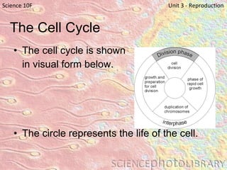
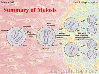
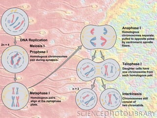
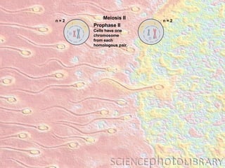
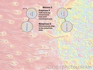
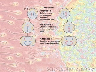
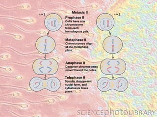
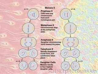
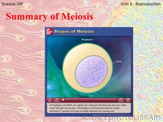
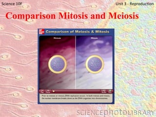
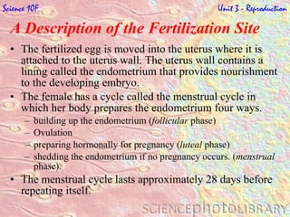
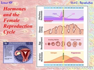
UNIT 3 Reproduction
Science 10F   Unit 3 - Reproduction 15 minutes
http://www.wiley.com/legacy/college/boyer/0470003790/animations/cell_structure/cell_structure.htm Science 10F   Unit 3 - Reproduction
Science 10F   Unit 3 - Reproduction
The Cell Cycle and Mitosis The  cell , known as the basic unit of life, is the fundamental building block for all living organisms. The  Cell Theory  was formulated after numerous observations.  The cell theory stated: All living things are made of cells.  Cells are the basic units of life.  Cells come only from other cells. The activity of an organism depends on the activity of its individual cells.  Science 10F   Unit 3 - Reproduction
The Cell Cycle and Mitosis A typical cell has numerous membrane-bounded organelles, which are specialized structures that perform specialized functions.  These organelles perform life functions such as, producing energy, eliminating wastes, building and repairing, storing information, detecting and responding to outside environment, and reproduction. Science 10F   Unit 3 - Reproduction
The Cell Cycle All cells come from preexisting cells. Any new cell must come from another cell through a process of reproduction. Cells needed for repair, growth or maintaining a species must use  cell division . Cells live through a sequence of steps called the  cell cycle .  Science 10F   Unit 3 - Reproduction
The Cell Cycle and Mitosis Science 10F   Unit 3 - Reproduction
The Cell Cycle The cell cycle is shown  in visual form below.   The circle represents the life of the cell.  Science 10F   Unit 3 - Reproduction
The Cell Cycle Science 10F   Unit 3 - Reproduction
The Cell Cycle The division phase is a part of the cell cycle, but interphase occupies the larger portion of the cell cycle. Interphase can occupy a longer or shorter part of the cell cycle depending on the type of cell. Science 10F   Unit 3 - Reproduction
The Cell Cycle The cell cycle consists of a sequence of events which include: cell division phase a small part of the cell cycle that consists of  mitosis  and  cytokinesis interphase rapid growth  The cell takes in sugars and proteins for growth and repair. duplication of chromosomes The cell prepares for division by duplicating  chromosomes  in the nucleus. Duplication of genetic material must take place  before  cell division.  growth and preparation for cell division.  After chromosome duplication there is another time of cell growth and preparation for cell division. Science 10F   Unit 3 - Reproduction
Interphase   Science 10F   Unit 3 - Reproduction
Cell Division There are two parts to cell division. Mitosis The division of nuclear materials in such a way that both cells have a complete set of chromosomes is called  mitosis. Mitosis is responsible for the division of non-sex cells which we call body cells.   Cytokinesis Cell division is complete when the cytoplasm is shared between the two cells resulting from the division. The process of sharing cytoplasm is called  cytokinesis . Cytokinesis is different in plant and animal cells. In animal cells the cell membrane comes together in the middle of the cell and two new cells are formed. In plants a new cell wall grows down the middle of the cell, producing two new plant cells. Science 10F   Unit 3 - Reproduction
Stages of Mitosis   Science 10F   Unit 3 - Reproduction
Stages of Mitosis   Phase 1 – Prophase   At the beginning of prophase, the chromosomes are double stranded.  The nucleolus and the nuclear membrane disappear.  In animal cells, spindle fibres begin to form and stretch from the centrioles which have moved to opposite ends of the cell. These spindle fibres attach themselves to one side of each centromere.  Science 10F   Unit 3 - Reproduction
Stages of Mitosis   Science 10F   Unit 3 - Reproduction
Stages of Mitosis   Phase 2 – Metaphase   In metaphase the spindle fibres pull the double stranded chromosomes to the middle where they line up. Science 10F   Unit 3 - Reproduction
Stages of Mitosis   Science 10F   Unit 3 - Reproduction
Stages of Mitosis   Phase 3 – Anaphase   In anaphase, the spindle fibres shorten. This action causes the centromere to split, allowing one chromosome to move to opposite ends or poles. Science 10F   Unit 3 - Reproduction
Stages of Mitosis   Science 10F   Unit 3 - Reproduction
Stages of Mitosis   Phase 4 – Telophase   In the final stage, telophase, the spindle fibres disappear and a new nuclear membrane begins to form around the chromosomes. A nucleolus appears in each nucleus.  The single stranded chromosomes begin to unwind and turn into thin strands of chromatin  Science 10F   Unit 3 - Reproduction
Stages of Mitosis   Science 10F   Unit 3 - Reproduction
Science 10F   Unit 3 - Reproduction
Meiosis & Sexual Reproduction sexual reproduction  requires two parents each parent produces  gametes  (sex cells) that contain half as many chromosomes as the parent’s body cells a normal body cell has 46 chromosomes, or 23 pairs of  homologous chromosomes the 46 chromosomes found in body cells are referred to as the  diploid  number of chromosomes fertilization  occurs when two gamete cells combine to form a  zygote  with a complete set of chromosomes male gametes are called  sperm ; female gametes are called  eggs human gametes have a  haploid  number of chromosomes (23 – one of each pair) Science 10F   Unit 3 - Reproduction
Meiosis & Sexual Reproduction Some organisms reproduce sexually and as a result, the offspring receives chromosomes from both parents.  In order for the organism to survive it must only receive half the number of chromosomes from each parent.  The process that ensures that each gamete contains half the number of chromosomes is called meiosis. The process of meiosis is similar to mitosis, but has some major differences.  Meiosis occurs in two stages; Meiosis I and Meiosis II.  Both stages have 4 phases.  Science 10F   Unit 3 - Reproduction
Meiosis Science 10F   Unit 3 - Reproduction
Summary of Meiosis Meiosis occurs in two parts.  Each part of meiosis involves cell division. The first part of meiosis results in two new cells, each having half as many chromosomes as the starting cell (but the chromosomes are double stranded) The second part of meiosis forms four new cells, each having half as many chromosomes as the original cell.  Science 10F   Unit 3 - Reproduction
nucleolus Summary of Meiosis Science 10F   Unit 3 - Reproduction centromere 2n = 4 DNA replication 2n = 4 synapsis sister chromatids n = 2 n = 2 Meiosis II Sister chromatids  separate, becoming  daughter chromosomes. Meiosis I Homologous pairs separate. n = 2 n = 2
Meiosis I Science 10F   Unit 3 - Reproduction
Meiosis I Prophase I Chromosomes are double stranded and are paired with their homologs Chromosomes continue to condense Crossing over occurs Spindle fibres form between centrioles Metaphase I Homologous pairs align at equator Spindle fibres attach to homologs Science 10F   Unit 3 - Reproduction
Meiosis I Anaphase I The homologs separate and the double stranded Chromosomes are pulled to opposite ends by the shortening spindle fibres Telophase I Chromosomes gather at opposite ends to form two clusters of chromosomes There are two nuclei with double stranded chromosomes Science 10F   Unit 3 - Reproduction
Homologous pairs align at the metaphase plate. Metaphase I DNA Replication Homologous chromosomes separate, pulled to opposite poles by centromeric spindle fibers. Anaphase I Daughter cells have one chromosome from each homologous pair. Telophase I Homologous chromosomes pair during synapsis. Prophase I Meiosis I 2n = 4 Chromosomes still consist of two chromatids. Interkinesis n = 2
DNA Replication
DNA Replication Homologous chromosomes pair during synapsis. Prophase I Meiosis I 2n = 4
Homologous pairs align at the metaphase plate. Metaphase I DNA Replication Homologous chromosomes pair during synapsis. Prophase I Meiosis I 2n = 4
Homologous pairs align at the metaphase plate. Metaphase I DNA Replication Homologous chromosomes separate, pulled to opposite poles by centromeric spindle fibers. Anaphase I Homologous chromosomes pair during synapsis. Prophase I Meiosis I 2n = 4
Homologous pairs align at the metaphase plate. Metaphase I DNA Replication Homologous chromosomes separate, pulled to opposite poles by centromeric spindle fibers. Anaphase I Daughter cells have one chromosome from each homologous pair. Telophase I Homologous chromosomes pair during synapsis. Prophase I Meiosis I 2n = 4
Homologous pairs align at the metaphase plate. Metaphase I DNA Replication Homologous chromosomes separate, pulled to opposite poles by centromeric spindle fibers. Anaphase I Daughter cells have one chromosome from each homologous pair. Telophase I Homologous chromosomes pair during synapsis. Prophase I Meiosis I 2n = 4 Chromosomes still consist of two chromatids. Interkinesis n = 2
Meiosis II Science 10F   Unit 3 - Reproduction
Meiosis II Prophase II The chromosomes again condense Centrioles move to opposite poles Metaphase II The spindle fibres attach to double stranded chromosomes and they align along the equator Science 10F   Unit 3 - Reproduction
Meiosis II Anaphase II The sister chromatids separate and are pulled to opposite ends of the cell Telophase II The chromosome gather at opposite ends Nuclear membranes form Cell division begins Science 10F   Unit 3 - Reproduction
Metaphase II Chromosomes align at the metaphase plate. Anaphase II Daughter chromosomes move toward the poles. Telophase II Spindle disappears, nuclei form, and  cytokinesis takes place. Meiosis II Prophase II Cells have one chromosome from each  homologous pair. n = 2 n = 2 Daughter Cells Meiosis results in four haploid daughter cells. n = 2 n = 2
Meiosis II Prophase II Cells have one chromosome from each  homologous pair. n = 2 n = 2
Metaphase II Chromosomes align at the metaphase plate. Meiosis II Prophase II Cells have one chromosome from each  homologous pair. n = 2 n = 2
Metaphase II Chromosomes align at the metaphase plate. Anaphase II Daughter chromosomes move toward the poles. Meiosis II Prophase II Cells have one chromosome from each  homologous pair. n = 2 n = 2
Metaphase II Chromosomes align at the metaphase plate. Anaphase II Daughter chromosomes move toward the poles. Telophase II Spindle disappears, nuclei form, and  cytokinesis takes place. Meiosis II Prophase II Cells have one chromosome from each  homologous pair. n = 2 n = 2
Metaphase II Chromosomes align at the metaphase plate. Anaphase II Daughter chromosomes move toward the poles. Telophase II Spindle disappears, nuclei form, and  cytokinesis takes place. Meiosis II Prophase II Cells have one chromosome from each  homologous pair. n = 2 n = 2 Daughter Cells Meiosis results in four haploid daughter cells. n = 2 n = 2
Summary of Meiosis Science 10F   Unit 3 - Reproduction
Comparison Mitosis and Meiosis Science 10F   Unit 3 - Reproduction
Male & Female Reproductive Systems Science 10F   Unit 3 - Reproduction
Male Reproductive System Science 10F   Unit 3 - Reproduction 6:48
Female Reproductive System Science 10F   Unit 3 - Reproduction 1:04
Female Reproductive System Science 10F   Unit 3 - Reproduction 5:35
Female Reproductive System Science 10F   Unit 3 - Reproduction 3:25
Male & Female Reproductive Systems Science 10F   Unit 3 - Reproduction
Developing the Male Sex Cell  Sperm is produced in the  seminiferous tubules.  Seminiferous tubules are  responsible for producing  huge amounts of sperm, up  to 500 million in a day.  Each sperm is a single cell containing 23 chromosomes (haploid number).  Only one sperm is needed to unite with an egg cell in the female to cause fertilization.  Science 10F   Unit 3 - Reproduction
Developing the Male Sex Cell  A sperm has three parts, a head, a middle, and a tail. The head contains the genetic material that is to be transferred to the egg. The head also contains an entry capsule made of chemicals that are capable of allowing the sperm to enter the egg.  The tail provides mobility the sperm needs to travel to the egg. The tail moves back and forth, propelling the cell to its destination.  Science 10F   Unit 3 - Reproduction
Science 10F   Unit 3 - Reproduction
Male Hormones in Reproduction   Male sex hormones are essential for proper functioning of the reproductive system.  The male and female embryos are identical until the seventh week following fertilization. At the seventh week a hormone is released in the embryo instructing it to produce male (or female) sexual structures.  Once the offspring is born, there are only small amounts of sex hormones released. These sex hormones help regulate the male sex organs but they remain nonfunctioning until the offspring reach  puberty .  At puberty, the brain instructs the body to begin rapid growth and to reach sexual maturity.  Science 10F   Unit 3 - Reproduction
Male Hormones in Reproduction   At this time, the brain also begins the production of male sex hormones called  androgens .  The part of the brain responsible for androgens is called the  hypothalamus .  The hypothalamus releases a hormone to the  pituitary gland .  The pituitary gland, in turn, releases two hormones abbreviated  LH  and  FSH .  LH travels to the testes and stimulates cells to produce  testosterone . Testosterone is the most abundant male sex hormone and is responsible for male characteristics. Science 10F   Unit 3 - Reproduction
Male Hormones in Reproduction   These characteristics begin to show at puberty. Onset of sperm production  Sexual organs grow and develop.  Voice changes; muscle and bone grow FSH combines with testosterone to stimulate the production of sperm. Science 10F   Unit 3 - Reproduction
The Process of Ovulation   The diagram below is an ovary. The formation of an egg cell is illustrated in the stages numbered from 1 to 6 with stages 7 and 8 illustrating the corpus luteum.  Science 10F   Unit 3 - Reproduction
The Process of Ovulation   Ovulation and Menstrual Cycle The human female differs from the male in that she receives all the egg cells ( oocytes ) she will ever have during her embryonic development inside her mother’s womb.  A Female receives around 7 million  follicle cells  between the 14th and 20th week of her development.  Follicle cells surround oocytes and provide nourishment for them. The number of follicle cells from that moment decrease until her birth where she will have about 2 million and to the time of  puberty  when she will have about 400,000.  Science 10F   Unit 3 - Reproduction
A Description of the Fertilization Site  The fertilized egg is moved into the uterus where it is attached to the uterus wall. The uterus wall contains a lining called the endometrium that provides nourishment to the developing embryo.  The female has a cycle called the menstrual cycle in which her body prepares the endometrium four ways. building up the endometrium ( follicular  phase) Ovulation preparing hormonally for pregnancy ( luteal  phase) shedding the endometrium if no pregnancy occurs. ( menstrual  phase). The menstrual cycle lasts approximately 28 days before repeating itself. Science 10F   Unit 3 - Reproduction
A Description of the Fertilization Site  If there is no fertilization of the egg cell, the endometrium is discarded through menstruation. Menstruation is a flow of liquids that clean out the egg and endometrium which have not been used for embryo development. If a pregnancy has taken place, there is no menstrual flow as the egg is developing into an embryo and the endometrium is needed to sustain the embryo.  Science 10F   Unit 3 - Reproduction
Hormones and the  Female Reproduction Cycle  Once the female reaches puberty, the pituitary gland secretes two hormones ( FSH  and  LH) . During this time, development of the external and internal reproductive organs takes place as the body prepares for reproduction.  The interaction of hormones in the reproductive cycle for females is shown below. The pituitary gland secretes FSH into the blood and stimulates follicles to develop in the ovary.  Follicle cells surrounding the developing oocyte secrete  estrogen , which is responsible for the thickening of the uterine lining. Science 10F   Unit 3 - Reproduction
Hormones and the  Female Reproduction Cycle  Estrogen travels to the pituitary and causes it to release LH. LH triggers ovulation.  Following ovulation, follicle cells produce a corpus luteum that secretes increasing amounts of estrogen and  progesterone . As the level of progesterone increases, it travels to the pituitary and signals the pituitary to decrease the production of LH and FSH. The decreasing levels of LH and FSH prevent the production of egg cells until the next cycle when levels of LH and FSH increase again. Science 10F   Unit 3 - Reproduction
Hormones  and the  Female  Reproduction  Cycle  Science 10F   Unit 3 - Reproduction
It Takes 2   Science 10F   Unit 3 - Reproduction
A New Life Begins   Science 10F   Unit 3 - Reproduction
Copulation Sexual intercourse for reproduction Fertilization (Conception) Union of  gametes  produced by different individuals Implantation The burrowing of the developing embryo into the uterus A New Life Begins   Science 10F   Unit 3 - Reproduction
Human Embryonic Development   Science 10F   Unit 3 - Reproduction
Human Embryonic Development   Human embryonic development begins at the time of conception when the egg is fertilized by a sperm. The egg and sperm join nuclei to produce a single diploid cell called a  zygote . Once the egg has been fertilized and the  zygote  is formed, there are two stages of development from fertilization to birth. Embryo  stage takes place over the first eight weeks Fetus  stage takes place from eight weeks to birth Science 10F   Unit 3 - Reproduction
Human Embryonic Development   Embryo Stage Immediately after fertilization, the zygote undergoes  cleavage  (Rapids cell division by mitosis) to produced many new cells called  blastomeres . As the cleaving embryo passes down the oviduct the blastomeres form   a hollow ball of cells, called a  blastocyst . Implantation  occurs in this stage. Science 10F   Unit 3 - Reproduction
Human Embryonic Development   Science 10F   Unit 3 - Reproduction
Human Embryonic Development   Fetus Stage During the fetal development there is further growth and development of the fetus. During this stage, differentiation of the major body systems occurs. Science 10F   Unit 3 - Reproduction
Human Embryonic Development   Science 10F   Unit 3 - Reproduction
Differentiation & Birth   During  gastrulation , 3 layers of cells begin to develop into different parts of the body.  Differentiation  describes how cells begin to become specialized in their function.  The human gestation period (period of differentiation) lasts about 38 weeks.  These weeks are broken down into 3 blocks of time called  trimesters . First trimester (weeks 1 – 12) Second Trimester (weeks 12 – 24) Third Trimester (weeks 24 – 38) Science 10F   Unit 3 - Reproduction
Differentiation & Birth   1st Trimester Science 10F   Unit 3 - Reproduction
Differentiation & Birth   First Trimester (weeks 1 – 12) all body systems begin to form, sex can be determined by formation of sex organs the embyo's movements are not perceptible to the woman, she may not know that she is pregnant by the third month the fetus weighs about 28 grams and is about 7.5 cm long the fetus is particularly sensitive to alcohol, smoking and other drugs  Science 10F   Unit 3 - Reproduction
Differentiation & Birth   2nd Trimester Science 10F   Unit 3 - Reproduction
Differentiation & Birth   Second Trimester (weeks 12 - 24) period of growth the fetus’s movements are perceptible to the woman; the fetus can suck its thumb by the sixth month the fetus weighs about 650 grams and is about 30 cm long if born prematurely, the fetus might survive with great medical assistance for breathing and maintinaing body temperature. Most do not survive at this stage.  Science 10F   Unit 3 - Reproduction
Differentiation & Birth   3nd Trimester Science 10F   Unit 3 - Reproduction
Differentiation & Birth   Third Trimester (weeks 24 - 38) period of growth the fetus kicks, stretches and moves freely; eyes open during the eight month by the ninth month the fetus has tripled its mass to 3300 grams and is about 35 cm long (from top of head to buttocks) dangers can include complications related lifestyle of the mother (use of drugs) and to complications during delivery  Science 10F   Unit 3 - Reproduction
Birth   Birth (parturition) usually takes place about 39 weeks after fertilization by a process of “labor” involving stimuli from oxytocin, a hormone secreted by the pituitary gland of the mother. Three stages of labor are recognized: Birth begins with contractions of the uterine muscles; the cervical canal dilates; the amniotic sac ruptures The fetus is forcefully expelled, accompanied by fluid and blood The umbilical cord is severed; the afterbirth (placenta) is expelled. Science 10F   Unit 3 - Reproduction

Reproduction Notes

  • 1.
  • 2.
    Science 10F Unit 3 - Reproduction 15 minutes
  • 3.
  • 4.
    Science 10F Unit 3 - Reproduction
  • 5.
    The Cell Cycleand Mitosis The cell , known as the basic unit of life, is the fundamental building block for all living organisms. The Cell Theory was formulated after numerous observations. The cell theory stated: All living things are made of cells. Cells are the basic units of life. Cells come only from other cells. The activity of an organism depends on the activity of its individual cells. Science 10F Unit 3 - Reproduction
  • 6.
    The Cell Cycleand Mitosis A typical cell has numerous membrane-bounded organelles, which are specialized structures that perform specialized functions. These organelles perform life functions such as, producing energy, eliminating wastes, building and repairing, storing information, detecting and responding to outside environment, and reproduction. Science 10F Unit 3 - Reproduction
  • 7.
    The Cell CycleAll cells come from preexisting cells. Any new cell must come from another cell through a process of reproduction. Cells needed for repair, growth or maintaining a species must use cell division . Cells live through a sequence of steps called the cell cycle . Science 10F Unit 3 - Reproduction
  • 8.
    The Cell Cycleand Mitosis Science 10F Unit 3 - Reproduction
  • 9.
    The Cell CycleThe cell cycle is shown in visual form below.   The circle represents the life of the cell. Science 10F Unit 3 - Reproduction
  • 10.
    The Cell CycleScience 10F Unit 3 - Reproduction
  • 11.
    The Cell CycleThe division phase is a part of the cell cycle, but interphase occupies the larger portion of the cell cycle. Interphase can occupy a longer or shorter part of the cell cycle depending on the type of cell. Science 10F Unit 3 - Reproduction
  • 12.
    The Cell CycleThe cell cycle consists of a sequence of events which include: cell division phase a small part of the cell cycle that consists of mitosis and cytokinesis interphase rapid growth The cell takes in sugars and proteins for growth and repair. duplication of chromosomes The cell prepares for division by duplicating chromosomes in the nucleus. Duplication of genetic material must take place before cell division. growth and preparation for cell division. After chromosome duplication there is another time of cell growth and preparation for cell division. Science 10F Unit 3 - Reproduction
  • 13.
    Interphase Science 10F Unit 3 - Reproduction
  • 14.
    Cell Division Thereare two parts to cell division. Mitosis The division of nuclear materials in such a way that both cells have a complete set of chromosomes is called mitosis. Mitosis is responsible for the division of non-sex cells which we call body cells.   Cytokinesis Cell division is complete when the cytoplasm is shared between the two cells resulting from the division. The process of sharing cytoplasm is called cytokinesis . Cytokinesis is different in plant and animal cells. In animal cells the cell membrane comes together in the middle of the cell and two new cells are formed. In plants a new cell wall grows down the middle of the cell, producing two new plant cells. Science 10F Unit 3 - Reproduction
  • 15.
    Stages of Mitosis Science 10F Unit 3 - Reproduction
  • 16.
    Stages of Mitosis Phase 1 – Prophase   At the beginning of prophase, the chromosomes are double stranded. The nucleolus and the nuclear membrane disappear. In animal cells, spindle fibres begin to form and stretch from the centrioles which have moved to opposite ends of the cell. These spindle fibres attach themselves to one side of each centromere. Science 10F Unit 3 - Reproduction
  • 17.
    Stages of Mitosis Science 10F Unit 3 - Reproduction
  • 18.
    Stages of Mitosis Phase 2 – Metaphase   In metaphase the spindle fibres pull the double stranded chromosomes to the middle where they line up. Science 10F Unit 3 - Reproduction
  • 19.
    Stages of Mitosis Science 10F Unit 3 - Reproduction
  • 20.
    Stages of Mitosis Phase 3 – Anaphase   In anaphase, the spindle fibres shorten. This action causes the centromere to split, allowing one chromosome to move to opposite ends or poles. Science 10F Unit 3 - Reproduction
  • 21.
    Stages of Mitosis Science 10F Unit 3 - Reproduction
  • 22.
    Stages of Mitosis Phase 4 – Telophase   In the final stage, telophase, the spindle fibres disappear and a new nuclear membrane begins to form around the chromosomes. A nucleolus appears in each nucleus. The single stranded chromosomes begin to unwind and turn into thin strands of chromatin Science 10F Unit 3 - Reproduction
  • 23.
    Stages of Mitosis Science 10F Unit 3 - Reproduction
  • 24.
    Stages of Mitosis Science 10F Unit 3 - Reproduction
  • 25.
    Science 10F Unit 3 - Reproduction
  • 26.
    Meiosis & SexualReproduction sexual reproduction requires two parents each parent produces gametes (sex cells) that contain half as many chromosomes as the parent’s body cells a normal body cell has 46 chromosomes, or 23 pairs of homologous chromosomes the 46 chromosomes found in body cells are referred to as the diploid number of chromosomes fertilization occurs when two gamete cells combine to form a zygote with a complete set of chromosomes male gametes are called sperm ; female gametes are called eggs human gametes have a haploid number of chromosomes (23 – one of each pair) Science 10F Unit 3 - Reproduction
  • 27.
    Meiosis & SexualReproduction Some organisms reproduce sexually and as a result, the offspring receives chromosomes from both parents. In order for the organism to survive it must only receive half the number of chromosomes from each parent. The process that ensures that each gamete contains half the number of chromosomes is called meiosis. The process of meiosis is similar to mitosis, but has some major differences. Meiosis occurs in two stages; Meiosis I and Meiosis II. Both stages have 4 phases. Science 10F Unit 3 - Reproduction
  • 28.
    Meiosis Science 10F Unit 3 - Reproduction
  • 29.
    Summary of MeiosisMeiosis occurs in two parts. Each part of meiosis involves cell division. The first part of meiosis results in two new cells, each having half as many chromosomes as the starting cell (but the chromosomes are double stranded) The second part of meiosis forms four new cells, each having half as many chromosomes as the original cell. Science 10F Unit 3 - Reproduction
  • 30.
    nucleolus Summary ofMeiosis Science 10F Unit 3 - Reproduction centromere 2n = 4 DNA replication 2n = 4 synapsis sister chromatids n = 2 n = 2 Meiosis II Sister chromatids separate, becoming daughter chromosomes. Meiosis I Homologous pairs separate. n = 2 n = 2
  • 31.
    Meiosis I Science10F Unit 3 - Reproduction
  • 32.
    Meiosis I ProphaseI Chromosomes are double stranded and are paired with their homologs Chromosomes continue to condense Crossing over occurs Spindle fibres form between centrioles Metaphase I Homologous pairs align at equator Spindle fibres attach to homologs Science 10F Unit 3 - Reproduction
  • 33.
    Meiosis I AnaphaseI The homologs separate and the double stranded Chromosomes are pulled to opposite ends by the shortening spindle fibres Telophase I Chromosomes gather at opposite ends to form two clusters of chromosomes There are two nuclei with double stranded chromosomes Science 10F Unit 3 - Reproduction
  • 34.
    Homologous pairs alignat the metaphase plate. Metaphase I DNA Replication Homologous chromosomes separate, pulled to opposite poles by centromeric spindle fibers. Anaphase I Daughter cells have one chromosome from each homologous pair. Telophase I Homologous chromosomes pair during synapsis. Prophase I Meiosis I 2n = 4 Chromosomes still consist of two chromatids. Interkinesis n = 2
  • 35.
  • 36.
    DNA Replication Homologouschromosomes pair during synapsis. Prophase I Meiosis I 2n = 4
  • 37.
    Homologous pairs alignat the metaphase plate. Metaphase I DNA Replication Homologous chromosomes pair during synapsis. Prophase I Meiosis I 2n = 4
  • 38.
    Homologous pairs alignat the metaphase plate. Metaphase I DNA Replication Homologous chromosomes separate, pulled to opposite poles by centromeric spindle fibers. Anaphase I Homologous chromosomes pair during synapsis. Prophase I Meiosis I 2n = 4
  • 39.
    Homologous pairs alignat the metaphase plate. Metaphase I DNA Replication Homologous chromosomes separate, pulled to opposite poles by centromeric spindle fibers. Anaphase I Daughter cells have one chromosome from each homologous pair. Telophase I Homologous chromosomes pair during synapsis. Prophase I Meiosis I 2n = 4
  • 40.
    Homologous pairs alignat the metaphase plate. Metaphase I DNA Replication Homologous chromosomes separate, pulled to opposite poles by centromeric spindle fibers. Anaphase I Daughter cells have one chromosome from each homologous pair. Telophase I Homologous chromosomes pair during synapsis. Prophase I Meiosis I 2n = 4 Chromosomes still consist of two chromatids. Interkinesis n = 2
  • 41.
    Meiosis II Science10F Unit 3 - Reproduction
  • 42.
    Meiosis II ProphaseII The chromosomes again condense Centrioles move to opposite poles Metaphase II The spindle fibres attach to double stranded chromosomes and they align along the equator Science 10F Unit 3 - Reproduction
  • 43.
    Meiosis II AnaphaseII The sister chromatids separate and are pulled to opposite ends of the cell Telophase II The chromosome gather at opposite ends Nuclear membranes form Cell division begins Science 10F Unit 3 - Reproduction
  • 44.
    Metaphase II Chromosomesalign at the metaphase plate. Anaphase II Daughter chromosomes move toward the poles. Telophase II Spindle disappears, nuclei form, and cytokinesis takes place. Meiosis II Prophase II Cells have one chromosome from each homologous pair. n = 2 n = 2 Daughter Cells Meiosis results in four haploid daughter cells. n = 2 n = 2
  • 45.
    Meiosis II ProphaseII Cells have one chromosome from each homologous pair. n = 2 n = 2
  • 46.
    Metaphase II Chromosomesalign at the metaphase plate. Meiosis II Prophase II Cells have one chromosome from each homologous pair. n = 2 n = 2
  • 47.
    Metaphase II Chromosomesalign at the metaphase plate. Anaphase II Daughter chromosomes move toward the poles. Meiosis II Prophase II Cells have one chromosome from each homologous pair. n = 2 n = 2
  • 48.
    Metaphase II Chromosomesalign at the metaphase plate. Anaphase II Daughter chromosomes move toward the poles. Telophase II Spindle disappears, nuclei form, and cytokinesis takes place. Meiosis II Prophase II Cells have one chromosome from each homologous pair. n = 2 n = 2
  • 49.
    Metaphase II Chromosomesalign at the metaphase plate. Anaphase II Daughter chromosomes move toward the poles. Telophase II Spindle disappears, nuclei form, and cytokinesis takes place. Meiosis II Prophase II Cells have one chromosome from each homologous pair. n = 2 n = 2 Daughter Cells Meiosis results in four haploid daughter cells. n = 2 n = 2
  • 50.
    Summary of MeiosisScience 10F Unit 3 - Reproduction
  • 51.
    Comparison Mitosis andMeiosis Science 10F Unit 3 - Reproduction
  • 52.
    Comparison Mitosis andMeiosis Science 10F Unit 3 - Reproduction
  • 53.
    Male & FemaleReproductive Systems Science 10F Unit 3 - Reproduction
  • 54.
    Male Reproductive SystemScience 10F Unit 3 - Reproduction 6:48
  • 55.
    Female Reproductive SystemScience 10F Unit 3 - Reproduction 1:04
  • 56.
    Female Reproductive SystemScience 10F Unit 3 - Reproduction 5:35
  • 57.
    Female Reproductive SystemScience 10F Unit 3 - Reproduction 3:25
  • 58.
    Male & FemaleReproductive Systems Science 10F Unit 3 - Reproduction
  • 59.
    Developing the MaleSex Cell Sperm is produced in the seminiferous tubules. Seminiferous tubules are responsible for producing huge amounts of sperm, up to 500 million in a day. Each sperm is a single cell containing 23 chromosomes (haploid number). Only one sperm is needed to unite with an egg cell in the female to cause fertilization. Science 10F Unit 3 - Reproduction
  • 60.
    Developing the MaleSex Cell A sperm has three parts, a head, a middle, and a tail. The head contains the genetic material that is to be transferred to the egg. The head also contains an entry capsule made of chemicals that are capable of allowing the sperm to enter the egg. The tail provides mobility the sperm needs to travel to the egg. The tail moves back and forth, propelling the cell to its destination. Science 10F Unit 3 - Reproduction
  • 61.
    Science 10F Unit 3 - Reproduction
  • 62.
    Male Hormones inReproduction Male sex hormones are essential for proper functioning of the reproductive system. The male and female embryos are identical until the seventh week following fertilization. At the seventh week a hormone is released in the embryo instructing it to produce male (or female) sexual structures. Once the offspring is born, there are only small amounts of sex hormones released. These sex hormones help regulate the male sex organs but they remain nonfunctioning until the offspring reach puberty . At puberty, the brain instructs the body to begin rapid growth and to reach sexual maturity. Science 10F Unit 3 - Reproduction
  • 63.
    Male Hormones inReproduction At this time, the brain also begins the production of male sex hormones called androgens . The part of the brain responsible for androgens is called the hypothalamus . The hypothalamus releases a hormone to the pituitary gland . The pituitary gland, in turn, releases two hormones abbreviated LH and FSH . LH travels to the testes and stimulates cells to produce testosterone . Testosterone is the most abundant male sex hormone and is responsible for male characteristics. Science 10F Unit 3 - Reproduction
  • 64.
    Male Hormones inReproduction These characteristics begin to show at puberty. Onset of sperm production Sexual organs grow and develop. Voice changes; muscle and bone grow FSH combines with testosterone to stimulate the production of sperm. Science 10F Unit 3 - Reproduction
  • 65.
    The Process ofOvulation The diagram below is an ovary. The formation of an egg cell is illustrated in the stages numbered from 1 to 6 with stages 7 and 8 illustrating the corpus luteum. Science 10F Unit 3 - Reproduction
  • 66.
    The Process ofOvulation Ovulation and Menstrual Cycle The human female differs from the male in that she receives all the egg cells ( oocytes ) she will ever have during her embryonic development inside her mother’s womb. A Female receives around 7 million follicle cells between the 14th and 20th week of her development. Follicle cells surround oocytes and provide nourishment for them. The number of follicle cells from that moment decrease until her birth where she will have about 2 million and to the time of puberty when she will have about 400,000. Science 10F Unit 3 - Reproduction
  • 67.
    A Description ofthe Fertilization Site The fertilized egg is moved into the uterus where it is attached to the uterus wall. The uterus wall contains a lining called the endometrium that provides nourishment to the developing embryo. The female has a cycle called the menstrual cycle in which her body prepares the endometrium four ways. building up the endometrium ( follicular phase) Ovulation preparing hormonally for pregnancy ( luteal phase) shedding the endometrium if no pregnancy occurs. ( menstrual phase). The menstrual cycle lasts approximately 28 days before repeating itself. Science 10F Unit 3 - Reproduction
  • 68.
    A Description ofthe Fertilization Site If there is no fertilization of the egg cell, the endometrium is discarded through menstruation. Menstruation is a flow of liquids that clean out the egg and endometrium which have not been used for embryo development. If a pregnancy has taken place, there is no menstrual flow as the egg is developing into an embryo and the endometrium is needed to sustain the embryo. Science 10F Unit 3 - Reproduction
  • 69.
    Hormones and the Female Reproduction Cycle Once the female reaches puberty, the pituitary gland secretes two hormones ( FSH and LH) . During this time, development of the external and internal reproductive organs takes place as the body prepares for reproduction. The interaction of hormones in the reproductive cycle for females is shown below. The pituitary gland secretes FSH into the blood and stimulates follicles to develop in the ovary. Follicle cells surrounding the developing oocyte secrete estrogen , which is responsible for the thickening of the uterine lining. Science 10F Unit 3 - Reproduction
  • 70.
    Hormones and the Female Reproduction Cycle Estrogen travels to the pituitary and causes it to release LH. LH triggers ovulation. Following ovulation, follicle cells produce a corpus luteum that secretes increasing amounts of estrogen and progesterone . As the level of progesterone increases, it travels to the pituitary and signals the pituitary to decrease the production of LH and FSH. The decreasing levels of LH and FSH prevent the production of egg cells until the next cycle when levels of LH and FSH increase again. Science 10F Unit 3 - Reproduction
  • 71.
    Hormones andthe Female Reproduction Cycle Science 10F Unit 3 - Reproduction
  • 72.
    It Takes 2 Science 10F Unit 3 - Reproduction
  • 73.
    A New LifeBegins Science 10F Unit 3 - Reproduction
  • 74.
    A New LifeBegins Science 10F Unit 3 - Reproduction
  • 75.
    A New LifeBegins Science 10F Unit 3 - Reproduction
  • 76.
    Copulation Sexual intercoursefor reproduction Fertilization (Conception) Union of gametes produced by different individuals Implantation The burrowing of the developing embryo into the uterus A New Life Begins Science 10F Unit 3 - Reproduction
  • 77.
    Human Embryonic Development Science 10F Unit 3 - Reproduction
  • 78.
    Human Embryonic Development Human embryonic development begins at the time of conception when the egg is fertilized by a sperm. The egg and sperm join nuclei to produce a single diploid cell called a zygote . Once the egg has been fertilized and the zygote is formed, there are two stages of development from fertilization to birth. Embryo stage takes place over the first eight weeks Fetus stage takes place from eight weeks to birth Science 10F Unit 3 - Reproduction
  • 79.
    Human Embryonic Development Embryo Stage Immediately after fertilization, the zygote undergoes cleavage (Rapids cell division by mitosis) to produced many new cells called blastomeres . As the cleaving embryo passes down the oviduct the blastomeres form a hollow ball of cells, called a blastocyst . Implantation occurs in this stage. Science 10F Unit 3 - Reproduction
  • 80.
    Human Embryonic Development Science 10F Unit 3 - Reproduction
  • 81.
    Human Embryonic Development Fetus Stage During the fetal development there is further growth and development of the fetus. During this stage, differentiation of the major body systems occurs. Science 10F Unit 3 - Reproduction
  • 82.
    Human Embryonic Development Science 10F Unit 3 - Reproduction
  • 83.
    Differentiation & Birth During gastrulation , 3 layers of cells begin to develop into different parts of the body. Differentiation describes how cells begin to become specialized in their function. The human gestation period (period of differentiation) lasts about 38 weeks. These weeks are broken down into 3 blocks of time called trimesters . First trimester (weeks 1 – 12) Second Trimester (weeks 12 – 24) Third Trimester (weeks 24 – 38) Science 10F Unit 3 - Reproduction
  • 84.
    Differentiation & Birth 1st Trimester Science 10F Unit 3 - Reproduction
  • 85.
    Differentiation & Birth First Trimester (weeks 1 – 12) all body systems begin to form, sex can be determined by formation of sex organs the embyo's movements are not perceptible to the woman, she may not know that she is pregnant by the third month the fetus weighs about 28 grams and is about 7.5 cm long the fetus is particularly sensitive to alcohol, smoking and other drugs Science 10F Unit 3 - Reproduction
  • 86.
    Differentiation & Birth 2nd Trimester Science 10F Unit 3 - Reproduction
  • 87.
    Differentiation & Birth Second Trimester (weeks 12 - 24) period of growth the fetus’s movements are perceptible to the woman; the fetus can suck its thumb by the sixth month the fetus weighs about 650 grams and is about 30 cm long if born prematurely, the fetus might survive with great medical assistance for breathing and maintinaing body temperature. Most do not survive at this stage. Science 10F Unit 3 - Reproduction
  • 88.
    Differentiation & Birth 3nd Trimester Science 10F Unit 3 - Reproduction
  • 89.
    Differentiation & Birth Third Trimester (weeks 24 - 38) period of growth the fetus kicks, stretches and moves freely; eyes open during the eight month by the ninth month the fetus has tripled its mass to 3300 grams and is about 35 cm long (from top of head to buttocks) dangers can include complications related lifestyle of the mother (use of drugs) and to complications during delivery Science 10F Unit 3 - Reproduction
  • 90.
    Birth Birth (parturition) usually takes place about 39 weeks after fertilization by a process of “labor” involving stimuli from oxytocin, a hormone secreted by the pituitary gland of the mother. Three stages of labor are recognized: Birth begins with contractions of the uterine muscles; the cervical canal dilates; the amniotic sac ruptures The fetus is forcefully expelled, accompanied by fluid and blood The umbilical cord is severed; the afterbirth (placenta) is expelled. Science 10F Unit 3 - Reproduction

Editor's Notes

  • #66 Starting at 1 and going clockwise through the stages of egg cell production, you can see the follicle gives rise to an egg cell that breaks through the ovary wall to deposit a mature egg cell into the oviduct. The process of releasing an egg into the oviduct is called ovulation . The corpus luteum remains in the ovary, ready to form hormones if a pregnancy begins. If the egg cell is not fertilized, the corpus luteum disappears in about ten days.
  • #67 Starting at 1 and going clockwise through the stages of egg cell production, you can see the follicle gives rise to an egg cell that breaks through the ovary wall to deposit a mature egg cell into the oviduct. The process of releasing an egg into the oviduct is called ovulation . The corpus luteum remains in the ovary, ready to form hormones if a pregnancy begins. If the egg cell is not fertilized, the corpus luteum disappears in about ten days.
  • #68 Starting at 1 and going clockwise through the stages of egg cell production, you can see the follicle gives rise to an egg cell that breaks through the ovary wall to deposit a mature egg cell into the oviduct. The process of releasing an egg into the oviduct is called ovulation . The corpus luteum remains in the ovary, ready to form hormones if a pregnancy begins. If the egg cell is not fertilized, the corpus luteum disappears in about ten days.
  • #69 Starting at 1 and going clockwise through the stages of egg cell production, you can see the follicle gives rise to an egg cell that breaks through the ovary wall to deposit a mature egg cell into the oviduct. The process of releasing an egg into the oviduct is called ovulation . The corpus luteum remains in the ovary, ready to form hormones if a pregnancy begins. If the egg cell is not fertilized, the corpus luteum disappears in about ten days.
  • #70 Starting at 1 and going clockwise through the stages of egg cell production, you can see the follicle gives rise to an egg cell that breaks through the ovary wall to deposit a mature egg cell into the oviduct. The process of releasing an egg into the oviduct is called ovulation . The corpus luteum remains in the ovary, ready to form hormones if a pregnancy begins. If the egg cell is not fertilized, the corpus luteum disappears in about ten days.
  • #71 Starting at 1 and going clockwise through the stages of egg cell production, you can see the follicle gives rise to an egg cell that breaks through the ovary wall to deposit a mature egg cell into the oviduct. The process of releasing an egg into the oviduct is called ovulation . The corpus luteum remains in the ovary, ready to form hormones if a pregnancy begins. If the egg cell is not fertilized, the corpus luteum disappears in about ten days.
  • #72 Starting at 1 and going clockwise through the stages of egg cell production, you can see the follicle gives rise to an egg cell that breaks through the ovary wall to deposit a mature egg cell into the oviduct. The process of releasing an egg into the oviduct is called ovulation . The corpus luteum remains in the ovary, ready to form hormones if a pregnancy begins. If the egg cell is not fertilized, the corpus luteum disappears in about ten days.