This document discusses various anatomical structures and behaviors in humans that are considered vestigial, meaning they have lost most or all of their original function through evolution. It provides numerous examples of vestigial structures in humans such as the appendix, tailbone, wisdom teeth, inner corner of the eye, outer ear structures like Darwin's tubercle, and others. It also discusses vestigial behaviors like goose bumps and grasping reflex in infants. The document traces the history of studies on vestigial structures and provides details on specific structures like the vermiform appendix and its analogous structure in rabbits.
are worm-like parasites. The clinically relevant groups are separated according to their general external shape and the host organ they inhabit. There are both hermaphroditic and bisexual species.
The definitive classification is based on the external and internal morphology of egg, larval, and adult stages.
Helminth is a general term meaning worm. The helminths are invertebrates characterized by elongated, flat or round bodies.
In flatworms or platyhelminths (platy from the Greek root meaning “flat”) include flukes and tapeworms.
Roundworms are nematodes (nemato from the Greek root meaning “thread”).
Vittelogenesis is a word developed from Latin vitellus-yolk, and genero-produce
Vitellogenesis (also known as yolk deposition) is the process of yolk formation via nutrients being deposited in the oocyte, or female germ cell involved in reproduction of lecithotrophic organisms. In insects, it starts when the fat body stimulates the release of juvenile hormones and produces vitellogenin protein.
Yolks is the most usual form of food storage in the egg.
Yolks appear in the oocyte in the secondary period of their growth called vittelogenesis.
Thus,the formation and deposition of yolks is known as vittelogenesis
Characteristic
Yolks is a complex variable assembled component.
The principle component are protein,phospholipid and fats in different combination.
Depending upon these component yolks is distinguished into protein yolks and fatty acid
For eg- the avian contain 48.19% water , 16.6 % protein, 32.6% phospholipids and fats and 1% carbohydrates.
types of orientation- primary and secondary, different types of kinesis - orthokinesis and klinokinesis and taxis - tropotaxis, klinotaxis, menotaxis, transverse orientation, dosal light reaction and ventral light reaction
DENTITION IN MAMMALS
The study of arrangement structure and number of types of teeth collectively is called as dentition. Teeth are present in the foetal as well as in adults of mammals, based on the presence of teeth Mammals are two types.
Edentata : In some animals teeth are absent hence called as edentate. e.g., Echidna or spiny ant-eater (Tachyglossus) the teeth are absent in all stages of life.
Dentata : Teeth are present in all mammals though a secon¬dary toothless condition is found in some mammals. Modern turtles and birds lack teeth. The adult platypus (Ornithorhynchus) bears epidermal teeth but no true teeth are present. In platypus embryonic teeth are replaced by horny epidermal teeth in adult.
Classification According to the Shape and Size of the Teeth:
Homodont:
Homodont or Isodont type of teeth is a condition where the teeth are all alike in their shape and size in the toothed whales e.g., Pinnipedians. Fishes, amphibians, reptiles and in the extinct toothed birds.
Heterodont
Heterodont condition is the usual feature in mammals, i.e. the teeth are distinguished according to their shape, size and function. The function is also different at different parts of the tooth row.
According to the Mode of Attachment of Teeth:
Thecodont : The teeth are lodged in bony sockets or alveoli of the jaw bone and capillaries and nerves enter the pulp cavity through the open tips of the hollow roots e.g., mammals, crocodiles and in some fishes.
Acrodont: The teeth are fused to the surface of the underlying jawbone. They have no roots and are attached to the edge of the jawbone by fibrous membrane e.g., fishes, amphibians and some reptiles.
Pleurodont:
The teeth are attached to the inner-side of the jawbone. The tooth touches the bone only with the outer surface of its root. In acrodont and pleurodont types of dentition, there are no roots, and nerves and blood vessels do not enter the pulp cavity at the base, e.g., Necturus (Amphibia) and some reptiles.
According to the Succession or Replace¬ment of Teeth:
INTRODUCTION
The jaw (Upper and lower) is any opposable articulated structure at the entrance of the mouth.
It is typically used for grasping and manipulating food.
Jaw suspension means the fusion of upper jaw and lower jaw or skull for efficient biting.
There are different ways in which these attachments are attained depending upon the modifications in visceral arches in vertebrates.
In most vertebrates, the jaws are bony or cartilaginous and oppose vertically.
The vertebrate jaw is derived from the most anterior two pharyngeal arches supporting the gills, and usually bears numerous teeth.
The vertebrate jaw probably originally evolved in the Silurian period and appeared in the Placoderm fish which further diversified in the Devonian.
It is believed that the hyoid system suspends the jaw from the brain case of the skull, permitting great mobility of the jaws.
The original selective advantage offered by the jaw may not be related to feeding, but rather to increased respiration efficiency.
The jaws were used in the buccal pump (observable in modern fish and amphibians) that pumps water across the gills of fish or air into the lungs in the case of amphibians.
Over evolutionary time the more familiar use of jaws (to humans), in feeding, was selected for and became a very important function in vertebrates. Many teleost fish have substantially modified jaws for suction feeding and jaw protrusion, resulting in highly complex jaws with dozens of bones involved.
Jaw Suspension or Suspensoria:
The method by which the upper and lower jaws are suspended or attached from the chondrocranium is known as jaw suspension or suspensorium.
Amongst the visceral arches, the first (mandibular) arch consists of
= a dorsal palato pterygoquadrate bar forming the upper jaw,
= and ventral Meckel’s cartilage forms the lower jaw.
The second (hyoid) arch consists of = a dorsal hyomandibular supporting and suspending the jaws with the cranium, and a ventral hyoid.
The remaining visceral arches support the gills and are, hence, called branchial arches. Thus, splanchnocranium forms the jaws and suspends them with the chondrocranium.
In this presentation, Phylum Mesozoa is described. After watching this you will learn the characteristics and Examples Dicyema spp., Pseudicyema spp., and Dicyemennea The brief description of Phylum Mesozoa along with reproduction and fertilization also described. Its relationship with Phylum Rhombozoa and Orthonectida. It is part of BS Zoology Course, Animal diversity.
Briefly discuss the adaptive changes seen in the chordates over the o.pdfmonikajain201
Briefly discuss the adaptive changes seen in the chordates over the other animal phyla?
Solution
Animals in the phylum Chordata reveal four key features that appear at some point all through
their development.
a. Notochord - The chordates are called for the notochord: a flexible, rod-shaped design that is
found within the embryonic stage of most chordates and also in the adult stage of some chordate
species. It is found between the intestinal pipe and the nerve cord, providing skeletal help
through along the body. In some chordates, the notochord acts while the primary axial support of
the human body throughout the animal\'s lifetime. In vertebrates, the notochord is provide during
embryonic development, of which time it induces the development of the neural pipe which acts
as an assistance for the developing embryonic body. The notochord, however, is replaced by the
vertebral column (spine) in most adult vertebrates.
b.Dorsal Hollow Nerve Cord - The dorsal hollow nerve cord derives from ectoderm that sheets
into a worthless pipe all through development. In chordates, it is found dorsally (at the top of the
animal) to the notochord. In contrast to the chordates, other animal phyla are known by strong
nerve cables which are positioned either ventrally or laterally. The nerve cord within most
chordate embryos develops into the brain and spinal cord, which include the main anxious
system.
c.Pharyngeal Slits - Pharyngeal slits are openings in the pharynx (the place just posterior to the
mouth) that expand to the surface environment. In organisms that are now living in aquatic
surroundings, pharyngeal slits allow for the quit of water that enters the mouth all through
feeding. Some invertebrate chordates use the pharyngeal slits to filtration food out of the water
that enters the mouth. In vertebrate fishes, the pharyngeal slits develop into gill arches, the bony
or cartilaginous gill supports. In most terrestrial animals, including mammals and birds,
pharyngeal slits are providing just during embryonic development. In these animals, the
pharyngeal slits develop into the jaw and inner ear bones.
d.Post-anal Tail - The post-anal tail is a posterior elongation of the human body, increasing
beyond the anus. The trail contains skeletal things and muscles, which give a way to obtain
locomotion in aquatic species. In some terrestrial vertebrates, the trail also assists with stability,
courting, and signaling when risk is near. In individuals and other apes, the post-anal tail is
provide during embryonic development, but is vestigial as an adult..
Lecture on arthropods and echinoderms.pptEsayDawit
zoologist now what are arthropods, what are the distinguishing features and what are echinoderms with identifying their features from the rest other invertebrates.
Phylum Mollusca, Class Gastropoda, Torsion, Locomotion, Digestion,Reproductio...Dr. Muhammad Moosa
In this presentation, Phylum Mollusca Is described. After watching this you will learn Evolutionary Perspective of Mollusca and Relationships to Other Animals, Molluscan Characteristics, Class Gastropoda, Torsion, Shell Coiling, Locomotion, Feeding and Digestion, Other Maintenance Functions, Reproduction and Development, Gastropod Diversity, Class Bivalvia, Shell and Associated Structures Gas Exchange, Filter Feeding, and Digestion, Other Maintenance Functions Reproduction and Development, Bivalve Diversity, Class Cephalopoda, Shell, Locomotion, Feeding and Digestion, Other Maintenance Functions, Learning, Reproduction and Development, Class Polyplacophora, Class Scaphopoda, Class Monoplacophora, Class Solenogastres, Class Caudofoveata, Further Phylogenetic Considerations. It is part of BS Zoology Course, Animal diversity.
THIS PROJECT IS MADE BY NEHA SAHU.
ALUMNI STUDENT OF NEW ERA PROGESSIVE SCHOOL KORBA OF 2018.
IN THIS PPT FILE YOU CAN STUDY ABOUT VESTIGIAL STRUCTURE ONLY FEW OF THE MOST IMPORTANT ORGANS ARE THERE AND OTHER ARE IN ANOTHER PPT.
2. Introduction
In the context of human evolution, human vestigiality
involves those characters (such as organs or behaviors)
occurring in the human species that are considered
vestigial—in other words having lost all or most of their
original function through evolution.
Although structures usually called "vestigial" often
appear functionless, a vestigial structure may retain
lesser functions or develop minor new ones.
In some cases, structures once identified as vestigial
simply had an unrecognized function.
3. Examples of human vestigiality are numerous
Anatomical (such as human appendix,
tailbone, wisdom teeth,& inner corner of eye)
Behavioral (goose bumps & palmar grasp
reflex),
sensory (decreased olfaction), and
molecular (junk DNA).
Many human characteristics are also vestigial
in other primates and related animals.
4. History
In 1893, Robert Wiedersheim published a book
on human anatomy and its relevance to man's
evolutionary history.
This book contained a list of 86 human organs
that he considered vestigial, or as Wiedersheim
himself explained: "Organs having become
wholly or in part functionless, some appearing
in the Embryo alone, others present during Life
constantly or inconstantly. For the greater part
Organs which may be rightly termed Vestigial”
5. History Contd.
Historically there was a trend not only to dismiss
the vermiform appendix as being uselessly
vestigial, but an anatomical hazard, a liability to
dangerous inflammation.
As late as the mid 20th century many reputable
authorities conceded it no beneficial function.
This was a view supported, or perhaps inspired,
by Darwin himself in the 1874 edition of his book
The Descent of Man, and Selection in Relation to
Sex.
6. Appendix
In modern humans, the vermiform appendix is a
vestigial organ that in ancestral species had
digestive functions.
Some herbivorous animals, such as rabbits, have
a terminal vermiform appendix and cecum that
apparently bear patches of tissue with immune
functions and may also be important in
maintaining the composition of gut flora
• Some carnivorous animals may have appendices
too, but seldom have more than vestigial caeca
7. Ileum-Caecum-Colon_of_rabbit
As shown in
accompanying pictures
however, human
appendix typically is
about comparable to
that of rabbit in size,
though caecum is
reduced to a single
bulge where ileum
empties into colon
8. Appendix
• The human vermiform
appendix on the
vestigial caecum
9. Coccyx
The coccyx, or tailbone, is the remnant of a lost tail.
All mammals have a tail at one point in their development;
In humans, it is present for a period of 4 weeks, during
stages 14 to 22 of human embryogenesis.
This tail is most prominent in human embryos 31–35 days
old.
The tailbone, located at the end of the spine, has lost its
original function in assisting balance and mobility, though it
still serves some secondary functions, such as being an
attachment point for muscles, which explains why it has
not degraded further.
In rare cases congenital defect results in a short tail-like
structure being present at birth. Twenty-three cases of
human babies born with such a structure have been
reported in the medical literature since 1884.
10. Wisdom teeth
Wisdom teeth are vestigial 3rd molars that human ancestors used
to help in grinding down plant tissue.
The common postulation is that the skulls of human ancestors had
larger jaws with more teeth, which were possibly used to help chew
down foliage to compensate for a lack of ability to efficiently digest
the cellulose that makes up a plant cell wall.
As human diets changed, smaller jaws were selected by evolution,
yet the 3rd molars, or "wisdom teeth," still commonly develop in
human mouths.
Currently, wisdom teeth have become useless and even harmful to
the extent where surgical procedures are often done to remove
them.
Agenesis of wisdom teeth in human populations ranges from zero
in Tasmanian Aboriginals to nearly 100% in indigenous Mexicans.
The difference is related to the PAX9 gene (and perhaps other
genes).
11. Vomeronasal organ (VNO)
In some animals VNO is part of a 2nd, completely separate sense of
smell, known as the accessory olfactory system.
Many studies have been performed to find if there is an actual
presence of a VNO in adult human beings. Trotier et al. estimated
that around 92% of their subjects that had no septal surgery had at
least one intact VNO. Kjaer and Fisher Hansen, on the other hand,
stated that VNO structure disappeared during fetal development as
it does for some primates.However, Smith and Bhatnagar (2000)
asserted that Kjaer and Fisher Hansen simply missed the structure
in older fetuses. Won (2000) found evidence of a VNO in 13 of his
22 cadavers (59.1%) and in 22 of his 78 living patients (28.2%).
Given these findings, some scientists have argued that there is a
VNO in adult human beings.However, most investigators have
sought to identify the opening of the vomeronasal organ in
humans, rather than identify the tubular epithelial structure itself.
Thus it has been argued that such studies, employing macroscopic
observational methods, have sometimes missed or even
misidentified the vomeronasal organ.
12. VNO Contd.
Among studies that use microanatomical methods, there is
no reported evidence that human beings have active
sensory neurons like those in working vomeronasal systems
of other animals.
Furthermore, there is no evidence to date that suggests
there are nerve and axon connections between any existing
sensory receptor cells that may be in the adult human VNO
and the brain.
Likewise, there is no evidence for any accessory olfactory
bulb in adult human beings, and the key genes involved in
VNO function in other mammals have become
pseudogenes in human beings.
Therefore, while the presence of a structure in adult human
beings is debated, a review of the scientific literature by
Tristram Wyatt concluded, "most in the field ... are sceptical
about the likelihood of a functional VNO in adult human
beings on current evidence."[
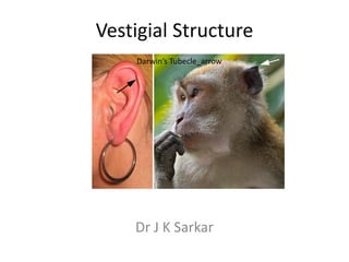
13. Ear
The ears of a Macaque monkey and most other monkeys have far
more developed muscles than those of humans, and therefore have
capability to move their ears to better hear potential threats.
Humans and other primates such as the orangutan and chimpanzee
however have ear muscles that are minimally developed and non-
functional, yet still large enough to be identifiable.
A muscle attached to the ear that cannot move the ear, for
whatever reason, can no longer be said to have any biological
function.
In humans there is variability in these muscles, such that some
people are able to move their ears in various directions, and it has
been said that it may be possible for others to gain such movement
by repeated trials.
In such primates the inability to move the ear is compensated
mainly by the ability to turn the head on a horizontal plane, an
ability which is not common to most monkeys—a function once
provided by one structure is now replaced by another.
14. The outer structure
Ear of the ear also
shows some
Darwin's tubercle vestigial features,
such as the node or
point on the helix of
the ear known as
Darwin's tubercle
which is found in
around 10% of the
population, this
feature is labelled
(arrow) in the
accompanying
figure.
15. Eye: plica semilunaris
• The plica semilunaris is a small fold of tissue on
the inside corner of the eye.
• It is the vestigial remnant of the nictitating
membrane, an organ that is fully functional in
some other species of mammals.
• Its associated muscles are also vestigial.
• The plica semilunaris of Africans and Indigenous
Australians are slightly larger than in other
peoples.
• Only one species of primate, the Calabar
Angwantibo, is known to have a functioning
nictitating membrane
16. The platysma is vestigial remnant of
panniculous carnosus of animals (which Platysma
allows the horse to flick a fly off its back)
& comes from the Greek work "plate"
It is a quadrangular sheet of muscle
which originates on the fascia of the
pectoralis major muscle and ascends to
three main points of insertion.
The most anterior fibers decussate the
midline at variable levels below chin and
insert into the mentum.
Central fibers insert into body of
mandible & the more posterior fibers
turn anteriorly & blend closely with
fibers of risorius muscle.
The platysma is innervated by cervical
branch of facial nerve. The main lower
branch exits the parotid & enters deep
surface of platysma. The upper twigs of
cervical branch & lower twigs of marginal
branch intermingle before marginal
nerve passes on to supply the depressor
anguli oris and risorius.
17. Head : Occipitalis Minor
• The Occipitalis Minor is a muscle in the back of
the head which normally joins to the auricular
muscles of the ear.
• This muscle is very sporadic in frequency—always
present in Malays, in 56% of Africans, 50% of
Japanese, 36% of Europeans, and is nonexistent
in the Khoikhoi people of southwestern Africa
and in Melanesians.
• Other small muscles in the head associated with
the occipital region and the post-auricular muscle
complex are often variable in their frequency.
18. Face
In many non-human mammals the upper lip and
sinus area is associated with whiskers or vibrissae
which serve a sensory function.
In humans these whiskers do not exist but there
are still sporadic cases where elements of the
associated vibrissal capsular muscles or Sinus hair
muscles can be found.
Based on histological studies of the upper lips of
20 cadavers, Tamatsu et al. found that structures
resembling such muscles were present in 35%
(7/20) of their specimens
19. Breasts
• Extra nipples or breasts sometimes appear
along the mammary lines of humans,
appearing as a remnant to mammalian
ancestors who possessed more than two
nipples or breasts.
20. Palmaris Longus
• The palmaris longus muscle is seen
as a small tendon between flexor
carpi radialis & flexor carpi ulnaris,
although it is not always present.
• The muscle is absent in about 14%
of population, however this varies
greatly with ethnicity.
• One study has shown prevalence
of palmaris longus agenesis in 500
Indian patients to be 17.2% (8%
bilateral and 9.2% unilateral).
• The palmaris is a popular source of
tendon material for grafts and this
has prompted studies which have
shown the absence of the palmaris
does not have any appreciable
effect on grip strength
21. Levator Claviculae
• The levator claviculae muscle in the posterior
triangle of the neck is a supernumerary
muscle present in only 2–3% of all people but
nearly always present in most mammalian
species, including gibbons and orangutans.
22. The pyramidalis muscle of
the abdomen is a small and Pyramidalis
triangular muscle, anterior
to the rectus abdominis,
and contained in the rectus
sheath.
It is absent in 20% of
humans and when absent
the lower end of the rectus
then becomes
proportionately increased
in size.
Anatomical studies suggest
that the forces generated
by the pyramidalis muscles
are relatively small
23. Plantaris
Plantaris muscle is composed of
a thin muscle belly & a long thin
tendon.
The muscle belly is approx. 2–4
inches long, and is absent in 7–
10% of the human population.
It has some weak functionality
in moving the knee & ankle but
is generally considered
redundant and is often used as
a source of tendon for grafts.
The long, thin tendon of the
plantaris is humorously called
"the freshman's nerve," as it is
often mistaken for a nerve by
first-year medical students.
24. Sensory
Although the sense of smell, or olfaction, is essential for many animals in
avoiding predators, finding food, and other functions, olfaction is greatly
decreased in humans as they have for the most part no predators and
obtain food mostly by agriculture.
There is great variation in olfactory sensitivity from person to person,
which is common in vestigial characteristics.
It has been observed that native South Americans, native North
Americans, and African peoples have a highly developed sense of smell,
such that they may be able to identify others in the dark by their odor
alone.
This does not mean that having any olfactory ability at all is vestigial, for
example it may save a person from inhaling toxic fumes.
A characteristic may degenerate despite being of some use if there is
very little or no selection pressure on the genes associated with it.
In other words, having a good sense of smell may be something a person
would desire, but unless those without such abilities have a lower
reproductive success or fitness, there is no barrier to its degeneration.
25. • Humans also bear some vestigial
behaviors and reflexes. E.g. the Behavioral
formation of goose bumps in
humans under stress is a
vestigial reflex; a possible Goose bump
function in human evolutionary
ancestors was to raise body's
hair, making ancestor appear
larger and scaring off predators.
• Raising the hair is also used to
trap an extra layer of air,
keeping an animal warm.
• Due to the diminished amount
of hair in humans, the reflex
formation of goose bumps when
cold is also vestigial.
26. Palmar grasp reflex is supported to be a
vestigial behavior in human infants.
When placing a finger or object to the Palmar grasp
palm of an infant, it will securely grasp
it. This grasp is found to be rather
strong.
reflex
Some infants—37% according to a 1932
study—are able to support their own
weight from a rod, although there is no
way they can cling to their mother.
The grasp is also evident in the feet too.
When a baby is sitting down, its
prehensile feet assume a curled-in
posture, much like what is observed in
an adult chimp.
An ancestral primate would have had
sufficient body hair to which an infant
could cling unlike modern humans, thus
allowing its mother to escape from
danger, such as climbing up a tree in
presence of a predator without having
to occupy her hands holding her baby.
27. Epoophoron_Paroophoron_Gartner’s
Cyst
Only parts
remaining from
the mesonephric
system are the
epoophoron,
paroophoron,
and Gartner's
cyst
28. • Excretory tubules
Genital Ducts in Male along the caudal pole
of the testis, the
paragenital tubules,
do not join the cords
of the rete testis.
Their vestiges are
collectively known as
paradidymis.
• The paramesonephric
ducts in male
degenerate except
for a small portion at
their cranial ends,
appendix testis.
29. Molecular
There are also vestigial molecular structures in
humans, which are no longer in use but may
indicate common ancestry with other species.
One example of this is L-gulonolactone oxidase, a
gene that is functional in most other mammals
and produces an enzyme that synthesizes Vitamin
C.
In humans and other members of the suborder
Haplorrhini, a mutation disabled the gene and
made it unable to produce the enzyme.
However, the remains of the gene are still present
in the human genome as a vestigial genetic
sequence called a pseudogene