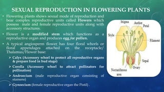
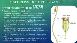
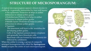
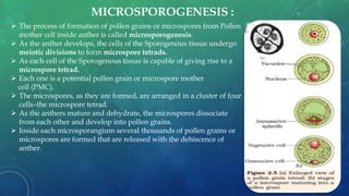
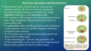
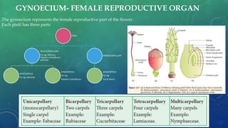
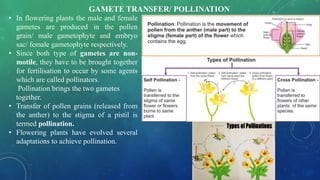
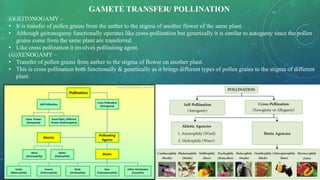
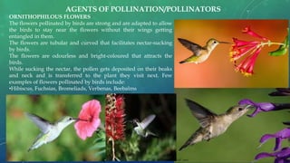
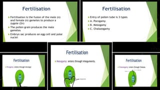
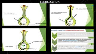
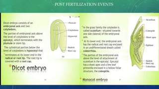
SEXUAL REPRODUCTION IN
FLOWERING PLANTS
CHAPTER-2
UNIT-I
PREPARED BY-
ANITA MISHRA
BIRLA BALIKA VIDYAPEETH,
PILANI
SEXUAL REPRODUCTION IN FLOWERING PLANTS
• Flowering plants shows sexual mode of reproduction and
bear complex reproductive units called Flowers which
possess male and female reproductive units along with
accessory structures.
• Flower is a modified stem which functions as a
reproductive organ and produces egg /or pollen.
• A typical angiosperm flower has four floral whorls or
floral appendages attached on the receptacle/
Thalamus/Flower base:
 Calyx (Accessory whorl to protect all reproductive organs
& prepare food in bud stage)
 Corolla (Accessory whorl to attract pollinators for
pollination)
 Androecium (male reproductive organ consisting of
stamens)
 Gynoecium (female reproductive organ the Pistil) .
MALE REPRODUCTIVE ORGAN OF
FLOWER
DETAILED STRUCTURE OF STAMEN:
A Typical stamen – It has 2 parts:
• Filament- The long and slender stalk the filament is
attached to the thalamus or the petal of the flower.
• Anther- The terminal generally bilobed structure.
• A typical angiosperm anther is bilobed with each lobe
having two theca, i.e., they are dithecous
• Often a longitudinal groove runs lengthwise separating
the theca.
• The anther is a four-sided (tetragonal) structure
consisting of four microsporangia located at the corners,
two in each lobe.
• The microsporangia develop further and become pollen
sacs.
• They extend longitudinally all through the length of an
anther and are packed with pollen grains.
STRUCTURE OF MICROSPORANGIUM:
A typical microsporangium appears almost circular in
outline & is generally surrounded by four wall layers:
The epidermis- Protective & helps in anther
dehiscence to release pollen grains
Endothecium-Protective & helps in anther
dehiscence to release pollen grains
Middle layers- Protective & helps in anther
dehiscence to release pollen grains.
Tapetum-
The innermost wall layer is the nourishes the
developing pollen grains.
Cells of the tapetum possess dense cytoplasm
and generally have binucleate cells.
Binucleate cells are formed by karyokinesis not
followed by cytokinesis.
When the anther is young, a group of compactly
arranged homogenous cells called the Sporogenous
tissue occupies the centre of each microsporangium.
MICROSPOROGENESIS :
 The process of formation of pollen grains or microspores from Pollen
mother cell inside anther is called microsporogenesis.
 As the anther develops, the cells of the Sporogenous tissue undergo
meiotic divisions to form microspore tetrads.
 As each cell of the Sporogenous tissue is capable of giving rise to a
microspore tetrad.
 Each one is a potential pollen grain or microspore mother
cell (PMC).
 The microspores, as they are formed, are arranged in a cluster of four
cells–the microspore tetrad.
 As the anthers mature and dehydrate, the microspores dissociate
from each other and develop into pollen grains.
 Inside each microsporangium several thousands of pollen grains or
microspores are formed that are released with the dehiscence of
anther.
• The haploid nuclei of pollen grain/ microspores
undergo mitotic cell division with an asymmetric
spindle formation leading to formation of a large
vegetative cell and a small generative cell .
• The vegetative cell is bigger, has abundant food reserve
and a large irregularly shaped nucleus & it later on
forms pollen tube.
• The generative cell is small and floats in the cytoplasm
of the vegetative cell. It is spindle shaped with dense
cytoplasm and a nucleus.
• In over 60 per cent of angiosperms, pollen grains are
shed at this 2-celled stage.
• In the remaining 40 % species, the generative cell
divides mitotically to give rise to the two male gametes
before pollen grains are shed (3-celled stage).
POLLEN GRAINS/ MICROSPORES
The pollen grains represent the male gametophytes which are generally
spherical measuring about 25-50µ in diameter.
It has a prominent two-layered wall.
Exine- The hard outer layer is made up of sporopollenin which is one of the
most resistant organic material .
It can withstand high temperatures and strong acids and alkali.
No enzyme that degrades sporopollenin is so far known.
Pollen grain exine has prominent apertures called germ pores where
sporopollenin is absent.
Pollen grains are well preserved as fossils due to presence of
sporopollenin. The exine exhibits a fascinating patterns & designs.
Intine- The inner wall of the pollen grain is a thin and continuous layer
made up of cellulose and pectin.
Plasma membrane- Inside the intine a lipid protein bilayered plasma
membrane is found surrounding the cytoplasm.
EXPERIMENT OF POLLEN TUBE GERMINATION: The first step involves the
preparation of a nutrient solution. This is done by dissolving 10g of sucrose as
well as 10mg of boric acid in 100ml of water.
Pour a few drops of this solution onto the cavity slide. Then, use a brush or
fingers to gently dust a few pollen grains from the stamen of mature flowers.
Let the slide set for 5 mins. Then, use the microscope to view the slides in 30-
minute intervals.
CHARACTERISTICS OF POLLEN GRAINS/ MICROSPORES
RICH IN NUTRIENTS.
• Pollen tablets & syrups are used as food supplements in western
countries.
• Pollen consumption has been claimed to increase the
performance of athletes and race horses.
VIABILITY PERIOD OF POLLENS-
• To bring about fertilisation the pollen grains have to land on the
stigma before they lose their viability.
• Pollen grains have variable viability in different species in some
it depends on the prevailing temperature and humidity.
• In some cereals such as rice and wheat, pollen grains lose
viability within 30 minutes of their release, and
• In some members of Rosaceae, Leguminoseae and Solanaceae
viability is for months.
POLLEN GRAINS AS ALLERGENS-
• Pollen grains of many species cause severe allergies and
bronchial inflammation in some it leads to chronic respiratory
disorders like asthma, bronchitis, etc.
for eg: Parthenium or carrot grass that came into India with
imported Mexica High Yielding Variety of wheat, causes severe
pollen allergy.
POLLEN PRESERVATION
• As sperms & semen of many animals including humans are preserved in sperm banks for artificial
insemination in the same way it is possible to store pollen
grains of a large number of species for years in liquid nitrogen
• At temperature -196°C it is called cryopreservation.
• Such stored pollen can be used as pollen banks, similar to
seed banks, in crop breeding programmes.
GYNOECIUM- FEMALE REPRODUCTIVE ORGAN
The gynoecium represents the female reproductive part of the flower.
Each pistil has three parts
PISTIL
MULTICARPELLARY
For eg: Hibiscus,
Papaver somniferum,
Michelia
APOCARPOUS
For eg: Michelia
SYNCARPOUS
For eg:
Papaver somniferum
MONOCARPELLARY
APOCARPOUS
For eg:
Pea & Beans
GYNOECIUM- FEMALE REPRODUCTIVE ORGAN
 STIGMA - Serves as a landing platform for pollen grains.
 STYLE- is the elongated slender part beneath the stigma.
 OVARY- The basal bulged part of the pistil is the ovary. Inside the ovary is the ovarian cavity (locule).
The placenta is located inside the ovarian cavity.
Megasporangia- From the placenta megasporangia arise which are commonly called ovules.
The number of ovules in an ovary may be one (wheat, paddy, mango) to many (papaya, water melon,
orchids).
Ovary
Style
Stigma
 FUNICLE- The ovule is a structure attached to the placenta
with a stalk called funicle.
 HILUM-The ovule fuses with funicle in the region called
hilum. Thus hilum represents the junction between ovule
and funicle.
 INTEGUMENT- Each ovule has one or two protective
envelopes/ layers called integuments.
 MICROPYLE- Integuments surround the ovule except at
the tip leaving a small opening called the micropyle which
serves for the entry & exit of substances into & outside the
ovule.
 CHALAZA- It is the end of ovule found opposite to the
micropylar end & it represents the base of the ovule.
 NUCELLUS- It is the mass of parenchymatous cells
enclosed within the integuments which serve for the nutrition
of embryo sac/ female gametophyte due to the presence of
reserve food..
 EMBRYO SAC OR FEMALE GAMETOPHYTE- It is
located in the centre of the nucellus. An ovule generally has
a single embryo sac formed from a megaspore through
reduction division.
THE MEGASPORANGIUM (OVULE)
TYPES OF OVULE BASED ON POSITION OF MICROPYLE & CHALAZA
DEFINITION- The process of formation of
megaspores from the megaspore mother cell is
called megasporogenesis.
STEPS OF MEGASPOROGENESIS-
 In the nucellus of ovule towards the
micropylar end a single megaspore mother
cell (MMC) is formed just contrary to
pollen/ microspore mother cell which are
produced several in no. in one
microsporangium.
 The Megaspore mother cell is a large cell
containing dense cytoplasm and a
prominent nucleus.
 The MMC undergoes meiotic division.
 resulting into the production of four
megaspores.
 One of the megaspores is functional while
the other three degenerate unlike the
microspores in which all the four
microspores formed in tetrad survive.
 The functional megaspore develops into the
female gametophyte (embryo sac).
MEGASPOROGENESIS
• The nucleus of the functional megaspore divides mitotically to form two nuclei which move to the opposite poles, forming
the bi-nucleate embryo sac.
• The two nuclei on the two opposite poles of embryo sac undergo two consecutive mitotic divisions leading to the
formation of 4 nuclei on each pole.
• The mitotic nuclear divisions result in the formation of the 4-nucleate and later the 8-nucleate stages of the embryo sac.
• These karyokinesis or nuclear divisions are not followed by cytokinesis or cytoplasmic division.
• One one nuclei from each of the poles migrate to the centre forming two Primary endosperm nuclei/PEN/Polar nuclei.
• Later the nuclei at the poles undergo cytokinesis forming 3 cells on each pole of embryo sac.
• There is a characteristic distribution of the cells within the embryo sac. Three cells are grouped together at the micropylar
end and constitute the egg apparatus.
• The egg apparatus, consists of two synergids and one egg cell/ female gamete.
• The synergids have special cellular thickenings at the micropylar tip called filiform apparatus play role in guiding the
pollen tubes into the synergid.
• The 3 cells at the chalazal end are called the antipodals which are nutritive in function.
• The embryo sac hence formed has a set of 3 cells- the egg apparatus at micropylar end, 3 antipodals atchalazal end and a
large central cell with a binucleate condition (polar nuclei) Thus, a typical angiosperm embryo sac, atmaturity, though 8-
nucleate is 7-celled.
DEVELOPMENT OF EMBRYO SAC/ FEMALE GAMETOPHYTE
• In flowering plants the male and female
gametes are produced in the pollen
grain/ male gametophyte and embryo
sac/ female gametophyte respectively.
• Since both type of gametes are non-
motile, they have to be brought together
for fertilisation to occur by some agents
which are called pollinators.
Pollination brings the two gametes
together.
• Transfer of pollen grains (released from
the anther) to the stigma of a pistil is
termed pollination.
• Flowering plants have evolved several
adaptations to achieve pollination.
GAMETE TRANSFER/ POLLINATION
(i) AUTOGAMY :
• Transfer of pollen grains from the anther to the stigma of the same flower.
• In a flower which opens and exposes the anthers and the stigma, both autogamy & Cross pollination (Xenogamy/
Geitenogamy is possible.
• Autogamy in such flowers requires synchrony in pollen release and stigma receptivity.
• The anthers and the stigma should lie close to each other so that self- pollination can occur.
GAMETE TRANSFER/ POLLINATION
ii)GEITONOGAMY –
• It is transfer of pollen grains from the anther to the stigma of another flower of the same plant.
• Although geitonogamy functionally operates like cross-pollination but genetically it is similar to autogamy since the pollen
grains come from the same plant are transferred.
• Like cross pollination it involves pollinating agent.
(iii)XENOGAMY –
• Transfer of pollen grains from anther to the stigma of flower on another plant.
• This is cross pollination both functionally & genetically as it brings different types of pollen grains to the stigma of different
plant.
GAMETE TRANSFER/ POLLINATION
• Majority of plants use biotic agents for pollination & a very few plants use
abiotic agents.
• Pollen grains coming in contact with the stigma is a chance factor in abiotic
transfer.
• To compensate the loss of pollen grains are produced in large enormous
numbers as compared to ovules.
FEATURES OF ANEMOPHILOUS FLOWERS-
• Pollination by wind is a type of abiotic pollinations.
• Flowers are non attractive, non fragrant.
• The pollen grains are light and non-sticky to be transported by wind currents.
• Well-exposed stamens (for easy dispersal of pollen)
• They have mostly feathery stigma to trap air-borne pollen grains.
• The ovary mostly has single ovule in each ovary.
• Flowers are borne in clusters in the inflorescence. For eg: is the corncob – the
tassels are stigma and style which trap pollen grains. Wind-pollination is quite
common in grasses.
AGENTS OF POLLINATION/POLLINATORS
FEATURES OF HYDROPHILOUS FLOWERS-
• Only around 30 species of flowering plants like Vallisneria/ Water Lily,
Zostera/ sea grass & Hydrilla are Hydrophilous while it is a prevalent
mode of gamete transfer in Bryophytes & Pteridophytes.
• In Vallisneria, the female flower reach the surface of water by the long
stalk and the male flowers which are borne on spiral stalk after maturity
the spiral opens & the pollen grains are released which float over the
surface of water.
• They are carried passively by water currents & some of them reach the
stigma of female flowers.
• In Zostera/seagrasses, female flowers are submerged in water and the
pollen grains are released inside the water.
• Pollen grains in such species are long, ribbon like and are carried inside the
water; to stigma for pollination.
• In most of the water-pollinated species, pollen grains are protected from
wetting by a mucilaginous covering.
Pollination in Zostera/ Sea grass.
Moss- Gamete transfer.
AGENTS OF POLLINATION/POLLINATORS
ENTAMOPHILOUS FLOWERS-The flowers pollinated by insects
are bright-coloured and produce nectar.
The fragrance of the flowers attracts the insects.
The pollen are sticky, large, heavy and rough so that stick to the
body of the insects.
The stigmas are also sticky so that the pollens depositing are not
dispersed.
Amorphophallus tallest flower is pollinated by moth, In Yucca
flower moth deposits its eggs in the ovarian locule, in turn, gets
pollinated by the moth. The larvae of the moth comes out of the
eggs as the seeds start developing.
Nectar guides are present on the petals. Few examples of the
flowers pollinated by insects are:
•Magnolia, Aster, Lithops
AGENTS OF POLLINATION/POLLINATORS
ORNITHOPHILOUS FLOWERS
The flowers pollinated by birds are strong and are adapted to allow
the birds to stay near the flowers without their wings getting
entangled in them.
The flowers are tubular and curved that facilitates nectar-sucking
by birds.
The flowers are odourless and bright-coloured that attracts the
birds.
While sucking the nectar, the pollen gets deposited on their beaks
and neck and is transferred to the plant they visit next. Few
examples of flowers pollinated by birds include:
•Hibiscus, Fuchsias, Bromeliads, Verbenas, Beebalms
AGENTS OF POLLINATION/POLLINATORS
Zoophily is a form of pollination whereby pollen is
transferred by animals, usually vertebrates but may
include invertebrates particularly by monkeys,
marsupials, lemurs, bears, rabbits, deer, rodents, lizards
like Gekco, and other animals.
Zoophilous are brightly colored or scented flowers,
nectar, and appealing shapes and patterns.
These plant-animal relationships are often mutually
beneficial because of the food source provided in
exchange for pollination.
Zoophilous species include Arctium, Acaena, and Galium
aparine.
CHIREPTEROPHILOUS FLOWERS-
The flowers are pollinated by bats.
They are mostly white, scented flowers (Fruity or musky
smell).
Flowers are large bell shaped.
For eg: Kigelia
Gecko Lizard
Kigelia
Chirepterophily
Zoophily
AGENTS OF POLLINATION/POLLINATORS
https://www.youtube.com/watch?v=GocKVMk-kSY
OUTBREEDING DEVICES IN FLOWERS TO DISCOURAGE INBREEDING
Inbreeding leads to deterioration of characters called inbreeding depression
FERTILIZATION
POST FERTILIZATION EVENTS
The remnants of Nucellus in few seeds is present in form of
perisperm
For eg; Back pepper, Beet root etc.
 Integuments of ovules harden to form tough protective seed coats-
i) Outer- Testa
ii) Inner Tegmen
 The micropyle remains as a small pore in the seed coat to facilitate
entry of oxygen and water into the seed during germination.
 On maturity the seed looses water having only (10-15 per cent moisture
by mass).
 The general metabolic activity of the embryo slows down due to lack of
moisture.
 The embryo may enter a state of inactivity called dormancy when seed
prepares itself for germination.
 When favourable conditions are available (adequate moisture, oxygen
and suitable temperature) & the reserve food they germinate.
 As ovules changes into seeds
 Ovary into fruit
 Ovarian wall forms Fruit wall/ Pericarp.
 Based on nature of Pericarp the fruits may be-
i) Fleshy/ Succulent (guava, orange, mango, plum,)
ii) Dry/ Non succulent (groundnut, and mustard, coconut,
almond)
 A fruit formed without fertilization is Parthenocarpic/ Seedless fruit
which is formed asexually.
POST FERTILIZATION CHANGES IN OVARY TO FORM FRUIT & SEEDS
SEED DORMANCY & SEED VIABILITY
• Dehydration and dormancy enable the seed storage/
preservation for favourable season, hence a seed is the basis of
our agriculture
•After maturity the seeds are dispersed through different agents
otherwise if they all fall under the same plant they won’t survive
due to lack of food & other resources.
•After dispersal the seeds have variable viability periods-
•For eg:
•The oldest is that of a lupine, Lupinus arcticus excavated from
Arctic Tundra The seed germinated and flowered after an
estimated record of 10,000 years of dormancy.
•A recent record of 2000 years old viable seed is of the date palm,
Phoenix dactylifera discovered during the archeological excavation
at King Herod’s palace near the Dead Sea.
STRUCTURE OF DICOT & MONOCOT SEEDS
a) Dicot Seed of Bean b) Dicot & albuminous Seed of Castor
c) Monocot Seed of Onion c) Monocot Seed of Maize
DIFFERENCE BETWEEN TRUE & FALSE FRUITS
STRUCTURE OF FALSE FRUITS- APPLE & STRAWBERRY
Developed from thalamus of flower instead of Ovary
S. No. CELLS OF PISTIL PLOIDY
1 Funiculus Diploid (2n)
2 Integument Diploid (2n)
3 Nucellus Diploid (2n)
4 Chalaza Diploid (2n)
5 Antipodals Haploid (n)
6 Synergids Haploid (n)
7 Egg cell Haploid (n)
8 Polar nuclei Haploid (n)
9 Zygote Diploid (2n)
10 Endosperm Triploid (3n)
11 ovary Diploid (2n)
12 Stigma Diploid (2n)
13 Style Diploid (2n)
14 Megaspore mother cell Diploid (2n)
S. No. CELLS OF STAMEN PLOIDY
1 Filament Diploid (2n)
2 Anther Diploid (2n)
3 Microsporangium/ Pollen
sac
Diploid (2n)
4 Microspore mother cell Diploid (2n)
5 Pollen Grain/ Microspores Haploid (n)
6 Generative cell Haploid (n)
7 Vegetative Cell Haploid (n)
8 Male gametes Haploid (n)
9 Pollen Tube Diploid (2n)
Reproduction in plants- CLASS XII

Reproduction in plants- CLASS XII

  • 1.
    SEXUAL REPRODUCTION IN FLOWERINGPLANTS CHAPTER-2 UNIT-I PREPARED BY- ANITA MISHRA BIRLA BALIKA VIDYAPEETH, PILANI
  • 2.
    SEXUAL REPRODUCTION INFLOWERING PLANTS • Flowering plants shows sexual mode of reproduction and bear complex reproductive units called Flowers which possess male and female reproductive units along with accessory structures. • Flower is a modified stem which functions as a reproductive organ and produces egg /or pollen. • A typical angiosperm flower has four floral whorls or floral appendages attached on the receptacle/ Thalamus/Flower base:  Calyx (Accessory whorl to protect all reproductive organs & prepare food in bud stage)  Corolla (Accessory whorl to attract pollinators for pollination)  Androecium (male reproductive organ consisting of stamens)  Gynoecium (female reproductive organ the Pistil) .
  • 3.
    MALE REPRODUCTIVE ORGANOF FLOWER DETAILED STRUCTURE OF STAMEN: A Typical stamen – It has 2 parts: • Filament- The long and slender stalk the filament is attached to the thalamus or the petal of the flower. • Anther- The terminal generally bilobed structure. • A typical angiosperm anther is bilobed with each lobe having two theca, i.e., they are dithecous • Often a longitudinal groove runs lengthwise separating the theca. • The anther is a four-sided (tetragonal) structure consisting of four microsporangia located at the corners, two in each lobe. • The microsporangia develop further and become pollen sacs. • They extend longitudinally all through the length of an anther and are packed with pollen grains.
  • 4.
    STRUCTURE OF MICROSPORANGIUM: Atypical microsporangium appears almost circular in outline & is generally surrounded by four wall layers: The epidermis- Protective & helps in anther dehiscence to release pollen grains Endothecium-Protective & helps in anther dehiscence to release pollen grains Middle layers- Protective & helps in anther dehiscence to release pollen grains. Tapetum- The innermost wall layer is the nourishes the developing pollen grains. Cells of the tapetum possess dense cytoplasm and generally have binucleate cells. Binucleate cells are formed by karyokinesis not followed by cytokinesis. When the anther is young, a group of compactly arranged homogenous cells called the Sporogenous tissue occupies the centre of each microsporangium.
  • 5.
    MICROSPOROGENESIS :  Theprocess of formation of pollen grains or microspores from Pollen mother cell inside anther is called microsporogenesis.  As the anther develops, the cells of the Sporogenous tissue undergo meiotic divisions to form microspore tetrads.  As each cell of the Sporogenous tissue is capable of giving rise to a microspore tetrad.  Each one is a potential pollen grain or microspore mother cell (PMC).  The microspores, as they are formed, are arranged in a cluster of four cells–the microspore tetrad.  As the anthers mature and dehydrate, the microspores dissociate from each other and develop into pollen grains.  Inside each microsporangium several thousands of pollen grains or microspores are formed that are released with the dehiscence of anther.
  • 6.
    • The haploidnuclei of pollen grain/ microspores undergo mitotic cell division with an asymmetric spindle formation leading to formation of a large vegetative cell and a small generative cell . • The vegetative cell is bigger, has abundant food reserve and a large irregularly shaped nucleus & it later on forms pollen tube. • The generative cell is small and floats in the cytoplasm of the vegetative cell. It is spindle shaped with dense cytoplasm and a nucleus. • In over 60 per cent of angiosperms, pollen grains are shed at this 2-celled stage. • In the remaining 40 % species, the generative cell divides mitotically to give rise to the two male gametes before pollen grains are shed (3-celled stage). POLLEN GRAINS/ MICROSPORES
  • 7.
    The pollen grainsrepresent the male gametophytes which are generally spherical measuring about 25-50µ in diameter. It has a prominent two-layered wall. Exine- The hard outer layer is made up of sporopollenin which is one of the most resistant organic material . It can withstand high temperatures and strong acids and alkali. No enzyme that degrades sporopollenin is so far known. Pollen grain exine has prominent apertures called germ pores where sporopollenin is absent. Pollen grains are well preserved as fossils due to presence of sporopollenin. The exine exhibits a fascinating patterns & designs. Intine- The inner wall of the pollen grain is a thin and continuous layer made up of cellulose and pectin. Plasma membrane- Inside the intine a lipid protein bilayered plasma membrane is found surrounding the cytoplasm. EXPERIMENT OF POLLEN TUBE GERMINATION: The first step involves the preparation of a nutrient solution. This is done by dissolving 10g of sucrose as well as 10mg of boric acid in 100ml of water. Pour a few drops of this solution onto the cavity slide. Then, use a brush or fingers to gently dust a few pollen grains from the stamen of mature flowers. Let the slide set for 5 mins. Then, use the microscope to view the slides in 30- minute intervals.
  • 8.
    CHARACTERISTICS OF POLLENGRAINS/ MICROSPORES RICH IN NUTRIENTS. • Pollen tablets & syrups are used as food supplements in western countries. • Pollen consumption has been claimed to increase the performance of athletes and race horses. VIABILITY PERIOD OF POLLENS- • To bring about fertilisation the pollen grains have to land on the stigma before they lose their viability. • Pollen grains have variable viability in different species in some it depends on the prevailing temperature and humidity. • In some cereals such as rice and wheat, pollen grains lose viability within 30 minutes of their release, and • In some members of Rosaceae, Leguminoseae and Solanaceae viability is for months. POLLEN GRAINS AS ALLERGENS- • Pollen grains of many species cause severe allergies and bronchial inflammation in some it leads to chronic respiratory disorders like asthma, bronchitis, etc. for eg: Parthenium or carrot grass that came into India with imported Mexica High Yielding Variety of wheat, causes severe pollen allergy.
  • 9.
    POLLEN PRESERVATION • Assperms & semen of many animals including humans are preserved in sperm banks for artificial insemination in the same way it is possible to store pollen grains of a large number of species for years in liquid nitrogen • At temperature -196°C it is called cryopreservation. • Such stored pollen can be used as pollen banks, similar to seed banks, in crop breeding programmes.
  • 10.
    GYNOECIUM- FEMALE REPRODUCTIVEORGAN The gynoecium represents the female reproductive part of the flower. Each pistil has three parts PISTIL MULTICARPELLARY For eg: Hibiscus, Papaver somniferum, Michelia APOCARPOUS For eg: Michelia SYNCARPOUS For eg: Papaver somniferum MONOCARPELLARY APOCARPOUS For eg: Pea & Beans
  • 11.
  • 12.
    GYNOECIUM- FEMALE REPRODUCTIVEORGAN  STIGMA - Serves as a landing platform for pollen grains.  STYLE- is the elongated slender part beneath the stigma.  OVARY- The basal bulged part of the pistil is the ovary. Inside the ovary is the ovarian cavity (locule). The placenta is located inside the ovarian cavity. Megasporangia- From the placenta megasporangia arise which are commonly called ovules. The number of ovules in an ovary may be one (wheat, paddy, mango) to many (papaya, water melon, orchids). Ovary Style Stigma
  • 13.
     FUNICLE- Theovule is a structure attached to the placenta with a stalk called funicle.  HILUM-The ovule fuses with funicle in the region called hilum. Thus hilum represents the junction between ovule and funicle.  INTEGUMENT- Each ovule has one or two protective envelopes/ layers called integuments.  MICROPYLE- Integuments surround the ovule except at the tip leaving a small opening called the micropyle which serves for the entry & exit of substances into & outside the ovule.  CHALAZA- It is the end of ovule found opposite to the micropylar end & it represents the base of the ovule.  NUCELLUS- It is the mass of parenchymatous cells enclosed within the integuments which serve for the nutrition of embryo sac/ female gametophyte due to the presence of reserve food..  EMBRYO SAC OR FEMALE GAMETOPHYTE- It is located in the centre of the nucellus. An ovule generally has a single embryo sac formed from a megaspore through reduction division. THE MEGASPORANGIUM (OVULE)
  • 14.
    TYPES OF OVULEBASED ON POSITION OF MICROPYLE & CHALAZA
  • 15.
    DEFINITION- The processof formation of megaspores from the megaspore mother cell is called megasporogenesis. STEPS OF MEGASPOROGENESIS-  In the nucellus of ovule towards the micropylar end a single megaspore mother cell (MMC) is formed just contrary to pollen/ microspore mother cell which are produced several in no. in one microsporangium.  The Megaspore mother cell is a large cell containing dense cytoplasm and a prominent nucleus.  The MMC undergoes meiotic division.  resulting into the production of four megaspores.  One of the megaspores is functional while the other three degenerate unlike the microspores in which all the four microspores formed in tetrad survive.  The functional megaspore develops into the female gametophyte (embryo sac). MEGASPOROGENESIS
  • 16.
    • The nucleusof the functional megaspore divides mitotically to form two nuclei which move to the opposite poles, forming the bi-nucleate embryo sac. • The two nuclei on the two opposite poles of embryo sac undergo two consecutive mitotic divisions leading to the formation of 4 nuclei on each pole. • The mitotic nuclear divisions result in the formation of the 4-nucleate and later the 8-nucleate stages of the embryo sac. • These karyokinesis or nuclear divisions are not followed by cytokinesis or cytoplasmic division. • One one nuclei from each of the poles migrate to the centre forming two Primary endosperm nuclei/PEN/Polar nuclei. • Later the nuclei at the poles undergo cytokinesis forming 3 cells on each pole of embryo sac. • There is a characteristic distribution of the cells within the embryo sac. Three cells are grouped together at the micropylar end and constitute the egg apparatus. • The egg apparatus, consists of two synergids and one egg cell/ female gamete. • The synergids have special cellular thickenings at the micropylar tip called filiform apparatus play role in guiding the pollen tubes into the synergid. • The 3 cells at the chalazal end are called the antipodals which are nutritive in function. • The embryo sac hence formed has a set of 3 cells- the egg apparatus at micropylar end, 3 antipodals atchalazal end and a large central cell with a binucleate condition (polar nuclei) Thus, a typical angiosperm embryo sac, atmaturity, though 8- nucleate is 7-celled. DEVELOPMENT OF EMBRYO SAC/ FEMALE GAMETOPHYTE
  • 18.
    • In floweringplants the male and female gametes are produced in the pollen grain/ male gametophyte and embryo sac/ female gametophyte respectively. • Since both type of gametes are non- motile, they have to be brought together for fertilisation to occur by some agents which are called pollinators. Pollination brings the two gametes together. • Transfer of pollen grains (released from the anther) to the stigma of a pistil is termed pollination. • Flowering plants have evolved several adaptations to achieve pollination. GAMETE TRANSFER/ POLLINATION
  • 19.
    (i) AUTOGAMY : •Transfer of pollen grains from the anther to the stigma of the same flower. • In a flower which opens and exposes the anthers and the stigma, both autogamy & Cross pollination (Xenogamy/ Geitenogamy is possible. • Autogamy in such flowers requires synchrony in pollen release and stigma receptivity. • The anthers and the stigma should lie close to each other so that self- pollination can occur. GAMETE TRANSFER/ POLLINATION
  • 20.
  • 21.
    ii)GEITONOGAMY – • Itis transfer of pollen grains from the anther to the stigma of another flower of the same plant. • Although geitonogamy functionally operates like cross-pollination but genetically it is similar to autogamy since the pollen grains come from the same plant are transferred. • Like cross pollination it involves pollinating agent. (iii)XENOGAMY – • Transfer of pollen grains from anther to the stigma of flower on another plant. • This is cross pollination both functionally & genetically as it brings different types of pollen grains to the stigma of different plant. GAMETE TRANSFER/ POLLINATION
  • 22.
    • Majority ofplants use biotic agents for pollination & a very few plants use abiotic agents. • Pollen grains coming in contact with the stigma is a chance factor in abiotic transfer. • To compensate the loss of pollen grains are produced in large enormous numbers as compared to ovules. FEATURES OF ANEMOPHILOUS FLOWERS- • Pollination by wind is a type of abiotic pollinations. • Flowers are non attractive, non fragrant. • The pollen grains are light and non-sticky to be transported by wind currents. • Well-exposed stamens (for easy dispersal of pollen) • They have mostly feathery stigma to trap air-borne pollen grains. • The ovary mostly has single ovule in each ovary. • Flowers are borne in clusters in the inflorescence. For eg: is the corncob – the tassels are stigma and style which trap pollen grains. Wind-pollination is quite common in grasses. AGENTS OF POLLINATION/POLLINATORS
  • 23.
    FEATURES OF HYDROPHILOUSFLOWERS- • Only around 30 species of flowering plants like Vallisneria/ Water Lily, Zostera/ sea grass & Hydrilla are Hydrophilous while it is a prevalent mode of gamete transfer in Bryophytes & Pteridophytes. • In Vallisneria, the female flower reach the surface of water by the long stalk and the male flowers which are borne on spiral stalk after maturity the spiral opens & the pollen grains are released which float over the surface of water. • They are carried passively by water currents & some of them reach the stigma of female flowers. • In Zostera/seagrasses, female flowers are submerged in water and the pollen grains are released inside the water. • Pollen grains in such species are long, ribbon like and are carried inside the water; to stigma for pollination. • In most of the water-pollinated species, pollen grains are protected from wetting by a mucilaginous covering. Pollination in Zostera/ Sea grass. Moss- Gamete transfer. AGENTS OF POLLINATION/POLLINATORS
  • 24.
    ENTAMOPHILOUS FLOWERS-The flowerspollinated by insects are bright-coloured and produce nectar. The fragrance of the flowers attracts the insects. The pollen are sticky, large, heavy and rough so that stick to the body of the insects. The stigmas are also sticky so that the pollens depositing are not dispersed. Amorphophallus tallest flower is pollinated by moth, In Yucca flower moth deposits its eggs in the ovarian locule, in turn, gets pollinated by the moth. The larvae of the moth comes out of the eggs as the seeds start developing. Nectar guides are present on the petals. Few examples of the flowers pollinated by insects are: •Magnolia, Aster, Lithops AGENTS OF POLLINATION/POLLINATORS
  • 25.
    ORNITHOPHILOUS FLOWERS The flowerspollinated by birds are strong and are adapted to allow the birds to stay near the flowers without their wings getting entangled in them. The flowers are tubular and curved that facilitates nectar-sucking by birds. The flowers are odourless and bright-coloured that attracts the birds. While sucking the nectar, the pollen gets deposited on their beaks and neck and is transferred to the plant they visit next. Few examples of flowers pollinated by birds include: •Hibiscus, Fuchsias, Bromeliads, Verbenas, Beebalms AGENTS OF POLLINATION/POLLINATORS
  • 26.
    Zoophily is aform of pollination whereby pollen is transferred by animals, usually vertebrates but may include invertebrates particularly by monkeys, marsupials, lemurs, bears, rabbits, deer, rodents, lizards like Gekco, and other animals. Zoophilous are brightly colored or scented flowers, nectar, and appealing shapes and patterns. These plant-animal relationships are often mutually beneficial because of the food source provided in exchange for pollination. Zoophilous species include Arctium, Acaena, and Galium aparine. CHIREPTEROPHILOUS FLOWERS- The flowers are pollinated by bats. They are mostly white, scented flowers (Fruity or musky smell). Flowers are large bell shaped. For eg: Kigelia Gecko Lizard Kigelia Chirepterophily Zoophily AGENTS OF POLLINATION/POLLINATORS https://www.youtube.com/watch?v=GocKVMk-kSY
  • 27.
    OUTBREEDING DEVICES INFLOWERS TO DISCOURAGE INBREEDING Inbreeding leads to deterioration of characters called inbreeding depression
  • 31.
  • 32.
  • 33.
  • 34.
  • 35.
    The remnants ofNucellus in few seeds is present in form of perisperm For eg; Back pepper, Beet root etc.
  • 37.
     Integuments ofovules harden to form tough protective seed coats- i) Outer- Testa ii) Inner Tegmen  The micropyle remains as a small pore in the seed coat to facilitate entry of oxygen and water into the seed during germination.  On maturity the seed looses water having only (10-15 per cent moisture by mass).  The general metabolic activity of the embryo slows down due to lack of moisture.  The embryo may enter a state of inactivity called dormancy when seed prepares itself for germination.  When favourable conditions are available (adequate moisture, oxygen and suitable temperature) & the reserve food they germinate.  As ovules changes into seeds  Ovary into fruit  Ovarian wall forms Fruit wall/ Pericarp.  Based on nature of Pericarp the fruits may be- i) Fleshy/ Succulent (guava, orange, mango, plum,) ii) Dry/ Non succulent (groundnut, and mustard, coconut, almond)  A fruit formed without fertilization is Parthenocarpic/ Seedless fruit which is formed asexually. POST FERTILIZATION CHANGES IN OVARY TO FORM FRUIT & SEEDS
  • 38.
    SEED DORMANCY &SEED VIABILITY • Dehydration and dormancy enable the seed storage/ preservation for favourable season, hence a seed is the basis of our agriculture •After maturity the seeds are dispersed through different agents otherwise if they all fall under the same plant they won’t survive due to lack of food & other resources. •After dispersal the seeds have variable viability periods- •For eg: •The oldest is that of a lupine, Lupinus arcticus excavated from Arctic Tundra The seed germinated and flowered after an estimated record of 10,000 years of dormancy. •A recent record of 2000 years old viable seed is of the date palm, Phoenix dactylifera discovered during the archeological excavation at King Herod’s palace near the Dead Sea.
  • 39.
    STRUCTURE OF DICOT& MONOCOT SEEDS a) Dicot Seed of Bean b) Dicot & albuminous Seed of Castor c) Monocot Seed of Onion c) Monocot Seed of Maize
  • 40.
    DIFFERENCE BETWEEN TRUE& FALSE FRUITS STRUCTURE OF FALSE FRUITS- APPLE & STRAWBERRY Developed from thalamus of flower instead of Ovary
  • 41.
    S. No. CELLSOF PISTIL PLOIDY 1 Funiculus Diploid (2n) 2 Integument Diploid (2n) 3 Nucellus Diploid (2n) 4 Chalaza Diploid (2n) 5 Antipodals Haploid (n) 6 Synergids Haploid (n) 7 Egg cell Haploid (n) 8 Polar nuclei Haploid (n) 9 Zygote Diploid (2n) 10 Endosperm Triploid (3n) 11 ovary Diploid (2n) 12 Stigma Diploid (2n) 13 Style Diploid (2n) 14 Megaspore mother cell Diploid (2n) S. No. CELLS OF STAMEN PLOIDY 1 Filament Diploid (2n) 2 Anther Diploid (2n) 3 Microsporangium/ Pollen sac Diploid (2n) 4 Microspore mother cell Diploid (2n) 5 Pollen Grain/ Microspores Haploid (n) 6 Generative cell Haploid (n) 7 Vegetative Cell Haploid (n) 8 Male gametes Haploid (n) 9 Pollen Tube Diploid (2n)