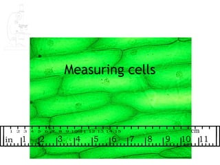
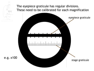
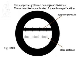
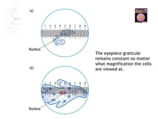
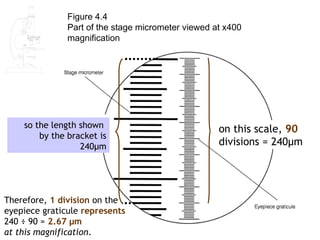
1) The document describes how to use an eyepiece graticule and stage micrometer to accurately measure the size of cellular structures under a microscope.
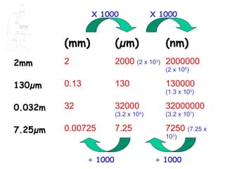
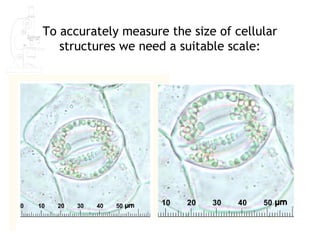
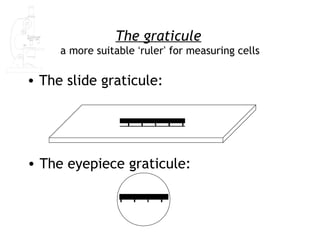
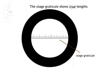
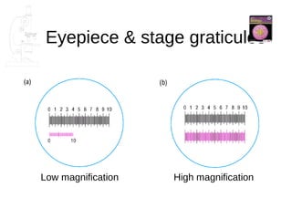
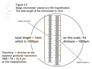
2) The stage graticule shows the true lengths, while the eyepiece graticule needs to be calibrated for each magnification using the stage graticule.
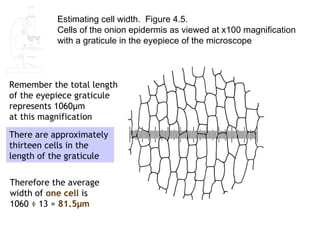
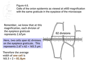
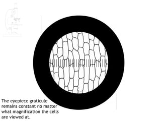
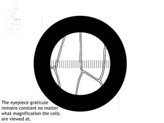
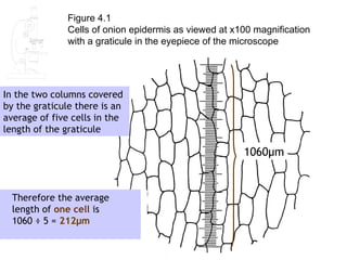
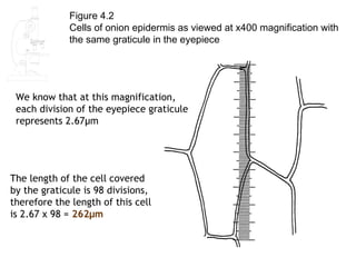
3) Measurements of onion epidermal cells at 100x and 400x magnification using the calibrated eyepiece graticule found average cell lengths of 212 μm and widths of 81.5 μm.
![Learning Objectives
- [PA] use an eyepiece graticule and stage
micrometer scale to measure cells and be
familiar with units (millimetre, micrometre,
nanometre) used in cell studies;
-[PA] calculate linear magnification of
drawings and photographs;
-(h) [PA] calculate actual sizes of specimens
from drawings and photographs;](https://image.slidesharecdn.com/lesson2-120829124948-phpapp02/85/AS-Biology-Lesson-2-Measuring-Cells-2-320.jpg)
![We now have two measurements for the length of an onion cell;
212μm and 262 μm.
Which of these is the more accurate estimate of the length of onion
epidermal cells?
• The answer from Q. 2 [212 μm]
• because this is a mean of several cells.
• Only one cell was measured in Q.3, and this one
may not be representative.](https://image.slidesharecdn.com/lesson2-120829124948-phpapp02/85/AS-Biology-Lesson-2-Measuring-Cells-17-320.jpg)