explains how cells create new cells through processes like mitosis and meiosis, ensuring growth, repair, and the continuation of life by passing on genetic information.
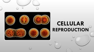
CELLULAR REPRODUCTION
Cellsreproduce by dividing into two in the process
called cell division.
Each dividing cell is called mother cell or parent cell,
and its descendants are called daughter cells.
The parent cell transmit copies of its hereditary
information (DNA) to its daughter cells which in turn,
pass it to their own daughter cells, becoming yet
another parent cell, and so on.
4.
CELLULAR REPRODUCTION
Ascell parent prepares to divide, the DNA inside the
nucleus becomes organized into chromosomes.
This is to ensure that both of the new cells get all of the
genetic information from the original cell.
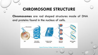
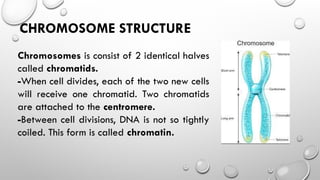
CHROMOSOME STRUCTURE
Chromosomes isconsist of 2 identical halves
called chromatids.
-When cell divides, each of the two new cells
will receive one chromatid. Two chromatids
are attached to the centromere.
-Between cell divisions, DNA is not so tightly
coiled. This form is called chromatin.
7.
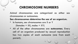
CHROMOSOME NUMBERS
Animal chromosomesare categorized as either sex
chromosomes or autosomes.
Sex chromosomes determine the sex of an organism.
• In humans, sex chromosomes are X or Y
(females = XX, males = XY)
• All of the other chromosomes are autosomes. Every
cell of an organism produced by sexual reproduction
has two copies of each autosome (one from each
parent).
8.
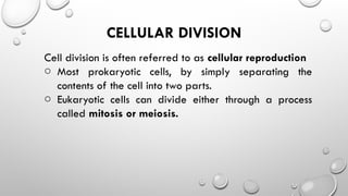
CELLULAR DIVISION
Cell divisionis often referred to as cellular reproduction
o Most prokaryotic cells, by simply separating the
contents of the cell into two parts.
o Eukaryotic cells can divide either through a process
called mitosis or meiosis.
9.
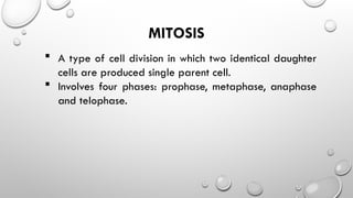
MITOSIS
A typeof cell division in which two identical daughter
cells are produced single parent cell.
Involves four phases: prophase, metaphase, anaphase
and telophase.
11.
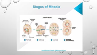
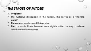
THE STAGES OFMITOSIS
1. Prophase
The nucleolus disappears in the nucleus. This serves as a “starting
signal”
The nuclear membrane disintegrates.
The chromatin fibers become more tightly coiled as they condense
into discrete chromosomes.
12.
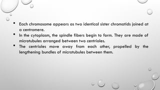
Each chromosomeappears as two identical sister chromatids joined at
a centromere.
In the cytoplasm, the spindle fibers begin to form. They are made of
microtubules arranged between two centrioles.
The centrioles move away from each other, propelled by the
lengthening bundles of microtubules between them.
14.
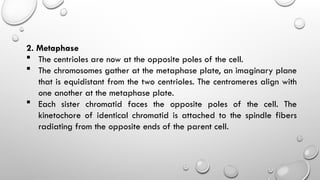
2. Metaphase
Thecentrioles are now at the opposite poles of the cell.
The chromosomes gather at the metaphase plate, an imaginary plane
that is equidistant from the two centrioles. The centromeres align with
one another at the metaphase plate.
Each sister chromatid faces the opposite poles of the cell. The
kinetochore of identical chromatid is attached to the spindle fibers
radiating from the opposite ends of the parent cell.
16.
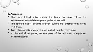
3. Anaphase
Theonce joined sister chromatids begin to move along the
microtubules toward the opposite poles of the cell.
The spindle fibers become shorter, pulling the chromosomes along
with them.
Each chromatid is now considered an individual chromosome.
At the end of anaphase, the two poles of the cell have an equal set
of chromosomes
18.
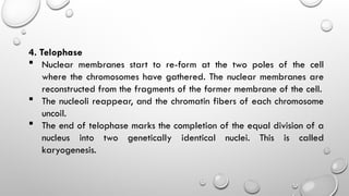
4. Telophase
Nuclearmembranes start to re-form at the two poles of the cell
where the chromosomes have gathered. The nuclear membranes are
reconstructed from the fragments of the former membrane of the cell.
The nucleoli reappear, and the chromatin fibers of each chromosome
uncoil.
The end of telophase marks the completion of the equal division of a
nucleus into two genetically identical nuclei. This is called
karyogenesis.
20.
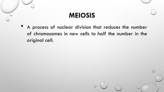
MEIOSIS
A processof nuclear division that reduces the number
of chromosomes in new cells to half the number in the
original cell.
21.
Two stages ofmeiosis:
First cell division = Meiosis I
Prophase I, Metaphase I, Anaphase I, Telophase I, and
Cytokinesis I
Second cell division = Meiosis II
Prophase II, Metaphase II, Anaphase II, Telophase II, and
Cytokinesis II
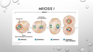
THE STAGES OFMEIOSIS I
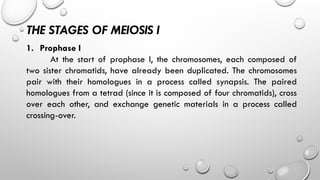
1. Prophase I
At the start of prophase I, the chromosomes, each composed of
two sister chromatids, have already been duplicated. The chromosomes
pair with their homologues in a process called synapsis. The paired
homologues from a tetrad (since it is composed of four chromatids), cross
over each other, and exchange genetic materials in a process called
crossing-over.
26.
2. Metaphase I
Whenspindle fibers are fully formed, the paired homologous
chromosomes align at the metaphase plate, with the homologues facing
the opposite poles.
28.
3. Anaphase I
Membersof homologous pairs separate from each other and
move toward opposite poles. The positioning of each pair of homologues
at the metaphase plate and their subsequent direction of movement are
random events. Their migration to the poles does not follow a specific
pattern.
30.
4. Telophase Iand Cytokinesis
In this stage, two daughter cells are formed. Each daughter cell
contains only one chromosome from each homologous pair. This means
that each daughter cell contains only a haploid number of chromosomes.
The result of telophase I is required for meiosis II.
32.
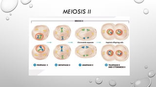
THE STAGES OFMEIOSIS II
1. Prophase II
There is no DNA replication in this stage. The sister chromatids of
each chromosome are still attached at the centromere.
33.
2. Metaphase II
Eachchromosome aligns at the metaphase plate with the sister
chromatids facing the opposite poles.
34.
3. Anaphase II
Thesister chromatids of each chromosome in this stage separate
and migrate toward opposite poles.
35.
4. Telophase IIand Cytokinesis
Cell division is completed in this stage. Four haploid cells are
formed. After cytokinesis, each gamete has a haploid number of
chromosomes.


![[7] CELL CYCLE _MITOSIS & MEIOSIS.ppt](https://cdn.slidesharecdn.com/ss_thumbnails/7cellcyclemitosismeiosis-221113164347-59a6d1ec-thumbnail.jpg?width=640&height=640&fit=bounds)


