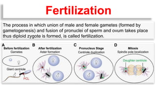
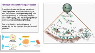
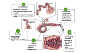
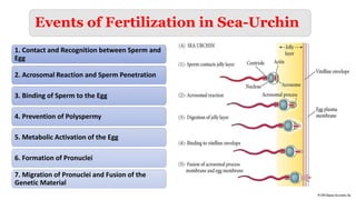
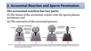
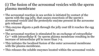
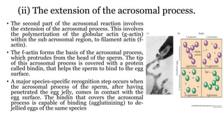
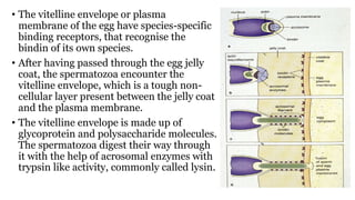
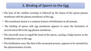
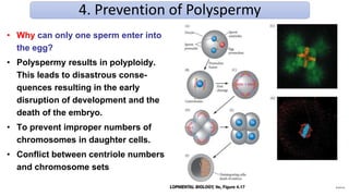
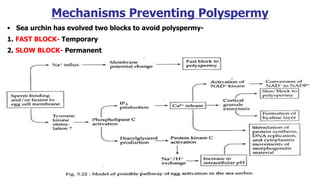
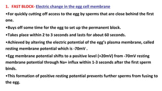
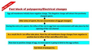
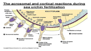
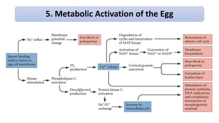
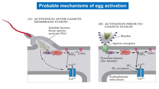
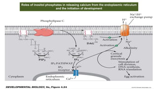
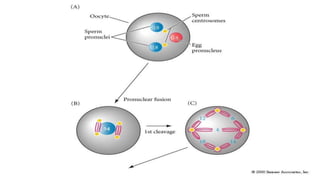
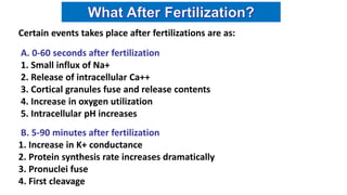
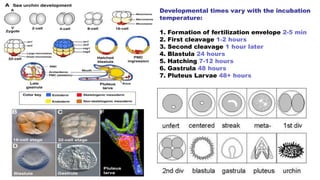
Prof. R. K. Lodha
The process in which union of male and female gametes (formed by
gametogenesis) and fusion of pronuclei of sperm and ovum takes place
thus diploid zygote is formed, is called fertilization.
Fertilization
Fertilization has following processes
The union of male and female gametes is
called Syngamy, where as intermixing of
their cytoplasm is called plasmogamy. The
fusion of pronuclei of sperm and ovum is
called karyogamy. The intermingling of their
chromosomes is called amphimixis.
Due to fertilization, a diploid zygote is
formed, by the union of two different types of
gametes.
.
Site of fertilization
(a) Internal fertilization –
Fertilization in the body (i.e., genital organs of animal) is
called internal fertilization. In this type of fertilization,
sperms are discharged by male directly into the genital
tract of female after coitus.
- Whole process of fertilization takes place within the
body of female. This is the most common adaptation in
terrestrial animals.
Examples :- Aschelminthes, reptiles, birds and
mammals.
(b) External Fertilization –
External fertilization takes place outside the body of
females i.e, in water.
Examples :- In most of the invertebrates, some
protochordates, amphibia and most of the fishes.
Types of fertilization
(a)Self fertilization –
This process takes place in the body of single animal i.e,
fusion of male and female gametes produced by male and
female organs of the same animal. This is called self-
fertilization. This is possible only in bisexual or hermaphrodite
animals.
Examples :- Taenia solium
(b)Cross-Fertilization –
Fertilization takes place between two (male & female)
different animals of same species. This is called cross
fertilization.
-This process is found in all unisexual animals. These
animals are also called dioecious animals
-Cross- fertilization is also found in most of the bisexual or
hermaphrodite animals because in these animals male
genital organs develop first. This condition is called
protandrous condition. In some of the species female organs
develop first, this condition is called protogynous condition
e.g. sponges.
Stages of Fertilization
The stages of fertilization can be divided into four processes:
i. sperm preparation,
ii. sperm-egg recognition and binding,
iii. sperm-egg fusion and
iv. fusion of sperm and egg pronuclei and activation of the zygote.
Fertilization Process in Echinoderms (Sea Urchin)
Why to study Sea Urchin?
• The egg is small enough to be seen under
the microscope.
• The gametes are produced in great
abundance.
• Fertilization occurs naturally outside the
body in sea water.
• The cytoplasm is relatively clear, so cleavage
and gastrulation are easily observed.
Sea Urchin
1. Contact and Recognition between Sperm and
Egg
2. Acrosomal Reaction and Sperm Penetration
3. Binding of Sperm to the Egg
4. Prevention of Polyspermy
5. Metabolic Activation of the Egg
6. Formation of Pronuclei
7. Migration of Pronuclei and Fusion of the
Genetic Material
Events of Fertilization in Sea-Urchin
• The mode of fertilization in sea urchin being external, the egg and spermatozoa are
released into the sea water.
• The first step is the encounter of spermatozoa and the egg. This encounter is brought
about by the swimming movement of the spermatozoa.
• To ensure the survival of the species, the gametes are produced in large numbers.
• A single female Arbacia releases about 4 million eggs, while the male releases about 100
billion spermatozoa, during a single spawning.
• Not-withstanding this, adult sea urchins improve the chances of a sperm meeting an egg
by moving into dense aggregates before spawning.
• However, a successful fertilization depends largely on coordinated timing in the release
of gametes and water conditions at that time.
1. CONTACT AND RECOGNITION BETWEEN SPERM AND EGG
CHEMOTAXIS
• Species-specific sperm attractants have been found in sea urchin.
• Sperms are attracted towards eggs of their own species by chemo-taxis, i.e., by
following a gradient of a chemical, secreted by the egg.
• One such chemotaxin is a 14-amino acid peptide called resect, has been isolated
from the egg jelly of a sea urchin Arbacia punctulata.
• Resact diffuses readily in sea-water.
• A very low concentration of resact in the sea water can attract sperm of its own
species.
• The sperms of A. punctulata have receptors in their plasma membranes that bind
resact.
• Resact also acts as a sperm-activating peptide that causes drastic and immediate
increase in mitochondrial respiration and sperm motility.
CHEMOTAXIS
Sperm diffuses to meet egg.
How can the sperm and egg meet in the diluted condition?
- by chemotaxis
- Resact: 14aa peptide chemoattractant expressed in sea urchin Arbacia punctulata
Speract: a chemoattractant in sea urchin Strongylocentrotus purpuratus
- Only A. punctulata has receptor for resact, which activates guanylyl cyclase activity and the
consequent elevation of cGMP to induce calcium entry into sperm. Resact also activate mitochondrial
respiration, which increase the motility.
Sperm chemotaxis in the sea urchin
Arbacia punctulata
In Short
The acrosomal reaction has two parts:
(i) The fusion of the acrosomal vesicles with the sperm plasma
membrane and
(ii) The extension of the acrosomal process.
2. Acrosomal Reaction and Sperm Penetration
(i) The fusion of the acrosomal vesicles with the sperm
plasma membrane
• The acrosomal reaction in sea urchin is initiated by contact of the
sperm with the egg jelly, that causes exocytosis of the sperm’s
acrosomal vesicle and the proteolytic enzyme present in the acrosome
gets released.
• This enzyme digests a path through the jelly coat till it reaches the egg
surface.
• The acrosomal reaction is stimulated by an exchange of extracellular
Ca++ with intracellular K+ by sperm plasma membrane resulting in the
increase of intracellular pH to more than 7-2.
• This initiates the localized fusion of the outer acrosomal membrane
with the plasma membrane.
• This releases the soluble enzymes located within the acrosomal vesicle.
(ii) The extension of the acrosomal process.
• The second part of the acrosomal reaction involves
the extension of the acrosomal process. This involves
the polymerization of the globular actin (g-actin)
within the sub acrosomal region, to filament actin (f-
actin).
• The f-actin forms the basis of the acrosomal process,
which protrudes from the head of the sperm. The tip
of this acrosomal process is covered with a protein
called bindin, that helps the sperm to bind to the egg
surface.
• A major species-specific recognition step occurs when
the acrosomal process of the sperm, after having
penetrated the egg jelly, comes in contact with the
egg surface. The bindin that covers the acrosomal
process is capable of binding (agglutinizing) to de-
jellied eggs of the same species
• The vitelline envelope or plasma
membrane of the egg have species-specific
binding receptors, that recognise the
bindin of its own species.
• After having passed through the egg jelly
coat, the spermatozoa encounter the
vitelline envelope, which is a tough non-
cellular layer present between the jelly coat
and the plasma membrane.
• The vitelline envelope is made up of
glycoprotein and polysaccharide molecules.
The spermatozoa digest their way through
it with the help of acrosomal enzymes with
trypsin like activity, commonly called lysin.
• The lysis of the vitelline envelope is followed by the fusion of the sperm plasma
membrane with the plasma membrane of the egg.
• This membrane fusion is a common feature of fertilization in all animals.
• The binding of sperm with egg membrane appears to cause the formation of
several microvilli in the egg plasma membrane.
• The microvilli seem to engulf the head of the sperm, causing a bulge known as the
fertilization cone in the egg.
• The fertilization cone, like that of the acrosomal process, appears to be extended by
the polymerization of actin.
3. Binding of Sperm to the Egg
• The plasma membrane of the sperm is antigenically different from that
of the egg. However, it becomes incorporated into the plasma
membrane of the egg as a mosaic patch (Shapiro et al., 1980), as the
sperm nucleus moves into the interior of the egg.
• In the sea urchin all region of the egg plasma membrane are capable of
fusing with sperm and this fusion is often mediated by specific
“fusogenic” proteins. Bindin, however, plays a second role as a
fusogenic protein.
4. Prevention of Polyspermy
• Why can only one sperm enter into
the egg?
• Polyspermy results in polyploidy.
This leads to disastrous conse-
quences resulting in the early
disruption of development and the
death of the embryo.
• To prevent improper numbers of
chromosomes in daughter cells.
• Conflict between centriole numbers
and chromosome sets
Mechanisms Preventing Polyspermy
• Sea urchin has evolved two blocks to avoid polyspermy-
1. FAST BLOCK- Temporary
2. SLOW BLOCK- Permanent
1. FAST BLOCK- Electric change in the egg cell membrane
•For quickly cutting off access to the egg by sperms that are close behind the first
one.
•Buys off some time for the egg to set up the permanent block.
•Takes place within 2 to 3 seconds and lasts for about 60 seconds.
•Achieved by altering the electric potential of the egg’s plasma membrane, called
resting membrane potential which is -70mV .
•Egg membrane potential shifts to a positive level (+20mV) from -70mV resting
membrane potential through Na+ influx within 1-3 seconds after the first sperm
binds.
•This formation of positive resting potential prevents further sperms from fusing to
the egg.
•
2. SLOW BLOCK- Exocytosis of the cortical granules
Steps-
1. Mobilization of Ca++ from the stores
Ca++ is first released at the site of sperm entry and a wave of free Ca++
passes through the egg. This wave of released Ca++ forces cortical granules to
move to the inner surface of the plasma membrane and to fuse with it
initiating cortical reaction.
2. Cortical granule reaction
Cortical granule fuse with egg cell membrane & release several proteins by
the exocytosis of the cortical granules which separate vitelline membrane from
egg membrane.
a) Cortical granule serine protease dissolves the protein linking the vitelline
membrane to egg membrane. They also clip off the bindin receptor and any
sperm attached to it.
b) Mucopolysaccharides from cortical granules produce an osmotic gradient,
that causes water to rush into the space between peri-vitelline envelope and
egg membrane which form a very sticky fertilization envelope and expand to
move away from the egg
c) Peroxidase hardens the fertilization envelope
d) Hyaline forms a coating around the egg below the fertilization membrane.
This hyaline layer provides support for the blastomeres at the time of
cleavage.
Cortical granule exocytosis
CGSP: cortical granule serine protease
TG(transglutamunases), OVOP/Dbdx; perosidase
5. Metabolic Activation of the Egg
Probable mechanisms of egg activation
(a) A three to five fold increase in oxygen consumption (probably related to the
formation of H2O2).
(b) Activation of the enzyme NAD+ kinase which converts NAD+ to NADP+. This
conversion may facilitate the biosynthesis of new membrane lipids, which is
important in the construction of many new cell membranes required during
cleavage.
(c) A second influx of Na+ coupled with an efflux of H+ from the cell, resulting in
the increase of intracellular pH. This increase of pH leads to an increase in protein
synthesis, the activation of transport systems and ultimately the initiation of DNA
synthesis, in preparation for the first cleavage division.
Other than the cortical reaction, the events that take place include:
Roles of inositol phosphates in releasing calcium from the endoplasmic reticulum
and the initiation of development
• Sperm’s nucleus and centriole separate from the mitochondria and flagellum.
• Shortly after the entry of the sperm into the egg cytoplasm (lifting the block to the
second meiotic division and releasing the second polar body), the nuclear membrane of
the sperm breaks down. The interaction between the nuclear contents of the sperm and
the cytoplasm of the egg results in de-condensation of the tightly packed nuclear
chromatin to form pronucleus.
• As the chromatin dispersion nears completion, a new nuclear membrane is formed. The
nucleus thus becomes vesicular and has an appearance like the interphase nucleus and is
called the male pro-nucleus.
• After completion of the second meiotic division, the haploid nucleus of the egg forms the
female pro-nucleus.
6. Formation of Pronuclei
7. Migration of Pronuclei and Fusion of the Genetic Material
• As the sperm moves inward, from the site of the fertilization cone, it
soon rotates 180° so that the centriole assumes the leading position
and is positioned between the sperm and egg pro-nucleuses.
• Pronuclear fusion - The two pronuclei come into contact with each
other, their membranes fuse, resulting in the formation of a single
membrane, which encloses both male and female chromosomes,
resulting in the formation of the diploid zygote nucleus.
• Soon after the pronuclear fusion, the chromosomes replicate their
DNA, for the first cleavage division completing fertilization.
Certain events takes place after fertilizations are as:
A. 0-60 seconds after fertilization
1. Small influx of Na+
2. Release of intracellular Ca++
3. Cortical granules fuse and release contents
4. Increase in oxygen utilization
5. Intracellular pH increases
B. 5-90 minutes after fertilization
1. Increase in K+ conductance
2. Protein synthesis rate increases dramatically
3. Pronuclei fuse
4. First cleavage
Developmental times vary with the incubation
temperature:
1. Formation of fertilization envelope 2-5 min
2. First cleavage 1-2 hours
3. Second cleavage 1 hour later
4. Blastula 24 hours
5. Hatching 7-12 hours
6. Gastrula 48 hours
7. Pluteus Larvae 48+ hours
Fertilization in sea urchin

Fertilization in sea urchin

  • 1.
  • 2.
    The process inwhich union of male and female gametes (formed by gametogenesis) and fusion of pronuclei of sperm and ovum takes place thus diploid zygote is formed, is called fertilization. Fertilization
  • 3.
    Fertilization has followingprocesses The union of male and female gametes is called Syngamy, where as intermixing of their cytoplasm is called plasmogamy. The fusion of pronuclei of sperm and ovum is called karyogamy. The intermingling of their chromosomes is called amphimixis. Due to fertilization, a diploid zygote is formed, by the union of two different types of gametes. .
  • 4.
    Site of fertilization (a)Internal fertilization – Fertilization in the body (i.e., genital organs of animal) is called internal fertilization. In this type of fertilization, sperms are discharged by male directly into the genital tract of female after coitus. - Whole process of fertilization takes place within the body of female. This is the most common adaptation in terrestrial animals. Examples :- Aschelminthes, reptiles, birds and mammals. (b) External Fertilization – External fertilization takes place outside the body of females i.e, in water. Examples :- In most of the invertebrates, some protochordates, amphibia and most of the fishes.
  • 5.
    Types of fertilization (a)Selffertilization – This process takes place in the body of single animal i.e, fusion of male and female gametes produced by male and female organs of the same animal. This is called self- fertilization. This is possible only in bisexual or hermaphrodite animals. Examples :- Taenia solium (b)Cross-Fertilization – Fertilization takes place between two (male & female) different animals of same species. This is called cross fertilization. -This process is found in all unisexual animals. These animals are also called dioecious animals -Cross- fertilization is also found in most of the bisexual or hermaphrodite animals because in these animals male genital organs develop first. This condition is called protandrous condition. In some of the species female organs develop first, this condition is called protogynous condition e.g. sponges.
  • 6.
    Stages of Fertilization Thestages of fertilization can be divided into four processes: i. sperm preparation, ii. sperm-egg recognition and binding, iii. sperm-egg fusion and iv. fusion of sperm and egg pronuclei and activation of the zygote.
  • 8.
    Fertilization Process inEchinoderms (Sea Urchin) Why to study Sea Urchin? • The egg is small enough to be seen under the microscope. • The gametes are produced in great abundance. • Fertilization occurs naturally outside the body in sea water. • The cytoplasm is relatively clear, so cleavage and gastrulation are easily observed. Sea Urchin
  • 9.
    1. Contact andRecognition between Sperm and Egg 2. Acrosomal Reaction and Sperm Penetration 3. Binding of Sperm to the Egg 4. Prevention of Polyspermy 5. Metabolic Activation of the Egg 6. Formation of Pronuclei 7. Migration of Pronuclei and Fusion of the Genetic Material Events of Fertilization in Sea-Urchin
  • 10.
    • The modeof fertilization in sea urchin being external, the egg and spermatozoa are released into the sea water. • The first step is the encounter of spermatozoa and the egg. This encounter is brought about by the swimming movement of the spermatozoa. • To ensure the survival of the species, the gametes are produced in large numbers. • A single female Arbacia releases about 4 million eggs, while the male releases about 100 billion spermatozoa, during a single spawning. • Not-withstanding this, adult sea urchins improve the chances of a sperm meeting an egg by moving into dense aggregates before spawning. • However, a successful fertilization depends largely on coordinated timing in the release of gametes and water conditions at that time. 1. CONTACT AND RECOGNITION BETWEEN SPERM AND EGG
  • 11.
    CHEMOTAXIS • Species-specific spermattractants have been found in sea urchin. • Sperms are attracted towards eggs of their own species by chemo-taxis, i.e., by following a gradient of a chemical, secreted by the egg. • One such chemotaxin is a 14-amino acid peptide called resect, has been isolated from the egg jelly of a sea urchin Arbacia punctulata. • Resact diffuses readily in sea-water. • A very low concentration of resact in the sea water can attract sperm of its own species. • The sperms of A. punctulata have receptors in their plasma membranes that bind resact. • Resact also acts as a sperm-activating peptide that causes drastic and immediate increase in mitochondrial respiration and sperm motility.
  • 12.
  • 13.
    Sperm diffuses tomeet egg. How can the sperm and egg meet in the diluted condition? - by chemotaxis - Resact: 14aa peptide chemoattractant expressed in sea urchin Arbacia punctulata Speract: a chemoattractant in sea urchin Strongylocentrotus purpuratus - Only A. punctulata has receptor for resact, which activates guanylyl cyclase activity and the consequent elevation of cGMP to induce calcium entry into sperm. Resact also activate mitochondrial respiration, which increase the motility. Sperm chemotaxis in the sea urchin Arbacia punctulata In Short
  • 14.
    The acrosomal reactionhas two parts: (i) The fusion of the acrosomal vesicles with the sperm plasma membrane and (ii) The extension of the acrosomal process. 2. Acrosomal Reaction and Sperm Penetration
  • 15.
    (i) The fusionof the acrosomal vesicles with the sperm plasma membrane • The acrosomal reaction in sea urchin is initiated by contact of the sperm with the egg jelly, that causes exocytosis of the sperm’s acrosomal vesicle and the proteolytic enzyme present in the acrosome gets released. • This enzyme digests a path through the jelly coat till it reaches the egg surface. • The acrosomal reaction is stimulated by an exchange of extracellular Ca++ with intracellular K+ by sperm plasma membrane resulting in the increase of intracellular pH to more than 7-2. • This initiates the localized fusion of the outer acrosomal membrane with the plasma membrane. • This releases the soluble enzymes located within the acrosomal vesicle.
  • 16.
    (ii) The extensionof the acrosomal process. • The second part of the acrosomal reaction involves the extension of the acrosomal process. This involves the polymerization of the globular actin (g-actin) within the sub acrosomal region, to filament actin (f- actin). • The f-actin forms the basis of the acrosomal process, which protrudes from the head of the sperm. The tip of this acrosomal process is covered with a protein called bindin, that helps the sperm to bind to the egg surface. • A major species-specific recognition step occurs when the acrosomal process of the sperm, after having penetrated the egg jelly, comes in contact with the egg surface. The bindin that covers the acrosomal process is capable of binding (agglutinizing) to de- jellied eggs of the same species
  • 17.
    • The vitellineenvelope or plasma membrane of the egg have species-specific binding receptors, that recognise the bindin of its own species. • After having passed through the egg jelly coat, the spermatozoa encounter the vitelline envelope, which is a tough non- cellular layer present between the jelly coat and the plasma membrane. • The vitelline envelope is made up of glycoprotein and polysaccharide molecules. The spermatozoa digest their way through it with the help of acrosomal enzymes with trypsin like activity, commonly called lysin.
  • 18.
    • The lysisof the vitelline envelope is followed by the fusion of the sperm plasma membrane with the plasma membrane of the egg. • This membrane fusion is a common feature of fertilization in all animals. • The binding of sperm with egg membrane appears to cause the formation of several microvilli in the egg plasma membrane. • The microvilli seem to engulf the head of the sperm, causing a bulge known as the fertilization cone in the egg. • The fertilization cone, like that of the acrosomal process, appears to be extended by the polymerization of actin. 3. Binding of Sperm to the Egg
  • 19.
    • The plasmamembrane of the sperm is antigenically different from that of the egg. However, it becomes incorporated into the plasma membrane of the egg as a mosaic patch (Shapiro et al., 1980), as the sperm nucleus moves into the interior of the egg. • In the sea urchin all region of the egg plasma membrane are capable of fusing with sperm and this fusion is often mediated by specific “fusogenic” proteins. Bindin, however, plays a second role as a fusogenic protein.
  • 20.
    4. Prevention ofPolyspermy • Why can only one sperm enter into the egg? • Polyspermy results in polyploidy. This leads to disastrous conse- quences resulting in the early disruption of development and the death of the embryo. • To prevent improper numbers of chromosomes in daughter cells. • Conflict between centriole numbers and chromosome sets
  • 21.
    Mechanisms Preventing Polyspermy •Sea urchin has evolved two blocks to avoid polyspermy- 1. FAST BLOCK- Temporary 2. SLOW BLOCK- Permanent
  • 22.
    1. FAST BLOCK-Electric change in the egg cell membrane •For quickly cutting off access to the egg by sperms that are close behind the first one. •Buys off some time for the egg to set up the permanent block. •Takes place within 2 to 3 seconds and lasts for about 60 seconds. •Achieved by altering the electric potential of the egg’s plasma membrane, called resting membrane potential which is -70mV . •Egg membrane potential shifts to a positive level (+20mV) from -70mV resting membrane potential through Na+ influx within 1-3 seconds after the first sperm binds. •This formation of positive resting potential prevents further sperms from fusing to the egg.
  • 23.
  • 25.
    2. SLOW BLOCK-Exocytosis of the cortical granules Steps- 1. Mobilization of Ca++ from the stores Ca++ is first released at the site of sperm entry and a wave of free Ca++ passes through the egg. This wave of released Ca++ forces cortical granules to move to the inner surface of the plasma membrane and to fuse with it initiating cortical reaction. 2. Cortical granule reaction Cortical granule fuse with egg cell membrane & release several proteins by the exocytosis of the cortical granules which separate vitelline membrane from egg membrane.
  • 26.
    a) Cortical granuleserine protease dissolves the protein linking the vitelline membrane to egg membrane. They also clip off the bindin receptor and any sperm attached to it. b) Mucopolysaccharides from cortical granules produce an osmotic gradient, that causes water to rush into the space between peri-vitelline envelope and egg membrane which form a very sticky fertilization envelope and expand to move away from the egg c) Peroxidase hardens the fertilization envelope d) Hyaline forms a coating around the egg below the fertilization membrane. This hyaline layer provides support for the blastomeres at the time of cleavage.
  • 27.
    Cortical granule exocytosis CGSP:cortical granule serine protease TG(transglutamunases), OVOP/Dbdx; perosidase
  • 30.
  • 31.
    Probable mechanisms ofegg activation
  • 32.
    (a) A threeto five fold increase in oxygen consumption (probably related to the formation of H2O2). (b) Activation of the enzyme NAD+ kinase which converts NAD+ to NADP+. This conversion may facilitate the biosynthesis of new membrane lipids, which is important in the construction of many new cell membranes required during cleavage. (c) A second influx of Na+ coupled with an efflux of H+ from the cell, resulting in the increase of intracellular pH. This increase of pH leads to an increase in protein synthesis, the activation of transport systems and ultimately the initiation of DNA synthesis, in preparation for the first cleavage division. Other than the cortical reaction, the events that take place include:
  • 33.
    Roles of inositolphosphates in releasing calcium from the endoplasmic reticulum and the initiation of development
  • 34.
    • Sperm’s nucleusand centriole separate from the mitochondria and flagellum. • Shortly after the entry of the sperm into the egg cytoplasm (lifting the block to the second meiotic division and releasing the second polar body), the nuclear membrane of the sperm breaks down. The interaction between the nuclear contents of the sperm and the cytoplasm of the egg results in de-condensation of the tightly packed nuclear chromatin to form pronucleus. • As the chromatin dispersion nears completion, a new nuclear membrane is formed. The nucleus thus becomes vesicular and has an appearance like the interphase nucleus and is called the male pro-nucleus. • After completion of the second meiotic division, the haploid nucleus of the egg forms the female pro-nucleus. 6. Formation of Pronuclei
  • 35.
    7. Migration ofPronuclei and Fusion of the Genetic Material • As the sperm moves inward, from the site of the fertilization cone, it soon rotates 180° so that the centriole assumes the leading position and is positioned between the sperm and egg pro-nucleuses. • Pronuclear fusion - The two pronuclei come into contact with each other, their membranes fuse, resulting in the formation of a single membrane, which encloses both male and female chromosomes, resulting in the formation of the diploid zygote nucleus. • Soon after the pronuclear fusion, the chromosomes replicate their DNA, for the first cleavage division completing fertilization.
  • 39.
    Certain events takesplace after fertilizations are as: A. 0-60 seconds after fertilization 1. Small influx of Na+ 2. Release of intracellular Ca++ 3. Cortical granules fuse and release contents 4. Increase in oxygen utilization 5. Intracellular pH increases B. 5-90 minutes after fertilization 1. Increase in K+ conductance 2. Protein synthesis rate increases dramatically 3. Pronuclei fuse 4. First cleavage
  • 41.
    Developmental times varywith the incubation temperature: 1. Formation of fertilization envelope 2-5 min 2. First cleavage 1-2 hours 3. Second cleavage 1 hour later 4. Blastula 24 hours 5. Hatching 7-12 hours 6. Gastrula 48 hours 7. Pluteus Larvae 48+ hours