

Recommended
More Related Content
What's hot
What's hot (20)
Similar to skin
Similar to skin (20)
More from JAYDIP NINAMA
More from JAYDIP NINAMA (20)
Recently uploaded
Recently uploaded (20)
skin
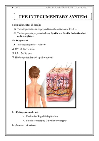
- 1. 1 | P a g e T H E I N T E G U M E N T A R Y S Y S T E M The integument as an organ: The integument as an organ, and is an alternative name for skin. The integumentary system includes the skin and the skin derivatives hair, nails, and glands. The Integument Is the largest system of the body 16% of body weight, 1.5 to 2m2 in area, The integument is made up of two parts: 1. Cutaneous membrane a. Epidermis– Superficial epithelium b. Dermis – underlying CT with blood supply 2. Accessory structures THE INTEGUMENTARY SYSTEM
- 2. 2 | P a g e T H E I N T E G U M E N T A R Y S Y S T E M a. Hair b. Nails c. Exocrine Glands INTRODUCTION The skin is an extensive organ which forms the outer covering of the body. It is continuous with mucous membrane lining the body orifices. The skin has 2 layers. 1. A superficial layer- the epidermis- made up of stratified squamous epithelium 2. A deeper layer-the dermis made up of connective tissue. 3. Hypodermis - subcutaneous tissue- loose connective tissue proper and adipose tissue The junction between these two layers is markedly wavy because of tee presence of finger like extensions from the dermis-the dermal papillae. SKIN STRUCTURE: EPIDERMIS
- 3. 3 | P a g e T H E I N T E G U M E N T A R Y S Y S T E M The Epidermis Is a vascular stratified squamous epithelium Nutrients and oxygen diffuse from capillaries in the dermis CELLS OF THE EPIDERMIS Keratinocytes Contain large amounts of keratin Are the most abundant cells in the epidermis Thin Skin Covers most of the body Has four layers of keratinocytes Thick Skin Covers the palms of the hands and soles of the feet
- 4. 4 | P a g e T H E I N T E G U M E N T A R Y S Y S T E M Has five layers of keratinocytes STRUCTURES OF THE EPIDERMIS The five strata of keratinocytes in thick skin From basal lamina to free surface Stratum basale Stratum spinosum Stratum granulosum Stratum lucidum Stratum corneum Stratum Basale Is attached to basement membrane by hemidesmosomes Forms a strong bond between epidermis and dermis Forms epidermal ridges (e.g., fingerprints)
- 5. 5 | P a g e T H E I N T E G U M E N T A R Y S Y S T E M Dermal papillae (tiny mounds) Increase the area of basement membrane Strengthen attachment between epidermis and dermis Has many basal cells or germinative cells Stratum Spinosum — the “spiny layer” Produced by division of stratum basale Eight to ten layers of keratinocytes bound by desmosomes Cells shrink until cytoskeletons stick out (spiny) Continue to divide, increasing thickness of epithelium Contain dendritic (Langerhans) cells, active in immune response Stratum Granulosum — the “grainy layer” Stops dividing, starts producing Keratin A tough, fibrous protein Makes up hair and nails Keratohyalin Dense granules Cross-link keratin fibers Stratum Lucidum — the “clear layer” Found only in thick skin Covers stratum granulosum Stratum Corneum — the “horn layer”
- 6. 6 | P a g e T H E I N T E G U M E N T A R Y S Y S T E M Exposed surface of skin 15 to 30 layers of keratinized cells Water resistant Shed and replaced every 2 weeks THE DERMIS Located between epidermis and subcutaneous layer Anchors epidermal accessory structures (hair follicles, sweat glands) Two components Outer papillary layer Deep reticular layer The Papillary Layer Consists of areolar tissue
- 7. 7 | P a g e T H E I N T E G U M E N T A R Y S Y S T E M Contains smaller capillaries, lymphatics, and sensory neurons Has dermal papillae projecting between epidermal ridges The Reticular Layer Consists of dense irregular connective tissue Contains larger blood vessels, lymphatic vessels, and nerve fibers Contains collagen and elastic fibers Contains connective tissue proper DERMATITIS An inflammation of the papillary layer Caused by infection, radiation, mechanical irritation, or chemicals (e.g., poison ivy) Characterized by itch or pain Characteristics Strong, due to collagen fibers Elastic, due to elastic fibers Flexible
- 8. 8 | P a g e T H E I N T E G U M E N T A R Y S Y S T E M THE HYPODERMIS (SUBCUTANEOUS LAYER) Lies below the integument Stabilizes the skin Allows separate movement Made of elastic areolar and adipose tissues Connected to the reticular layer of integument by connective tissue fibers Deposits of Subcutaneous Fat Distribution patterns determined by hormones Reduced by cosmetic liposuction (lipoplasty)
- 9. 9 | P a g e T H E I N T E G U M E N T A R Y S Y S T E M FUNCTIONS OF THE INTEGUMENTARY SYSTEM Protection First line of defense against Bacteria Viruses Protects underlying structures from Ultraviolet (UV) radiation Dehydration Vitamin D production Needed for calcium absorption Sensation Sensory receptors
- 10. 10 | P a g e T H E I N T E G U M E N T A R Y S Y S T E M Body temperature regulation If too hot Dermal blood vessels dilate Vessels carry more blood to surface so heat can escape If too cold Dermal blood vessels constrict Prevents heat from escaping Excretion Small amounts of waste products are lost through perspiration APPENDAGES OF THE SKIN THEY ARE: 1. HAIRS 2. NAIL 3. SWEAT GLANDS 4. SEBACEOUS GLAND STRUCTURE OF HAIR The Hair Follicle Hair follicles are the organs that form the hairs. Hair follicles are the organs that form the hairs. Located deep in dermis. Produces nonliving hairs. Wrapped in a dense connective tissue sheath.
- 11. 11 | P a g e T H E I N T E G U M E N T A R Y S Y S T E M Base is surrounded by sensory nerves (root hair plexus). Control bacteria Accessory Structures of Hair Arrector pili Involuntary smooth muscle Causes hairs to stand up Produces “goose bumps” Sebaceous glands Lubricate the hair Regions of the Hair Hair root Lower part of the hair
- 12. 12 | P a g e T H E I N T E G U M E N T A R Y S Y S T E M Attached to the integument Hair shaft Upper part of the hair Not attached to the integument Hair Shaft Structure Medulla Core, dead cells contain soft keratin and air to provide flexible Cortex Middle layer, dead cells contain hard keratin to provide stiffness Cuticle Outermost, overlapping dead keratinized cells form shiny surface
- 13. 13 | P a g e T H E I N T E G U M E N T A R Y S Y S T E M HAIR FUNCTION Head: UV protection Cushion from trauma Insulation Nostrils, Ear canals, Eyelashes: Prevent entry of foreign material Body Hair: sensory detection Root hair plexus: Sensory nerves at base of hair follicle that detect slight movement of hair Arrector pili muscle: Attached to every hair follicle Contract to stand hair perpendicular to skin surface STRUCTURE & FUNCTION OF NAIL Nails Protect fingers and toes Made of dead cells packed with keratin Metabolic disorders can change nail structure Nail Production Occurs in a deep epidermal fold near the bone called the nail root
- 14. 14 | P a g e T H E I N T E G U M E N T A R Y S Y S T E M STRUCTURE OF A NAIL Nail body The visible portion of the nail Covers the nail bed Lunula The pale crescent at the base of the nail Sides of nails Lie in lateral nail grooves Surrounded by lateral nail folds
- 15. 15 | P a g e T H E I N T E G U M E N T A R Y S Y S T E M SEBACEOUS GLAND They are seen in relation to the hair follicles. Each gland consists of a number of alveoli that are connected to a duct.
- 16. 16 | P a g e T H E I N T E G U M E N T A R Y S Y S T E M This duct opens in to the hair folicle. As mentioned earlier, the sebaceous gland is situated between the hair folicle and the arrector pilli muscle. When the muscle contracts,it sequeezess the gland,which facilitates the discharge of its secretions in to hair folicle. Functions of sebum 1. Its oily nature helps to keep the skin and hair soft 2. It prevents drying of skin 3. It makes the skin resistant to moisture. Modified sebaceous glands The tarsal glands or eyelids are modified sebaceous glands. They are called maibomian glands SWEAT GLANDS Sweat glands produce sweat or perspiration. There are 2 type of sweat glands in the body: 1. Typical 2. Atypical Typical sweat gland A typical sweat gland consists of a single long tube. The lower end of the tube is highly coiled forming the body of the gland.it usually lies in the reticular layer of the dermis. Atypical sweat gland Atypical sweat glands are found in the following regions: THE SECRETION OF SEBACEOUS GLAND IS CALLED “ SEBUM”
- 17. 17 | P a g e T H E I N T E G U M E N T A R Y S Y S T E M 1. Axilla 2. Nipple and areola 3. Perianal region 4. The glans penis 5. Some parts of female external genitallia. These glands are larger in size. They show branching. Their ducts open in to hair folicles.their secretions are viscous. The secretions of atypical glands are odorless but due to bacterial decomposition they give off body odors. Modified sweat gland 1. Ceruminous glands of external auditory meatus 2. Ciliary glands of eyelids 3. Mammary gland. BLOOD SUPPLY OF SKIN Skin is highly vascular organ. It derives its arterial blood from a number of plexuses.one plexus of arteries is present over the deep fascia, another plexus, just below the dermis, is called reticular plexus, the papillary plexus lies just below the dermal papilla. Capillary loops arising from this plexus pass in to each dermal papilla. The epidermis has no blood supply. It derives its nutrition entirely by diffusion from the capillary loops of the dermal papillae. There are numerous arteriovenous anastomoses in the skin, which have an important role in temperature regulation. CUTANEOUS RECEPTORS OR EXTEROCEPTIVE RECEPTORS These receptors present in the skin are concerned with touch , pain, temperature and pressure.
- 18. 18 | P a g e T H E I N T E G U M E N T A R Y S Y S T E M 1. Free nerve endings 2. Tactile corpusles of meissner 3. Lamellated corpuscles of pacini 4. Tactile menisci or merkel cell endings