

Recommended
More Related Content
What's hot
What's hot (20)
Similar to DIAGNOSIS OF PREGNANCY
Similar to DIAGNOSIS OF PREGNANCY (20)
More from JAYDIP NINAMA
More from JAYDIP NINAMA (20)
Recently uploaded
Recently uploaded (20)
DIAGNOSIS OF PREGNANCY
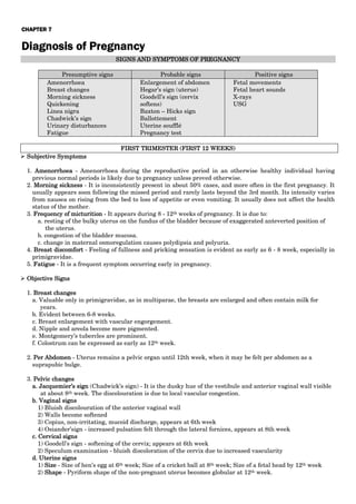
- 1. CHAPTER 7 Diagnosis of Pregnancy SIGNS AND SYMPTOMS OF PREGNANCY Presumptive signs Probable signs Positive signs Amenorrhoea Breast changes Morning sickness Quickening Linea nigra Chadwick’s sign Urinary disturbances Fatigue Enlargement of abdomen Hegar’s sign (uterus) Goodell’s sign (cervix softens) Baxton – Hicks sign Ballottement Uterine soufflé Pregnancy test Fetal movements Fetal heart sounds X-rays USG FIRST TRIMESTER (FIRST 12 WEEKS) Subjective Symptoms 1. Amenorrhoea - Amenorrhoea during the reproductive period in an otherwise healthy individual having previous normal periods is likely due to pregnancy unless proved otherwise. 2. Morning sickness - It is inconsistently present in about 50% cases, and more often in the first pregnancy. It usually appears soon following the missed period and rarely lasts beyond the 3rd month. Its intensity varies from nausea on rising from the bed to loss of appetite or even vomiting. It usually does not affect the health status of the mother. 3. Frequency of micturition - It appears during 8 - 12th weeks of pregnancy. It is due to: a. resting of the bulky uterus on the fundus of the bladder because of exaggerated anteverted position of the uterus. b. congestion of the bladder mucosa. c. change in maternal osmoregulation causes polydipsia and polyuria. 4. Breast discomfort - Feeling of fullness and pricking sensation is evident as early as 6 - 8 week, especially in primigravidae. 5. Fatigue - It is a frequent symptom occurring early in pregnancy. Objective Signs 1. Breast changes a. Valuable only in primigravidae, as in multiparae, the breasts are enlarged and often contain milk for years. b. Evident between 6-8 weeks. c. Breast enlargement with vascular engorgement. d. Nipple and areola become more pigmented. e. Montgomery’s tubercles are prominent. f. Colostrum can be expressed as early as 12th week. 2. Per Abdomen - Uterus remains a pelvic organ until 12th week, when it may be felt per abdomen as a suprapubic bulge. 3. Pelvic changes a. Jacquemier’s sign (Chadwick’s sign) - It is the dusky hue of the vestibule and anterior vaginal wall visible at about 8th week. The discolouration is due to local vascular congestion. b. Vaginal signs 1) Bluish discolouration of the anterior vaginal wall 2) Walls become softened 3) Copius, non-irritating, mucoid discharge, appears at 6th week 4) Osiander’sign - increased pulsation felt through the lateral fornices, appears at 8th week c. Cervical signs 1) Goodell’s sign - softening of the cervix; appears at 6th week 2) Speculum examination - bluish discoloration of the cervix due to increased vascularity d. Uterine signs 1) Size - Size of hen’s egg at 6th week; Size of a cricket ball at 8th week; Size of a fetal head by 12th week 2) Shape - Pyriform shape of the non-pregnant uterus becomes globular at 12th week.
- 2. 3) Consistency - Pregnant uterus feels soft and elastic 4) Hegar’s sign - It is present in two-thirds of cases. It can be demonstrated between 6 -10 weeks, a little earlier in multiparae. This sign is based on the fact that: a) Upper part of the body of the uterus is enlarged by the growing fetus b) Lower part of the body is empty and extremely soft c) Cervix is comparatively firm. Because of variation in consistency, on manual examination (two fingers in the anterior fornix and the abdominal fingers behind the uterus), the abdominal and vaginal fingers seem to appose below the body of the uterus. Examination must be gentle to avoid the risk of abortion. 5) Palmer’s sign - Regular and rhythmic uterine contraction can be elicited during bimanual examination as early as 4-8 weeks. Immunological Tests for Diagnosis of Pregnancy 1. Principle - Pregnancy tests depend on detection of the antigen (hCG) present in the maternal urine or serum with antibody, either polyclonal or monoclonal, available commercially. 2. Tests Used a. Immunoassays without radioisotopes 1) Agglutination inhibition tests - using latex (latex agglutination inhibition) 2) Direct agglutination test (hCG direct test) 3) ELISA 4) Fluoroimmunoassay (FIA). b. Immunoassays with radioisotopes 1. Radioimmunoassay (RIA) 2. Immuno-radiometric assay (IRMA). 3. Selection of time - Should be done by 8-11 days after conception. The test is not reliable after 12 weeks. 4. Collection of urine - First voided urine in the morning in a clean container. 5. Other uses of pregnancy tests a. Diagnosis of ectopic pregnancy b. Monitoring pregnancy following IVF and embryo transfer c. Follow-up of cases of hydatidiform mole and choriocarcinoma. 6. Limitations - Test accuracy is affected due to presence of haemoglobin, albumin, LH and immunological diseases. 7. Advantages - Speed, simplicity, accuracy and less cost. Ultrasonography 1. Intra-decidual gestational sac (GS) identified as early as 29-35 days of gestation 2. Fetal viability and gestational age is determined by detecting the following structures by transvaginal ultrasound: a) Gestational sac and yolk sac by 5 weeks b) Fetal pole and cardiac activity by 6 weeks c) Embryonic movements by 7 weeks 3. Fetal gestational age is best determined by measuring CRL between 7-12 weeks 4. Doppler ultrasound can pick up the fetal heart rate reliably by 10th week SECOND TRIMESTER (13-28 WEEKS) Subjective Symptoms 1. Nausea, vomiting, and frequency of micturition usually subside, while amenorrhoea continues. 2. Quickening - This is the first perception of the active fetal movements by the mother. It is usually felt between 18-20 weeks in primigravidae, and between 16-18 weeks in multiparae, due to experience from previous pregnancy. 3. Progressive enlargement of the lower abdomen by the growing uterus. Objective Signs General Examination a. Face - Chloasma gravidarum may appear at 24th week. b. Breast changes 1) More enlarged with prominent veins under the skin 2) Secondary areola become especially demarcated in primigravidae, appears at 20th week
- 3. 3) Montgomery’s tubercles are prominent and extend to the secondary areola 4) Colostrum becomes thick and yellowish by 16th week 5) Variable degree of striae may be visible with advancing weeks. Abdominal Examination a. Inspection 1) Linea nigra becomes visible at 20th week. 2) Striae (both pink and white) of varying degree are visible in the lower abdomen, more towards the flanks. b. Palpation 1) Fundal height is increased with progressive enlargement of the uterus. Approximate duration of pregnancy can be ascertained by noting the height of the uterus in relation to different levels of the abdomen. a) 16 weeks - midway between the symphysis pubis and umbilicus b) 24 weeks - at the level of umbilicus c) 28 weeks - junction of the lower third and upper two-third of the distance between the umbilicus and ensiform cartilage 2) Uterus feels soft and elastic, and becomes ovoid shape. 3) Braxton-Hicks contractions are evident. 4) Palpation of fetal parts can be made distinctly by 20th week. This is useful for diagnosis of pregnancy, and to identify the presentation and position of the fetus in later weeks. 5) Active fetal movements can be felt at intervals by placing the hand over the uterus as early as 20th week. It gives positive evidence of pregnancy and also a live fetus. 6) External ballottement is usually elicited as early as 20th week when the fetus is relatively smaller than the volume of the amniotic fluid. c. Auscultation - Fetal heart sound (FHS) is most conclusive sign of pregnancy. With an ordinary stethoscope, it can be detected between 18-20 weeks. The rate varies from 140-160 bpm but gradually settles down to 120- 140 bpm as the pregnancy advances. • Two other sounds confused with FHS are: 1. Uterine soufflé - soft blowing and systolic murmur heard low down at the sides of the uterus. The sound is synchronous with maternal pulse and is due to increased blood flow though the dilated uterine vessels. It can be heard in big uterine fibroid. 2. Funic or fetal soufflé - due to rush of blood through the umbilical arteries. It is a soft, blowing murmur synchronous with the FHS. Vaginal Examination a. Bluish discolouration of the vulva, vagina and cervix is much more evident b. Increased softening of the cervix c. Internal ballottement can be elicited between 16th - 28th week. The fetus is too small before 16th week and too large to displace after 28th week. Investigations 1. Sonography - Routine sonography at 18-20 weeks permits a detailed survey of fetal anatomy, placental localization and the integrity of the cervical canal. Gestational age is determined by measuring the BPD, AC and FL. It is most accurate when done between 12-20 weeks. 2. Fetal organ anatomy is surveyed to detect any malformation. 3. Fetal viability is determined by real-time ultrasound. Absence of fetal cardiac motion confirms fetal death. 4. Radiological evidence of fetal skeletal shadow may be visible at 16th week. Skiagram for diagnostic purpose is not undertaken because of radiation hazards. THIRD TRIMESTER (29-40 WEEKS) Subjective Symptoms 1. Amenorrhoea persists. 2. Enlargement of the abdomen is progressive which produces some mechanical discomfort to the patient such as palpitation or dyspnea following exertion. 3. Lightening - At about 38th week, especially in primigravida, a sense of relief of the pressure symptoms is obtained due to engagement of the presenting part. 4. Frequency of micturition reappears. 5. Fetal movements are more pronounced.
- 4. Objective Signs 1. Cutaneous changes are more prominent with increased pigmentation and striae. 2. Uterine shape is changed from cylindrical to spherical beyond 36th week. 3. Fundal height - The distance between the umbilicus and the ensiform cartilage is divided into 3 equal parts: a. 32 weeks - junction of the upper and middle third b. 36 weeks - at the level of ensiform cartilage c. 40 weeks - comes down to 32 week level due to engagement of the presenting part. * To determine whether the height of the uterus corresponds to 32 weeks or 40 weeks, engagement of the head should be tested. If the head is floating, it is of 32 weeks pregnancy and if the head is engaged, it is of 40 weeks. 4. Braxton-Hicks contractions are more evident. 5. Fetal movements are easily felt. 6. Palpation of the fetal parts and their identification become much easier. Lie, presentation and position of the fetus are determined. 7. FHS is heard distinctly in areas corresponding to the presentation and position of the fetus. FHS may not be audible in cases of maternal obesity, polyhydramnios, occipito-posterior position and IUD. Sonography Gestational age estimation by BPD, HC, AC and FL is less accurate. Fetal growth assessment can be made provide accurate dating scan has been done in first or second trimester. 1. Fetal AC at the level of the umbilical vein is used to assess gestational age and fetal growth profile (IUGR or macrosomia) 2. Amniotic fluid assessment is done to detect oligohydramnios (AFI < 5) or polyhydramnios (AFI>25) 3. Placental anatomy - location, thickness or other abnormalities 4. Other information - fetal life, number, presentation and organ anatomy are surveyed again. DIFFERENTIAL DIAGNOSIS OF PREGNANCY 1. Pseudocyesis (Phantom/Spurious/False pregnancy) — It is a psychological disorder where the woman has the false but firm belief that she is pregnant although no pregnancy exists. The woman often is infertile who has an intense desire to have a baby. The conspicuous feature is cessation of menstruation. Other confusing manifestations are gradual enlargement of the abdomen because of deposition of fat, secretion from the breasts and intestinal movement, imagining it to be fetal movement. In some cases, the condition continues until eventually spurious labour sets in. Obstetric examination reveals absence of positive signs of pregnancy. Examination with ultrasound and/or immunological tests for pregnancy may be required to negate the diagnosis. 2. Cystic ovarian tumor. 3. Fibroid. 4. Encysted peritonitis. 5. Distended urinary bladder. SIGNS OF PREVIOUS CHILD BIRTH 1. Breast: flabby with prominent nipples & striae 2. Abdominal wall: lax with white striae & linea alba 3. Uterine wall: less rigid 4. Perineum: lax 5. Vagina: more roomy 6. Cervix: cylindrical & external os is a transverse patulous slit
- 5. CALCULATION OF THE GESTATIONAL AGE AND EXPECTED DATE OF DELIVERY (1) Menstruation - Labour Interval (Naegele’s rule): Expected date of delivery (EDD) = 1st day of the last menstrual period + 7 days + 9 calendar months , or 1st day of LMP + 7 days - 3 calendar months + one year , or 1st day of LMP + 280 days (40 weeks). (Labour may occur one week before or after the calculated EDD) (2) Date of Single Coitus: EDD is calculated by adding 266 days to it. (3) Date of Quickening: EDD = date of quickening + 20 - 22 weeks in primigravida or + 22 - 24 weeks in multipara. (4) Size of the Uterus (fundal level): • During the first month, no clinically appreciable changes. • At the end of 8th week, uterus is 5 cm in diameter. • At the end of 12th week, uterus is 10 cm in diameter, globular in shape, felt at symphysis pubis in primigravida and a little higher in multigravida. • From the 16th week upwards it is pyriform in shape and felt at the levels shown in the figure. (5) Symphysio - Fundal Length (Mc Donald Formula): After 24th weeks, the distance from the symphysis to the fundus (in cms) multiplied by 8/7 gives the duration of pregnancy in weeks e.g. at full term distance = 35 X 8/7 = 40 weeks. (6) Radiology: a) Cephalometry: BPD is 7.5 cm at 32 weeks, 8.5 cm at 36 weeks and 9.5 cm at 40 weeks. b) Ossification centres: Talus at 26 weeks, distal femoral epiphyses at 36-37 weeks, proximal tibial epiphyses and femoral head at 38-40 weeks. (7) Ultrasound: Detection of gestational age and hence EDD by measuring of; • 5-6 weeks - by measurement of gestational sac, • 6-12 weeks - by measurement of the CRL, • 12-26 weeks - by measurement of the BPD (most convenient at this time). • 26 weeks onwards - BPD and/or FL measurement. CALCULATON OF FETAL WEIGHT Johnson’s formula Weight of fetus (in gms) = {Fundal height (in cms) – 12 } X 155 if vertex at or above the ischial spines (head not engaged) Or {Fundal height (in cms) – 11 } X 155 if vertex is below the ischial spines (head engaged) PREVIOUS EXAMINATION QUESTIONS FROM THIS CHAPTER LONG ESSAY 1. What are the subjective symptoms and objective signs of 1st and 2nd trimester pregnancy? 2. Differential diagnosis of pregnancy. SHORT ESSAY 1. Mention the methods of diagnosis of early pregnancy. Mention three important complications in early pregnancy. 2. How to calculate estimated date of delivery? 3. Hegar’s sign. 4. Diagnosis of pregnancy. SHORT ANSWERS 1.Signs and symptoms of early pregnancy.