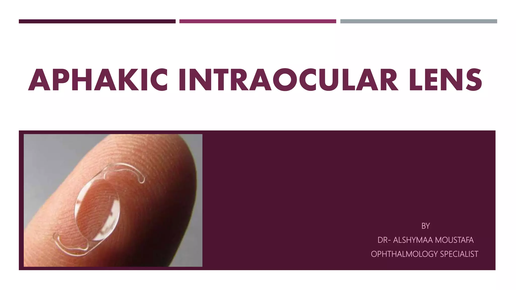
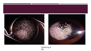
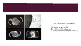
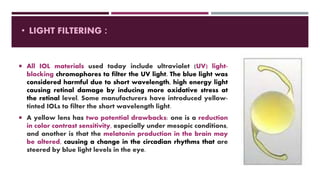
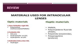
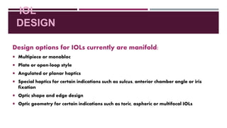
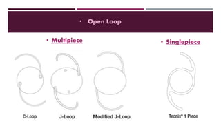
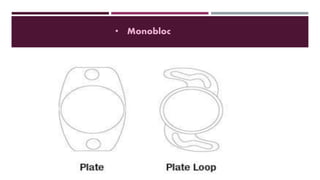
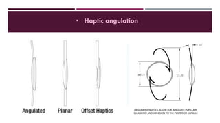
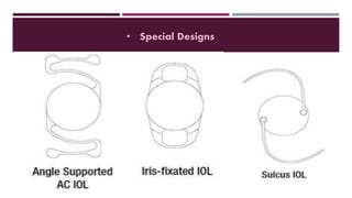
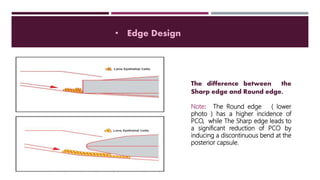
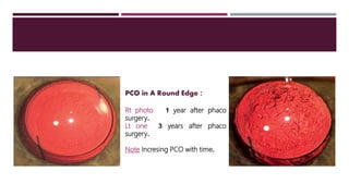
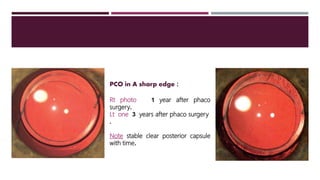
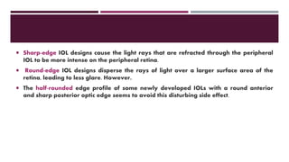
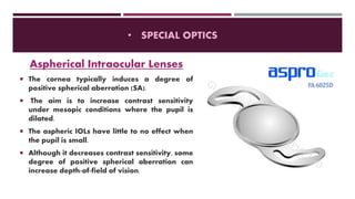
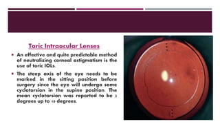
APHAKIC INTRAOCULAR LENS
BY
DR- ALSHYMAA MOUSTAFA
OPHTHALMOLOGY SPECIALIST
INTRAOCULAR LENS (IOL )
 Several different factors affect choosing an IOL for cataract surgery.
 Materials and design should result in a low degree of postoperative inflammation by
being as inert as possible and have a good track record concerning long-term
complications such as posterior capsule opacification (PCO).
 IOL materials should also be easy to handle concerning folding and implantation.
 In special cases, such as those with incomplete capsule support, high myopia, or with a
history of uveitis, the IOL choice may differ from the usual one.
 Additionally, in eyes with a cornea that either has astigmatism and/or
spherical aberrations (SA), special IOLs may be indicated.
 Patients who want to be less dependent on spectacles for near work or
intermediate distance vision after surgery may be considered for
multifocal IOL designs. Use of these lenses should be dissuaded in the
presence of corneal or macular pathology.
 Patient’s should be thoroughly counseled regarding both the benefits
and drawbacks of multifocal lenses (eg. glare, haloes, decreased
contrast sensitivity) prior to their insertion.
IOL MATERIALS
 The earliest IOLs were made of polymethylmethacrylate (PMMA), the
plastic that IOL inventor Harold Ridley had noticed to be inert in eyes of
World War II aviators struck by flying plastic during combat.
 With the introduction of phacoemulsification and the possibility to
remove the cataract through smaller incisions, foldable materials were
developed for IOLs such as hydrophobic acrylic, hydrophilic acrylic (or
hydrogel), and hydrophobic silicone, the three main material groups in
use today.
• POLYMETHYLMETHACRYLATE (PMMA):
 PMMA today plays little role mainly because of large wound size, it still plays an
important role in extracapsular cataract extraction (ECCE)
 PMMA IOLs with a sharp optic edge have been shown to result in relatively low (PCO)
rates, and heparin-surface modified PMMA IOLs have been used in uveitis patients.
 Currently, PMMA is still used for sulcus-placed IOLs due to their overall rigidity, which
results in good centration and resistance to tilt, as well as in sulcus-sutured IOLs for
the same reasons.
 Anterior chamber IOLs as well as iris-fixated IOLs are also made of PMMA and known
to be very inert concerning the uveal inflammatory reaction.
 PMMA lenses have a long track record of maintaining clarity with no glistenings or
deposits.
PC IOL Sulcus fixated IOL
AC IOL
 Currently the most commonly used material
group, these polymers of acrylate are
foldable under room temperature. The
materials have very low water content, a
high refractive index, and usually a high
memory, which also makes the material
suitable for the haptics of a monobloc open-
loop IOL.
 This group of material unfolds in a
controlled fashion and has been shown to
have a good uveal and excellent capsular
biocompatibility .
• FOLDABLE HYDROPHOBIC ACRYLIC :
 The drawbacks of this material group has been intralenticular changes. Small water inclusions in
the optic material called glistenings can occur in hydrophobic materials. Over time, the glistenings
can increase. Although controversial, evidence to this date does not indicate an effect on visual
function.
 The other drawback has been dysphotopsias reported with this high refractive index material.
The most common positive dysphotopsia was edge glare, which was due to internal reflections at
the rectangular edge of the Acrys of IOL under mesopic conditions with a large pupil, typically
induced by a light source from the side and reported as a peripheral arc of light by patients.
 As a result of changes in optic geometry, these dysphotopsias have been reduced significantly
with newer hydrophobic acrylic models.
Glistening of
IOL
• HYDROPHILIC ACRYLIC :
 Hydrophilic acrylic is a quite heterogeneous material group and has a high
water content.
 These lenses are cut in the dehydrated state and then hydrated and stored in
solution. The water content between IOLs varies widely and can be as high as
38%.
 The hydrophilic acrylic lenses are more prone to develop PCO than hydrophobic
acrylic lenses or silicone lenses. This may be due to the high water content
being more “inviting” to lens epithelial cells (LEC) ingrowth or the fact that the
optic edge of IOLs in this group is never as sharp as with the hydrophobic
materials, therefore inducing a less sharp bend of the capsule at the edge and
being a less effective barrier to regenerating LECs.
IOL calcification ( hydrophillic):
A,B: on the surface of IOL.
C : within the IOL substance.
D: on the posterior surface of silicon IOL
.
• SILICONE :
 Silicone was the first material available for foldable IOLs.
In the past decade, we have been seeing a continuous
decline in the use of silicone IOLs. While silicone is a very
good IOL material, especially concerning its PCO blocking
effect, it cannot be used for a monobloc open-loop lens.
 This lens design is the preferred choice for use with
preloaded injectors that allow implantation through
incisions smaller than 2.8 mm, which appears to be the
current trend. When using an injector for small incisions,
there is a risk of tearing of the optic at the optic-haptic
junction or kinking of the haptics during injection with
multipiece open-loop IOLs.
 All IOL materials used today include ultraviolet (UV) light-
blocking chromophores to filter the UV light. The blue light was
considered harmful due to short wavelength, high energy light
causing retinal damage by inducing more oxidative stress at
the retinal level. Some manufacturers have introduced yellow-
tinted IOLs to filter the short wavelength light.
 A yellow lens has two potential drawbacks: one is a reduction
in color contrast sensitivity, especially under mesopic conditions,
and another is that the melatonin production in the brain may
be altered, causing a change in the circadian rhythms that are
steered by blue light levels in the eye.
• LIGHT FILTERING :
REVIEW
IOL
DESIGN
Design options for IOLs currently are manifold:
 Multipiece or monobloc
 Plate or open-loop style
 Angulated or planar haptics
 Special haptics for certain indications such as sulcus, anterior chamber angle or iris
fixation
 Optic shape and edge design
 Optic geometry for certain indications such as toric, aspheric or multifocal IOLs
• Open Loop
• Multipiece • Singlepiece
• Monobloc
• Haptic angulation
• Special Designs
Rt and Lt eyes of same
patient 3years after phaco
surgery showing :
Rt: Multipiece Acrysoft IOL.
Lt: Single piece Acrysoft
IOL.
Note : eht
ssel
OCP
ni
esac
esac
fo
eceipitluM
LOI
.
• Edge Design
The difference between the
Sharp edge and Round edge.
Note: The Round edge ( lower
photo ) has a higher incidence of
PCO, while The Sharp edge leads to
a significant reduction of PCO by
inducing a discontinuous bend at the
posterior capsule.
PCO in A Round Edge :
Rt photo 1 year after phaco
surgery.
Lt one 3 years after phaco
surgery.
Note Incresing PCO with time.
PCO in A sharp edge :
Rt photo 1 year after phaco
surgery.
Lt one 3 years after phaco surgery
.
Note stable clear posterior capsule
with time.
 Sharp-edge IOL designs cause the light rays that are refracted through the peripheral
IOL to be more intense on the peripheral retina.
 Round-edge IOL designs disperse the rays of light over a larger surface area of the
retina, leading to less glare. However.
 The half-rounded edge profile of some newly developed IOLs with a round anterior
and sharp posterior optic edge seems to avoid this disturbing side effect.
 Most IOLs on the market have a
symmetrically biconvex optic, meaning that
the radius of curvature of the front and back
surface are identical.
 There are asymmetrically biconvex optic
lenses, This causes a slight shift of the
principal optical plane of the IOL and also
implies that the lens should not be implanted
front-to-back (reversed) in the eye, apart
from the angulation of the haptics being
backward as well. In a symmetrically
biconvex lens with no angulation, the IOL
could be implanted front-to-back (reversed)
without a change in optical power.
• Optic Geometry
Bioconvexity Optical Zone
 Most IOLs have a full-size effective
optical zone of 6 mm in the main
range of IOL powers.
 Therefore, the higher powered IOLs
will have a thicker optic than the
lower powers.
 This has the advantage of a full optic
zone but can make folding of the IOL
(or injecting with an injector) variable
depending on IOL power.
 Some IOLs keep a constant center
thickness of the optic and vary the
effective optical zone, thereby varying
the curvature of the optic and
therefore, optic power.
• SPECIAL OPTICS
Aspherical Intraocular Lenses
 The cornea typically induces a degree of
positive spherical aberration (SA).
 The aim is to increase contrast sensitivity
under mesopic conditions where the pupil is
dilated.
 The aspheric IOLs have little to no effect when
the pupil is small.
 Although it decreases contrast sensitivity, some
degree of positive spherical aberration can
increase depth-of-field of vision.
Toric Intraocular Lenses
 An effective and quite predictable method
of neutralizing corneal astigmatism is the
use of toric IOLs.
 The steep axis of the eye needs to be
marked in the sitting position before
surgery since the eye will undergo some
cyclotorsion in the supine position. The
mean cyclotorsion was reported to be 2
degrees up to 10 degrees.
Multifocal Intraocular Lenses
 MIOL are designed to overcome the
postoperative lack of accommodation by
dividing the incoming light on two or more focal
points.
 One of these is used for distance vision, the
other for near or intermediate vision. These IOLs
have shown to reduce the need for spectacle
correction in daily life.
 However, good refractive outcome and low
residual astigmatism after surgery are key to
success. Therefore, meticulous biometry and
power calculation are needed.
THANKS

APHACIC IOL

  • 1.
    APHAKIC INTRAOCULAR LENS BY DR-ALSHYMAA MOUSTAFA OPHTHALMOLOGY SPECIALIST
  • 2.
    INTRAOCULAR LENS (IOL)  Several different factors affect choosing an IOL for cataract surgery.  Materials and design should result in a low degree of postoperative inflammation by being as inert as possible and have a good track record concerning long-term complications such as posterior capsule opacification (PCO).  IOL materials should also be easy to handle concerning folding and implantation.  In special cases, such as those with incomplete capsule support, high myopia, or with a history of uveitis, the IOL choice may differ from the usual one.
  • 3.
     Additionally, ineyes with a cornea that either has astigmatism and/or spherical aberrations (SA), special IOLs may be indicated.  Patients who want to be less dependent on spectacles for near work or intermediate distance vision after surgery may be considered for multifocal IOL designs. Use of these lenses should be dissuaded in the presence of corneal or macular pathology.  Patient’s should be thoroughly counseled regarding both the benefits and drawbacks of multifocal lenses (eg. glare, haloes, decreased contrast sensitivity) prior to their insertion.
  • 4.
    IOL MATERIALS  Theearliest IOLs were made of polymethylmethacrylate (PMMA), the plastic that IOL inventor Harold Ridley had noticed to be inert in eyes of World War II aviators struck by flying plastic during combat.  With the introduction of phacoemulsification and the possibility to remove the cataract through smaller incisions, foldable materials were developed for IOLs such as hydrophobic acrylic, hydrophilic acrylic (or hydrogel), and hydrophobic silicone, the three main material groups in use today.
  • 5.
    • POLYMETHYLMETHACRYLATE (PMMA): PMMA today plays little role mainly because of large wound size, it still plays an important role in extracapsular cataract extraction (ECCE)  PMMA IOLs with a sharp optic edge have been shown to result in relatively low (PCO) rates, and heparin-surface modified PMMA IOLs have been used in uveitis patients.  Currently, PMMA is still used for sulcus-placed IOLs due to their overall rigidity, which results in good centration and resistance to tilt, as well as in sulcus-sutured IOLs for the same reasons.  Anterior chamber IOLs as well as iris-fixated IOLs are also made of PMMA and known to be very inert concerning the uveal inflammatory reaction.  PMMA lenses have a long track record of maintaining clarity with no glistenings or deposits.
  • 6.
    PC IOL Sulcusfixated IOL AC IOL
  • 7.
     Currently themost commonly used material group, these polymers of acrylate are foldable under room temperature. The materials have very low water content, a high refractive index, and usually a high memory, which also makes the material suitable for the haptics of a monobloc open- loop IOL.  This group of material unfolds in a controlled fashion and has been shown to have a good uveal and excellent capsular biocompatibility . • FOLDABLE HYDROPHOBIC ACRYLIC :
  • 8.
     The drawbacksof this material group has been intralenticular changes. Small water inclusions in the optic material called glistenings can occur in hydrophobic materials. Over time, the glistenings can increase. Although controversial, evidence to this date does not indicate an effect on visual function.  The other drawback has been dysphotopsias reported with this high refractive index material. The most common positive dysphotopsia was edge glare, which was due to internal reflections at the rectangular edge of the Acrys of IOL under mesopic conditions with a large pupil, typically induced by a light source from the side and reported as a peripheral arc of light by patients.  As a result of changes in optic geometry, these dysphotopsias have been reduced significantly with newer hydrophobic acrylic models.
  • 9.
  • 10.
    • HYDROPHILIC ACRYLIC:  Hydrophilic acrylic is a quite heterogeneous material group and has a high water content.  These lenses are cut in the dehydrated state and then hydrated and stored in solution. The water content between IOLs varies widely and can be as high as 38%.  The hydrophilic acrylic lenses are more prone to develop PCO than hydrophobic acrylic lenses or silicone lenses. This may be due to the high water content being more “inviting” to lens epithelial cells (LEC) ingrowth or the fact that the optic edge of IOLs in this group is never as sharp as with the hydrophobic materials, therefore inducing a less sharp bend of the capsule at the edge and being a less effective barrier to regenerating LECs.
  • 11.
    IOL calcification (hydrophillic): A,B: on the surface of IOL. C : within the IOL substance. D: on the posterior surface of silicon IOL .
  • 12.
    • SILICONE : Silicone was the first material available for foldable IOLs. In the past decade, we have been seeing a continuous decline in the use of silicone IOLs. While silicone is a very good IOL material, especially concerning its PCO blocking effect, it cannot be used for a monobloc open-loop lens.  This lens design is the preferred choice for use with preloaded injectors that allow implantation through incisions smaller than 2.8 mm, which appears to be the current trend. When using an injector for small incisions, there is a risk of tearing of the optic at the optic-haptic junction or kinking of the haptics during injection with multipiece open-loop IOLs.
  • 13.
     All IOLmaterials used today include ultraviolet (UV) light- blocking chromophores to filter the UV light. The blue light was considered harmful due to short wavelength, high energy light causing retinal damage by inducing more oxidative stress at the retinal level. Some manufacturers have introduced yellow- tinted IOLs to filter the short wavelength light.  A yellow lens has two potential drawbacks: one is a reduction in color contrast sensitivity, especially under mesopic conditions, and another is that the melatonin production in the brain may be altered, causing a change in the circadian rhythms that are steered by blue light levels in the eye. • LIGHT FILTERING :
  • 14.
  • 15.
    IOL DESIGN Design options forIOLs currently are manifold:  Multipiece or monobloc  Plate or open-loop style  Angulated or planar haptics  Special haptics for certain indications such as sulcus, anterior chamber angle or iris fixation  Optic shape and edge design  Optic geometry for certain indications such as toric, aspheric or multifocal IOLs
  • 16.
    • Open Loop •Multipiece • Singlepiece
  • 17.
  • 18.
  • 19.
  • 20.
    Rt and Lteyes of same patient 3years after phaco surgery showing : Rt: Multipiece Acrysoft IOL. Lt: Single piece Acrysoft IOL. Note : eht ssel OCP ni esac esac fo eceipitluM LOI .
  • 21.
    • Edge Design Thedifference between the Sharp edge and Round edge. Note: The Round edge ( lower photo ) has a higher incidence of PCO, while The Sharp edge leads to a significant reduction of PCO by inducing a discontinuous bend at the posterior capsule.
  • 22.
    PCO in ARound Edge : Rt photo 1 year after phaco surgery. Lt one 3 years after phaco surgery. Note Incresing PCO with time.
  • 23.
    PCO in Asharp edge : Rt photo 1 year after phaco surgery. Lt one 3 years after phaco surgery . Note stable clear posterior capsule with time.
  • 24.
     Sharp-edge IOLdesigns cause the light rays that are refracted through the peripheral IOL to be more intense on the peripheral retina.  Round-edge IOL designs disperse the rays of light over a larger surface area of the retina, leading to less glare. However.  The half-rounded edge profile of some newly developed IOLs with a round anterior and sharp posterior optic edge seems to avoid this disturbing side effect.
  • 25.
     Most IOLson the market have a symmetrically biconvex optic, meaning that the radius of curvature of the front and back surface are identical.  There are asymmetrically biconvex optic lenses, This causes a slight shift of the principal optical plane of the IOL and also implies that the lens should not be implanted front-to-back (reversed) in the eye, apart from the angulation of the haptics being backward as well. In a symmetrically biconvex lens with no angulation, the IOL could be implanted front-to-back (reversed) without a change in optical power. • Optic Geometry Bioconvexity Optical Zone  Most IOLs have a full-size effective optical zone of 6 mm in the main range of IOL powers.  Therefore, the higher powered IOLs will have a thicker optic than the lower powers.  This has the advantage of a full optic zone but can make folding of the IOL (or injecting with an injector) variable depending on IOL power.  Some IOLs keep a constant center thickness of the optic and vary the effective optical zone, thereby varying the curvature of the optic and therefore, optic power.
  • 27.
    • SPECIAL OPTICS AsphericalIntraocular Lenses  The cornea typically induces a degree of positive spherical aberration (SA).  The aim is to increase contrast sensitivity under mesopic conditions where the pupil is dilated.  The aspheric IOLs have little to no effect when the pupil is small.  Although it decreases contrast sensitivity, some degree of positive spherical aberration can increase depth-of-field of vision.
  • 28.
    Toric Intraocular Lenses An effective and quite predictable method of neutralizing corneal astigmatism is the use of toric IOLs.  The steep axis of the eye needs to be marked in the sitting position before surgery since the eye will undergo some cyclotorsion in the supine position. The mean cyclotorsion was reported to be 2 degrees up to 10 degrees.
  • 29.
    Multifocal Intraocular Lenses MIOL are designed to overcome the postoperative lack of accommodation by dividing the incoming light on two or more focal points.  One of these is used for distance vision, the other for near or intermediate vision. These IOLs have shown to reduce the need for spectacle correction in daily life.  However, good refractive outcome and low residual astigmatism after surgery are key to success. Therefore, meticulous biometry and power calculation are needed.
  • 30.