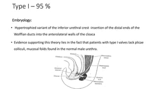
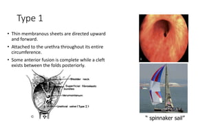
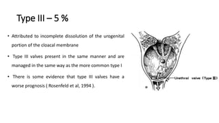
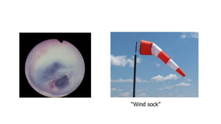
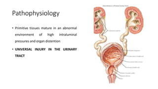
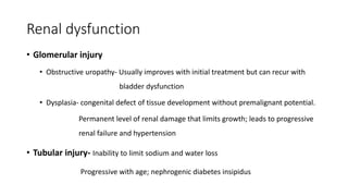
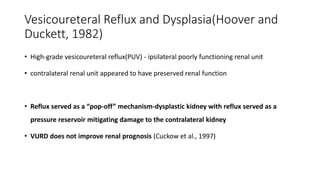
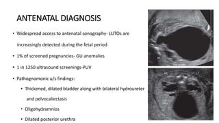
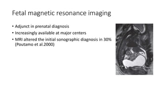
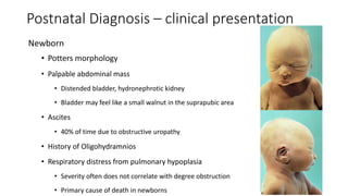
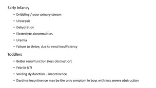
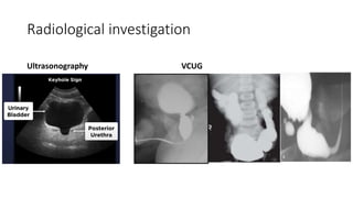
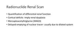
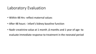
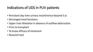
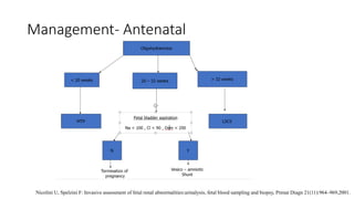
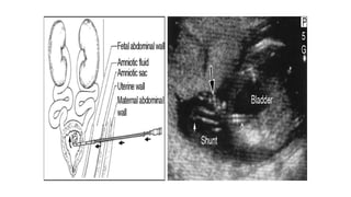
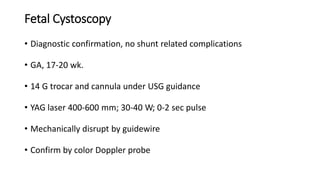
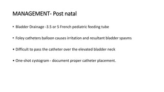
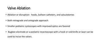
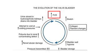
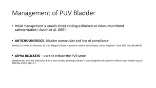
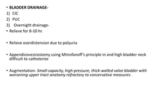
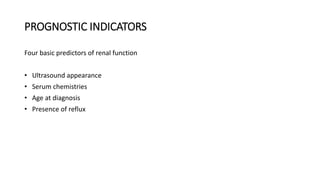
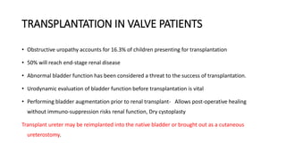
Posterior urethral valve
Dr Santosh K
Senior Resident
All India Institute of Medical Sciences
History
 Morgagni - 1769
 Langenbeck-1802
 Hugh Hampton Young- defined and named it posterior urethral valves*
 The term “valves” implies function- not true
 The obstructive membrane
 No active function
 Not a developmental stage in the embryologic development of the urethra
congenital obstruction of the posterior
urethra by valve-like leaflets
*Young HH, Frontz WA, Baldwin JC. J Urol. 2002 Jan;167(1):265-7
Prevalence
• 1 in 5,000 to 8,000 pregnancies and make up 10% of urinary obstructions diagnosed in utero
• 2.10 in 10,000 total births( live + stillbirths)1
• 1.28 per 10,000 live births2
• Prevalence in Indian population not known
1. Malin G, Tonks AM, Morris RK, et al: Congenital lower urinary tract obstruction: a population-based epidemiological study, BJOG 119(12):1455–
1464, 2012.
2. Thakkar D, Deshpande AV, Kennedy SE: Epidemiology and demography of recently diagnosed cases of posterior urethral valves, Pediatr Res
76(6):560– 563, 2014
CLASSIFICATION -Young
Type I – 95 %
Embryology:
• Hypertrophied variant of the inferior urethral crest -insertion of the distal ends of the
Wolffian ducts into the anterolateral walls of the cloaca
• Evidence supporting this theory lies in the fact that patients with type I valves lack plicae
colliculi, mucosal folds found in the normal male urethra.
Type 1
• Thin membranous sheets are directed upward
and forward.
• Attached to the urethra throughout its entire
circumference.
• Some anterior fusion is complete while a cleft
exists between the folds posteriorly.
“ spinnaker sail”
Pathology
• Rigid thick tissue / thin translucent membrane
• Fibrous stroma covered on each side by transitional epithelium
• There is no muscle
• The degree of obstruction varies
Type III – 5 %
• Attributed to incomplete dissolution of the urogenital
portion of the cloacal membrane
• Type III valves present in the same manner and are
managed in the same way as the more common type I
• There is some evidence that type III valves have a
worse prognosis ( Rosenfeld et al, 1994 ).
“Wind sock”
Pathophysiology
• Primitive tissues mature in an abnormal
environment of high intraluminal
pressures and organ distention
• UNIVERSAL INJURY IN THE URINARY
TRACT
Renal dysplasia
• Defined as a congenital defect of tissue development without premalignant potential
• Histological diagnosis
• Cause ?
• 1- high pelvic pressure during nephrogenesis.
• 2- primary embryologic abnormality from abnormal position of ureteric bud
Lower Urinary tract
• Storage and voiding of urine at high pressures
• Partial bladder outlet obstruction-Increased bladder compliance (Kirsch et al., 2003)
• More compliant bladders - more upper tract dilation
• Increased bladder dilation places the upper urinary tract at risk
Bladder dysfunction
• Last trimester- 30 cycles/24 hr
• If Obstruction; Decreases-
1) Total collagen
2) Collagen 3/1 ratio
3) Smooth muscles
• Attainment of Day & Night time continence (Smith etal)-
19%- 5yr; 46%-10 yr
Urinary bladder
• Deposition in the extracellular matrix
• Reduction in detrusor blood flow – ischemia
• Alteration of the phenotype of myosin bundles and actin filaments
• Poor sensation, hypercontractility, low compliance, and eventual myogenic failure
• Incontinence and poor emptying
• Bladder problems are lifelong and change with age and management
Bladder neck and Posterior Urethra
• Marked dilation of the posterior urethra
• Hypertrophy of the bladder neck, and
• Flattening of the verumontanum
• Dilation of ejaculatory ducts
• Changes are reversible and return to normal after valve ablation
Upper urinary tract
• Transmission of pressure to the ureter , renal pelvis and, ultimately, the glomerular units-
architectural and functional changes
ureteral dilation –Back pressure and Polyuria
• Ureteral wall thickening
• Loss of peristalsis
• Loss of mucosal coaptation
• Increasing the risk for urine stasis
• Infection
• Increased pressures in the renal units
Renal dysfunction
• Glomerular injury
• Obstructive uropathy- Usually improves with initial treatment but can recur with
bladder dysfunction
• Dysplasia- congenital defect of tissue development without premalignant potential.
Permanent level of renal damage that limits growth; leads to progressive
renal failure and hypertension
• Tubular injury- Inability to limit sodium and water loss
Progressive with age; nephrogenic diabetes insipidus
Vesicoureteral Reflux and Dysplasia(Hoover and
Duckett, 1982)
• High-grade vesicoureteral reflux(PUV) - ipsilateral poorly functioning renal unit
• contralateral renal unit appeared to have preserved renal function
• Reflux served as a “pop-off” mechanism-dysplastic kidney with reflux served as a
pressure reservoir mitigating damage to the contralateral kidney
• VURD does not improve renal prognosis (Cuckow et al., 1997)
ANTENATAL DIAGNOSIS
• Widespread access to antenatal sonography- LUTOs are
increasingly detected during the fetal period
• 1% of screened pregnancies- GU anomalies
• 1 in 1250 ultrasound screenings-PUV
• Pathognomonic u/s findings:
• Thickened, dilated bladder along with bilateral hydroureter
and pelvocaliectasis
• Oligohydramnios
• Dilated posterior urethra
Fetal magnetic resonance imaging
• Adjunct in prenatal diagnosis
• Increasingly available at major centers
• MRI altered the initial sonographic diagnosis in 30%
(Poutamo et al.2000)
Postnatal Diagnosis – clinical presentation
Newborn
• Potters morphology
• Palpable abdominal mass
• Distended bladder, hydronephrotic kidney
• Bladder may feel like a small walnut in the suprapubic area
• Ascites
• 40% of time due to obstructive uropathy
• History of Oligohydramnios
• Respiratory distress from pulmonary hypoplasia
• Severity often does not correlate with degree obstruction
• Primary cause of death in newborns
Early Infancy
• Dribbling / poor urinary stream
• Urosepsis
• Dehydration
• Electrolyte abnormalities
• Uremia
• Failure to thrive; due to renal insufficiency
Toddlers
• Better renal function (less obstruction)
• Febrile UTI
• Voiding dysfunction – incontinence
• Daytime incontinence may be the only symptom in boys with less severe obstruction
Radiological investigation
Ultrasonography VCUG
Radionuclide Renal Scan
• Quantification of differential renal function
• Cortical deficits -imply renal dysplasia
• Mercaptoacetyltriglycine (MAG3)
• Delayed emptying of nuclear tracer- usually due to dilated system
Laboratory Evaluation
• Within 48 Hrs- reflect maternal values
• After 48 hours - infant’s kidney baseline function
• Nadir creatinine value at 1 month ,6 months and 1 year of age- to
evaluate immediate response to treatment in the neonatal period
Indications of UDS in PUV patients
• Persistant day time urinary incontinence beyond 5 yr.
• Derranged renal functions
• Upper tract dilatation in absence of outflow obstruction.
• Prior to transplant
• To know efficacy of treatment
• Research tool
Management- Antenatal
Nicolini U, Spelzini F: Invasive assessment of fetal renal abnormalities:urinalysis, fetal blood sampling and biopsy, Prenat Diagn 21(11):964–969,2001.
Management- Antenatal
• Poor prognosis group – may offer termination because infants ultimately die of pulmonary
hypoplasia
• Fetuses with good prognosis –
Placement of vesicoamniotic shunt (double pigtail)
• Placement- midway b/w pubic ramus and umbilical cord
• Follow with serial u/s to confirm placement
• Complication rate of 21-59%
• Dislocation of the shunt -44%,
• Mortality :33% -43%
• Renal insufficiency -50%
Fetal Cystoscopy
• Diagnostic confirmation, no shunt related complications
• GA, 17-20 wk.
• 14 G trocar and cannula under USG guidance
• YAG laser 400-600 mm; 30-40 W; 0-2 sec pulse
• Mechanically disrupt by guidewire
• Confirm by color Doppler probe
MANAGEMENT- Post natal
• Bladder Drainage -3.5 or 5 French pediatric feeding tube
• Foley catheters balloon causes irritation and resultant bladder spasms
• Difficult to pass the catheter over the elevated bladder neck
• One-shot cystogram - document proper catheter placement.
Valve Ablation
• Ablation or disruption- hooks, balloon catheters, and valvulotomes
• Both retrograde and antegrade approach
• Smaller pediatric cystoscopes with improved optics are favored
• Bugbee electrode or a pediatric resectoscope with a hook or cold knife or laser can be
used to incise the valves.
7.5 F storz Bugbee
Cold knife Hook
Ablation
Incision
Valve Ablation
• Initially surgeons attempted to completely resect the valves
• Now-Goal is not to remove the valves
Incise them so that they are not suspended across the urethra
• 12-o'clock position
• 5- and 7-o'clock
• All three locations
• Valve remnants resolve after incision- no evidence of them on later cystoscopy
Post Valve ablation followup-
• VCUG- Repeated within 1 month
• 25% residual posterior urethral dilatation persists.
• Post ablative urethral ratio P/A 2.5 to 3 acceptable at 12 wk
• Reflux ceases spontaneously in 20-53%
• USG- If by 4-6 wk HDN persists/increases-
1) Continued reflux
2) Distal ureteral obstruction
3) Valve bladder syndrome
4) Incomplete valve ablation
5) Iatrogenic urethral stricture
• Radionuclides- Abnormal upper tracts/persistent HDN- 3 monthly
Cutaneous vesicostomy
• Infant is too small for safe instrumentation
• Adequate drainage of the upper tracts in more than 90% of cases
• Does not significantly affect bladder capacity
• Primary ablation is the preferred and vesicostomy is reserved for very small or
very ill infants
Upper Tract Diversion
• Indication - renal dilation and renal function fail to improve despite maximal bladder drainage
• VUJ obstruction - tortuous intramural ureter passing through a thickened bladder
• If the S.cr falls to < 2.0-continue bladder decompression
• If remains above 2.0 after 10 days of adequate bladder decompression and if hydronephrosis is
unimproved- consider upper tract diversion
A, Vesicostomy. B, Distal ureterostomy. C, Proximal loop
ureterostomy. D, Cutaneous pyelostomy. E, Ring
ureterostomy. F, Sober Y ureterostomy
Reversal
• Delayed until the bladder and upper tracts have improved as much as can be
expected
• Undivert the child at the age of 2 or 3 years
• Fully evaluate the bladder function before the need for transplantation
Other procedures
• Circumcision-
 Risk of UTI in children with PUV is 50% to 60%
 Circumcision reduces that risk by 83% to 92%- reduction to a level of risk
similar to that for unaffected boys
• Nephroureterectomy
 Frequent urinary tract infections localizing to the non functioning renal unit
Management of VUR
• Between 50% and 70%
• Reflux resolves after valve ablation in between 20% and 32% of refluxing ureters
• Inadequate emptying and high storage pressures are the usual causes of persistent reflux
• First step in evaluation - Repeat VCUG to look for persistent valve remnants
• If no remnants found-urodynamic study to evaluate the bladder
• Ureteroneocystostomy is usually limited to the situation in which infections cannot be
controlled and bladder function is adequate
Hydronephrosis
• Severe hydroureteronephrosis at diagnosis
• Scott (1985) found hydronephrosis in 96.5% of valve patients, and it was bilateral in 78%
• Hydronephrosis is a secondary - resolves once obstruction is relieved
• 25% of valve patients have dilated upper tracts 5 to 15 years after successful valve
ablation
• Majority of patients with persistent hydronephrosis do not have obstruction
VALVE BLADDER SYNDROME
• valve bladder and full valve bladder were coined by Mitchell,1982
• Chronic condition in patients with valves in which, despite successful valve ablation,
intrinsic bladder dysfunction leads to deterioration of the upper urinary tracts and
incontinence
• Poor sensation, high bladder volumes, and poor compliance produces storage pressures
high enough to prevent adequate drainage of the upper tracts
• Video urodynamics are necessary to diagnose this condition
Management of PUV Bladder
• Initial management is usually timed voiding,α blockers or clean intermittent
catheterization ( Austin et al, 1999 )
• ANTICHOLINERGICS- Bladder overactivity and loss of compliance
(Misseri, R, Combs, AJ, Horowitz, M, et al. Myogenic failure in posterior urethral valve disease: real or imagined? J Urol 2002 Oct;168:1844-8)
• AlPHA BLOCKERS – used to reduce the PVR urine
(Abraham MK, Nasir AR, Sudarsanan B, et al. Role of alpha adrenergic blocker in the management of posterior urethral valves. Pediatr Surg Int
2009 Dec;25(12):1113-5.)
• BLADDER DRAINAGE-
1) CIC
2) PUC
3) Overnight drainage-
• Relieve for 8-10 hr.
• Relieve overdistension due to polyuria
• Appendicovesicostomy using Mitrofanoff’s principle in and high bladder neck
difficult to catheterize
• Augmentation- Small-capacity, high-pressure, thick-walled valve bladder with
worsening upper tract anatomy refractory to conservative measures.
PROGNOSTIC INDICATORS
Four basic predictors of renal function
• Ultrasound appearance
• Serum chemistries
• Age at diagnosis
• Presence of reflux
USG
• Method of estimating the amount of dysplasia
• Appears more echogenic on ultrasound
• Kidneys that are considerably brighter than the liver usually contain significant
dysplasia
• Loss of the normal corticomedullary diffrentiation correlates with poor long-term
renal function
Age
• Infants presenting before 1 year of age had a worse prognosis than those who
present later in life
• Older boys considered to have better prognosis
• Boys who presented after 5 years of age revealed that 35% had renal insufficiency
and 10% reached end-stage renal disease
Reflux
• Reflux in valve patients carries a poor prognosis
• Mortality 57% in patients with bilateral reflux, 17% with unilateral reflux, and 9%
with no reflux.
• The incompetent ureterovesical junction - transmits the high pressures generated
by the bladder
Serum Chemistries
• Nadir creatinine of 0.8 mg/dL or less during the first year of life -good predictor of
long-term renal function
TRANSPLANTATION IN VALVE PATIENTS
• Obstructive uropathy accounts for 16.3% of children presenting for transplantation
• 50% will reach end-stage renal disease
• Abnormal bladder function has been considered a threat to the success of transplantation.
• Urodynamic evaluation of bladder function before transplantation is vital
• Performing bladder augmentation prior to renal transplant- Allows post-operative healing
without immuno-suppression risks renal function, Dry cystoplasty
Transplant ureter may be reimplanted into the native bladder or brought out as a cutaneous
ureterostomy.
Posterior urethral valve

Posterior urethral valve

  • 1.
    Posterior urethral valve DrSantosh K Senior Resident All India Institute of Medical Sciences
  • 2.
    History  Morgagni -1769  Langenbeck-1802  Hugh Hampton Young- defined and named it posterior urethral valves*  The term “valves” implies function- not true  The obstructive membrane  No active function  Not a developmental stage in the embryologic development of the urethra congenital obstruction of the posterior urethra by valve-like leaflets *Young HH, Frontz WA, Baldwin JC. J Urol. 2002 Jan;167(1):265-7
  • 3.
    Prevalence • 1 in5,000 to 8,000 pregnancies and make up 10% of urinary obstructions diagnosed in utero • 2.10 in 10,000 total births( live + stillbirths)1 • 1.28 per 10,000 live births2 • Prevalence in Indian population not known 1. Malin G, Tonks AM, Morris RK, et al: Congenital lower urinary tract obstruction: a population-based epidemiological study, BJOG 119(12):1455– 1464, 2012. 2. Thakkar D, Deshpande AV, Kennedy SE: Epidemiology and demography of recently diagnosed cases of posterior urethral valves, Pediatr Res 76(6):560– 563, 2014
  • 4.
  • 5.
    Type I –95 % Embryology: • Hypertrophied variant of the inferior urethral crest -insertion of the distal ends of the Wolffian ducts into the anterolateral walls of the cloaca • Evidence supporting this theory lies in the fact that patients with type I valves lack plicae colliculi, mucosal folds found in the normal male urethra.
  • 6.
    Type 1 • Thinmembranous sheets are directed upward and forward. • Attached to the urethra throughout its entire circumference. • Some anterior fusion is complete while a cleft exists between the folds posteriorly. “ spinnaker sail”
  • 7.
    Pathology • Rigid thicktissue / thin translucent membrane • Fibrous stroma covered on each side by transitional epithelium • There is no muscle • The degree of obstruction varies
  • 8.
    Type III –5 % • Attributed to incomplete dissolution of the urogenital portion of the cloacal membrane • Type III valves present in the same manner and are managed in the same way as the more common type I • There is some evidence that type III valves have a worse prognosis ( Rosenfeld et al, 1994 ).
  • 9.
  • 10.
    Pathophysiology • Primitive tissuesmature in an abnormal environment of high intraluminal pressures and organ distention • UNIVERSAL INJURY IN THE URINARY TRACT
  • 11.
    Renal dysplasia • Definedas a congenital defect of tissue development without premalignant potential • Histological diagnosis • Cause ? • 1- high pelvic pressure during nephrogenesis. • 2- primary embryologic abnormality from abnormal position of ureteric bud
  • 12.
    Lower Urinary tract •Storage and voiding of urine at high pressures • Partial bladder outlet obstruction-Increased bladder compliance (Kirsch et al., 2003) • More compliant bladders - more upper tract dilation • Increased bladder dilation places the upper urinary tract at risk
  • 13.
    Bladder dysfunction • Lasttrimester- 30 cycles/24 hr • If Obstruction; Decreases- 1) Total collagen 2) Collagen 3/1 ratio 3) Smooth muscles • Attainment of Day & Night time continence (Smith etal)- 19%- 5yr; 46%-10 yr
  • 14.
    Urinary bladder • Depositionin the extracellular matrix • Reduction in detrusor blood flow – ischemia • Alteration of the phenotype of myosin bundles and actin filaments • Poor sensation, hypercontractility, low compliance, and eventual myogenic failure • Incontinence and poor emptying • Bladder problems are lifelong and change with age and management
  • 15.
    Bladder neck andPosterior Urethra • Marked dilation of the posterior urethra • Hypertrophy of the bladder neck, and • Flattening of the verumontanum • Dilation of ejaculatory ducts • Changes are reversible and return to normal after valve ablation
  • 16.
    Upper urinary tract •Transmission of pressure to the ureter , renal pelvis and, ultimately, the glomerular units- architectural and functional changes ureteral dilation –Back pressure and Polyuria • Ureteral wall thickening • Loss of peristalsis • Loss of mucosal coaptation • Increasing the risk for urine stasis • Infection • Increased pressures in the renal units
  • 17.
    Renal dysfunction • Glomerularinjury • Obstructive uropathy- Usually improves with initial treatment but can recur with bladder dysfunction • Dysplasia- congenital defect of tissue development without premalignant potential. Permanent level of renal damage that limits growth; leads to progressive renal failure and hypertension • Tubular injury- Inability to limit sodium and water loss Progressive with age; nephrogenic diabetes insipidus
  • 18.
    Vesicoureteral Reflux andDysplasia(Hoover and Duckett, 1982) • High-grade vesicoureteral reflux(PUV) - ipsilateral poorly functioning renal unit • contralateral renal unit appeared to have preserved renal function • Reflux served as a “pop-off” mechanism-dysplastic kidney with reflux served as a pressure reservoir mitigating damage to the contralateral kidney • VURD does not improve renal prognosis (Cuckow et al., 1997)
  • 19.
    ANTENATAL DIAGNOSIS • Widespreadaccess to antenatal sonography- LUTOs are increasingly detected during the fetal period • 1% of screened pregnancies- GU anomalies • 1 in 1250 ultrasound screenings-PUV • Pathognomonic u/s findings: • Thickened, dilated bladder along with bilateral hydroureter and pelvocaliectasis • Oligohydramnios • Dilated posterior urethra
  • 20.
    Fetal magnetic resonanceimaging • Adjunct in prenatal diagnosis • Increasingly available at major centers • MRI altered the initial sonographic diagnosis in 30% (Poutamo et al.2000)
  • 21.
    Postnatal Diagnosis –clinical presentation Newborn • Potters morphology • Palpable abdominal mass • Distended bladder, hydronephrotic kidney • Bladder may feel like a small walnut in the suprapubic area • Ascites • 40% of time due to obstructive uropathy • History of Oligohydramnios • Respiratory distress from pulmonary hypoplasia • Severity often does not correlate with degree obstruction • Primary cause of death in newborns
  • 22.
    Early Infancy • Dribbling/ poor urinary stream • Urosepsis • Dehydration • Electrolyte abnormalities • Uremia • Failure to thrive; due to renal insufficiency Toddlers • Better renal function (less obstruction) • Febrile UTI • Voiding dysfunction – incontinence • Daytime incontinence may be the only symptom in boys with less severe obstruction
  • 23.
  • 24.
    Radionuclide Renal Scan •Quantification of differential renal function • Cortical deficits -imply renal dysplasia • Mercaptoacetyltriglycine (MAG3) • Delayed emptying of nuclear tracer- usually due to dilated system
  • 25.
    Laboratory Evaluation • Within48 Hrs- reflect maternal values • After 48 hours - infant’s kidney baseline function • Nadir creatinine value at 1 month ,6 months and 1 year of age- to evaluate immediate response to treatment in the neonatal period
  • 26.
    Indications of UDSin PUV patients • Persistant day time urinary incontinence beyond 5 yr. • Derranged renal functions • Upper tract dilatation in absence of outflow obstruction. • Prior to transplant • To know efficacy of treatment • Research tool
  • 27.
    Management- Antenatal Nicolini U,Spelzini F: Invasive assessment of fetal renal abnormalities:urinalysis, fetal blood sampling and biopsy, Prenat Diagn 21(11):964–969,2001.
  • 28.
    Management- Antenatal • Poorprognosis group – may offer termination because infants ultimately die of pulmonary hypoplasia • Fetuses with good prognosis – Placement of vesicoamniotic shunt (double pigtail) • Placement- midway b/w pubic ramus and umbilical cord • Follow with serial u/s to confirm placement • Complication rate of 21-59% • Dislocation of the shunt -44%, • Mortality :33% -43% • Renal insufficiency -50%
  • 30.
    Fetal Cystoscopy • Diagnosticconfirmation, no shunt related complications • GA, 17-20 wk. • 14 G trocar and cannula under USG guidance • YAG laser 400-600 mm; 30-40 W; 0-2 sec pulse • Mechanically disrupt by guidewire • Confirm by color Doppler probe
  • 31.
    MANAGEMENT- Post natal •Bladder Drainage -3.5 or 5 French pediatric feeding tube • Foley catheters balloon causes irritation and resultant bladder spasms • Difficult to pass the catheter over the elevated bladder neck • One-shot cystogram - document proper catheter placement.
  • 32.
    Valve Ablation • Ablationor disruption- hooks, balloon catheters, and valvulotomes • Both retrograde and antegrade approach • Smaller pediatric cystoscopes with improved optics are favored • Bugbee electrode or a pediatric resectoscope with a hook or cold knife or laser can be used to incise the valves.
  • 33.
    7.5 F storzBugbee Cold knife Hook Ablation Incision
  • 35.
    Valve Ablation • Initiallysurgeons attempted to completely resect the valves • Now-Goal is not to remove the valves Incise them so that they are not suspended across the urethra • 12-o'clock position • 5- and 7-o'clock • All three locations • Valve remnants resolve after incision- no evidence of them on later cystoscopy
  • 36.
    Post Valve ablationfollowup- • VCUG- Repeated within 1 month • 25% residual posterior urethral dilatation persists. • Post ablative urethral ratio P/A 2.5 to 3 acceptable at 12 wk • Reflux ceases spontaneously in 20-53% • USG- If by 4-6 wk HDN persists/increases- 1) Continued reflux 2) Distal ureteral obstruction 3) Valve bladder syndrome 4) Incomplete valve ablation 5) Iatrogenic urethral stricture • Radionuclides- Abnormal upper tracts/persistent HDN- 3 monthly
  • 37.
    Cutaneous vesicostomy • Infantis too small for safe instrumentation • Adequate drainage of the upper tracts in more than 90% of cases • Does not significantly affect bladder capacity • Primary ablation is the preferred and vesicostomy is reserved for very small or very ill infants
  • 38.
    Upper Tract Diversion •Indication - renal dilation and renal function fail to improve despite maximal bladder drainage • VUJ obstruction - tortuous intramural ureter passing through a thickened bladder • If the S.cr falls to < 2.0-continue bladder decompression • If remains above 2.0 after 10 days of adequate bladder decompression and if hydronephrosis is unimproved- consider upper tract diversion
  • 39.
    A, Vesicostomy. B,Distal ureterostomy. C, Proximal loop ureterostomy. D, Cutaneous pyelostomy. E, Ring ureterostomy. F, Sober Y ureterostomy
  • 40.
    Reversal • Delayed untilthe bladder and upper tracts have improved as much as can be expected • Undivert the child at the age of 2 or 3 years • Fully evaluate the bladder function before the need for transplantation
  • 41.
    Other procedures • Circumcision- Risk of UTI in children with PUV is 50% to 60%  Circumcision reduces that risk by 83% to 92%- reduction to a level of risk similar to that for unaffected boys • Nephroureterectomy  Frequent urinary tract infections localizing to the non functioning renal unit
  • 42.
    Management of VUR •Between 50% and 70% • Reflux resolves after valve ablation in between 20% and 32% of refluxing ureters • Inadequate emptying and high storage pressures are the usual causes of persistent reflux • First step in evaluation - Repeat VCUG to look for persistent valve remnants • If no remnants found-urodynamic study to evaluate the bladder • Ureteroneocystostomy is usually limited to the situation in which infections cannot be controlled and bladder function is adequate
  • 43.
    Hydronephrosis • Severe hydroureteronephrosisat diagnosis • Scott (1985) found hydronephrosis in 96.5% of valve patients, and it was bilateral in 78% • Hydronephrosis is a secondary - resolves once obstruction is relieved • 25% of valve patients have dilated upper tracts 5 to 15 years after successful valve ablation • Majority of patients with persistent hydronephrosis do not have obstruction
  • 44.
    VALVE BLADDER SYNDROME •valve bladder and full valve bladder were coined by Mitchell,1982 • Chronic condition in patients with valves in which, despite successful valve ablation, intrinsic bladder dysfunction leads to deterioration of the upper urinary tracts and incontinence • Poor sensation, high bladder volumes, and poor compliance produces storage pressures high enough to prevent adequate drainage of the upper tracts • Video urodynamics are necessary to diagnose this condition
  • 46.
    Management of PUVBladder • Initial management is usually timed voiding,α blockers or clean intermittent catheterization ( Austin et al, 1999 ) • ANTICHOLINERGICS- Bladder overactivity and loss of compliance (Misseri, R, Combs, AJ, Horowitz, M, et al. Myogenic failure in posterior urethral valve disease: real or imagined? J Urol 2002 Oct;168:1844-8) • AlPHA BLOCKERS – used to reduce the PVR urine (Abraham MK, Nasir AR, Sudarsanan B, et al. Role of alpha adrenergic blocker in the management of posterior urethral valves. Pediatr Surg Int 2009 Dec;25(12):1113-5.)
  • 47.
    • BLADDER DRAINAGE- 1)CIC 2) PUC 3) Overnight drainage- • Relieve for 8-10 hr. • Relieve overdistension due to polyuria • Appendicovesicostomy using Mitrofanoff’s principle in and high bladder neck difficult to catheterize • Augmentation- Small-capacity, high-pressure, thick-walled valve bladder with worsening upper tract anatomy refractory to conservative measures.
  • 48.
    PROGNOSTIC INDICATORS Four basicpredictors of renal function • Ultrasound appearance • Serum chemistries • Age at diagnosis • Presence of reflux
  • 49.
    USG • Method ofestimating the amount of dysplasia • Appears more echogenic on ultrasound • Kidneys that are considerably brighter than the liver usually contain significant dysplasia • Loss of the normal corticomedullary diffrentiation correlates with poor long-term renal function
  • 50.
    Age • Infants presentingbefore 1 year of age had a worse prognosis than those who present later in life • Older boys considered to have better prognosis • Boys who presented after 5 years of age revealed that 35% had renal insufficiency and 10% reached end-stage renal disease
  • 51.
    Reflux • Reflux invalve patients carries a poor prognosis • Mortality 57% in patients with bilateral reflux, 17% with unilateral reflux, and 9% with no reflux. • The incompetent ureterovesical junction - transmits the high pressures generated by the bladder
  • 52.
    Serum Chemistries • Nadircreatinine of 0.8 mg/dL or less during the first year of life -good predictor of long-term renal function
  • 53.
    TRANSPLANTATION IN VALVEPATIENTS • Obstructive uropathy accounts for 16.3% of children presenting for transplantation • 50% will reach end-stage renal disease • Abnormal bladder function has been considered a threat to the success of transplantation. • Urodynamic evaluation of bladder function before transplantation is vital • Performing bladder augmentation prior to renal transplant- Allows post-operative healing without immuno-suppression risks renal function, Dry cystoplasty Transplant ureter may be reimplanted into the native bladder or brought out as a cutaneous ureterostomy.